anatomy exam 2
 Call Kai
 Learn
 Practice Test
 Spaced Repetition
 Match
 Flashcards
 Knowt Play
1/123

There's no tags or description
Looks like no tags are added yet.
124 Terms
function of the skeletal system
support→ body and soft organs
protection → brain, spinal cord, and vital organs
movement → levers for muscle action
mineral storage
blood cell formation
triglyceride (fat) storage
hormone production
206 bones
minerals of bones
calcium & phosphorus → hydroxyapatite
Ca2 (PO4 )3 → Ca₁₀(PO₄)₆(OH)₂
blood cell formation
hematopoiesis in red marrow cavities of certain bones
hormone production in bones
Osteocalcin → secreted by osteoblasts
regulates bone formation
protects against obesity, glucose intolerance, diabetes mellitus
Leydig cells(stimulates testosterone) and pancreatic cells
all skeletal cartilages contain
chondrocytes in lacunae and extracellular matrix
axial skeleton
long axis of body
skull and vertebral column, rib cage
appendicular skeleton
bones of upper and lower limbs
girdles attaching limbs to axial skeleton'
pelvic girdles and pectoral gridles
long bones
longer than they are wide
function: leverage
→ scapula
→ clavicle
short bones
cube shaped bone, equal in length& width & thickness
functions: provide stability, support, while allowing for motion
→tarsals or wrist
outer & inner region of compact bone, in between is spongy bone. The outer layer of compact bone is covered in thin periosteum, the inside spongy bone is covered in endosteum.
sesamoid bones
small and round embedded in tendons
functions: protect tendons from compressive forces
→ patellae, pisiform, first metatarsal
irregular bones
complex shape
functions: protect internal organs
→ vertebrae, facial bones
outer & inner region of compact bone, in between is spongy bone. The outer layer of compact bone is covered in thin periosteum, the inside spongy bone is covered in endosteum.
flat bones
thin and curved
functions: points of attachment for muscles, protectors of internal organs
→ sternum, ribs, scapulae, cranial bones
outer & inner region of compact bone, in between is spongy bone. The outer layer of compact bone is covered in thin periosteum, the inside spongy bone is covered in endosteum.
compact bone(lamellar bone) characteristics
→ dense outer layer, smooth and solid
→osteocytes are in concentric layers around a Haversian canal(located in osteon)
→ lamella in concentric layers
→ bears weight
→ lamellaeL hollo tubes of bone matrix
collagen fibers in adjacent rings run in different directions (withstands stress)
hyaline cartilage cover articular surfaces
spongy bone
→ lamella not in osteons (no osteons)
→ honeycomb of flat pieces of bone = trabeculae
→ red bone marrow for blood cell formation in between holes
→ yellow marrow for adipose/ energy reserve in between holes
→ capillaries in endosteum supply nutrients
→ not as thick/dense as its a location without stress, makes the bone lighter

structure of long bones (ex. femur, tibia, radius)
Diaphysis
tubular shaft forms long axis
compact bones surrounding medullary cavity (filled with) yellow bone marrow
Epiphyses
bone ends
external compact bone, internal spongy bone
articular cartilage covers articular surfaces
between an epiphyseal line: remnant if childhood bone growth at epiphyseal plate
Metaphysis
epiphyseal line also known as growth( epiphyseal) plate, thicker in childhood, turns to line after puberty, used to track growth
→ made of compact bone

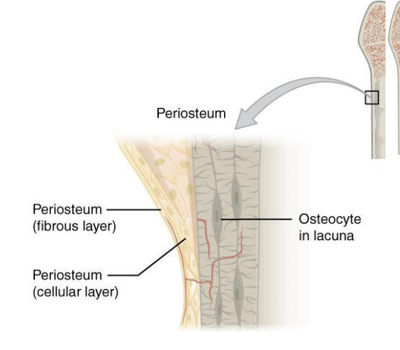
Periosteum (membrane)
Where: covers external surfaces except joint tissues
white, double layer
Made of: outer fibrous layer of dense irregular connective tissue. Cellular layer or (osteogenic layer) : primitive stem cells- osteogenic cells and osteoblasts
function: anchoring points for tendons and ligaments

think of osteoblasts..
helping to build bone
endosteum
Where: delicate connective tissue membranes covering internal bone surface
Made of: osteogenic cells that can differentiate into other bone cells, osteoclasts, osteoblasts, bone lining cells
function: lines canals that pass through compact bone, covers trabeculae of spongy bone

red bone marrow
found in trabecular cavities of spongy bone and diploe of flat bones
in medullary cavities and spongy bone of newborns
adult long bones have little red marrow (heads of femur and humerus only )
yellow marrow can convert to red if necessary
(intro to bone markings) process
→ general description: elevations or projections
any projection or bump
(intro to bone markings) ramus
→ general description: elevations or projections
an extension of a bone making an angle with the rest if the sturcture
(intro to bone markings) trochanter
→ general description: process formed where tendons or ligaments attach
a large, rough projection
(intro to bone markings) Tuberosity
→ general description: process formed where tendons or ligaments attach
a smaller , rough projection
(intro to bone markings) Tubercle
→ general description: process formed where tendons or ligaments attach
a small , rounded projection
(intro to bone markings) crest
→ general description: process formed where tendons or ligaments attach
a prominent ridge
(intro to bone markings) line
→ general description: process formed where tendons or ligaments attach
a low ridge
(intro to bone markings) spine
→ general description: process formed where tendons or ligaments attach
a pointed or narrow process
(intro to bone markings) head
→ general description: process formed for articulation with adjacent bones
the expanded articular end of an epiphysis, separated from the shaft by a neck
(intro to bone markings) neck
→ general description: process formed for articulation with adjacent bones
a narrow connection between the epiphysis and the diaphysis
(intro to bone markings) condyle
→ general description: process formed for articulation with adjacent bones
a smooth, rounded articular process
(intro to bone markings) trochlea
→ general description: process formed for articulation with adjacent bones
a smooth, grooved articular process shaped like a pulley
(intro to bone markings) facet
→ general description: process formed for articulation with adjacent bones
a small, flat articular surface
(intro to bone markings) fossa
→ general description: depressions
a shallow depression
(intro to bone markings) sulcus
→ general description: depressions
a narrow groove
(intro to bone markings) foramen
→ general description: openings
a rounded passageway for blood vessels or nerves
(intro to bone markings) canal
→ general description: openings
a duct or channel
(intro to bone markings) meatus
→ general description: openings
a passageway through a bone
(intro to bone markings) fissure
→ general description: openings
an elongated cleft or slit
(intro to bone markings) sinus
→ general description: openings
a chamber within a bone, normally filled with air
(major bone cell types) osteogenic cells: also called osteoprogenitor cells
precursor cells→ undifferentiated and develop into osteoblasts
mitotically active stem cells in periosteum and endosteum
when stimulated differentiate unto osteoblasts or bone lining cells
(major bone cell types) osteocytes
mature bone cells ( mature osteoblast) located in lacunae surrounded by higher levels of bony matrix
respond to and communicate mechanical stimuli to osteoblasts and osteoclasts for bone remodeling
(major bone cell types) osteoclasts
break down bone
→ osteoclast-inducing cytokines convert monocytes and macrophages to osteoclasts
appearance: large, multi nucleated cells for bone resorption
at the site of active bone reabsorption, the osteoclast forms specialized cell membrane that secrete acid and collgaenase

(major bone cell types) osteoblasts
bone forming cells
→ secrete unmineralized bone matrix or osteoid
includes collagen(90%) of bone and calcium -binding protiens
(major bone cell types) bone lining cells
→ flat cells on bone surfaces believed to help maintain matrix
on external bone surface called: periosteal cells
lining internal surfaces called: endosteal
differentiate into osteoblasts, help maintain outer layer of compact bone
canaliculi?
hairlike canals that connect lacunae to each other and the central canal

canaliculi formation
osteoblasts secreting bone matrix maintain contact with each other and osteocytes via cell projections with gap junctions
when the matrix hardens and cells are trapped the canaliculi forms allowing for→ communication b, permit nutrients and waste to be relayed from one osteocyte to another throughout osteon
osteoid (chemical composition of bones)
organic bone matrix ( collagen and other non collagen proteins) secreted by osteoblasts.
Function: contributes to tensile strength and flexibilty
→ unmineralized organic portion of the bone matrix that forms prior to the maturation of bone tissue
Made up of:
ground substance (proteoglycans and glycoprotiens)
collagen fibers, osteocalcin
→ when osteoid become mineralized, it and adjacent bones cells develop into new bone tissue
hydroxyapatite- mineral salts (chemical composition of bones)
→ 65% of bone by mass
→ mainly tiny calcium phosphate crystals in and around collagen fibers
→ harness and resistance to compression
ossification [ osteogenesis] - bone development
process of bone tissue formation
→ formation of bony skeleton : begins in 2nd month of development
→ postnatal bone growth : until early childhood
→ bone remodeling and repair: lifelong
endochondral & intramembranous ossification
endochondral
→bone develops from replacing hyaline cartilage
→bones called cartilage ( endochondral ) bones
→ forms most of the skeleton inferior to base of skull
→ begins late in 2nd month of devolpment
→ breakdown of hyaline cartilage need prior to ossifcation
intramembranous
→bone develops from fibrous membrane
→ bones called membrane bones
→ forms flat bones ex. clavicles and crainal bones
→ begins in utero during fetal development, last bones to undergo intramembranous ossification are the bones of the face
intramembranous ossification method
forms from fibrous membrane: forms frontal, parietal, occipital, temporal bones and clavicles
1.This is the first type of ossification that occurs. Where bone is formed from a fibrous
membrane. The process begins before week 8. This includes cranial bones and the
clavicle.
2. Mesenchymal stem cells aggregate and differentiate into osteoblasts. the osteoblasts will group together to form ossification centers., the starting point for bone formation. Osteoblasts begin secreting osteoid inward, towards the ossification center. After a few days you get the binding of calcium to osteoid results in the hardening of the matrix into form bone.
3. Osteoblasts become trapped by the osteoid they secrete, transforming/differentiate into osteocytes which are involved in maintaining the bony matrix.
4. As osteoid continues to be secreted by osteoblasts, it surrounds blood vessels, forming trabecular/cancellous/spongy bone. These vessels will eventually form red bone marrow, which is where all blood cells are formed.
5. Mesenchymal cells on the outer surface of the bone come together to and change into the periosteum. Cells on the inner surface of the periosteum differentiate into osteoblasts and secrete osteoid parallel to that of the existing matrix, thus forming layers. These layers differentiate into compact bone.
endochondral ossification method
Mesenchymal cells differentiate into chondrocytes → hyaline cartilage
The perichondrium becomes vascularized → blood vessels → brings nutrients to the area → nutrients stimulate mesenchymal cells to become → osteoblasts → osteoblasts line the diaphysis wall → lay down superficial layer of bone → periosteal (bony) collar develops. This happens in the primary ossification center.
Chondrocytes that remain in the central cavity enlarge, sending a signal to the cartilage to calcify.
Bony collar → impermeability to the developing bone → cells in the area not receiving nutrients → chondrocytes cell death → central clearing → remodeling to form medullary cavity.
Cartilage and chondrocytes at distal end of bone continue to grow → bone elongation → blood vessels grow into the epiphysis.
interstitial growth
→ growth in length of long bones
requires presence of epiphyseal cartilage
five zones within cartilage
resting/reserve (quiescent) zone : relatively inactive
proliferation (growth) zone: rapidly divides pushing away epiphysis from diaphysis (lengthening)
hypertrophic zone : older chondrocytes closer to diaphysis and their lacune enlarge (interconnecting spaces)
calcification zone : surrounding cartilage matrix calcifies, chondrocytes die and deteriorate
ossification (osteogenic) zone : capillaries and osteoblasts from diaphysis move into zone. osteoblast secret bone tissue on the remaining calcified cartilage
appositional growth
→ growth in width, allows lengthening bones to widen
occurs throughout life
osteoblasts beneath periosteum secrete bon matrix on external bone
osteoclasts remove bone on endosteal surface
growth hormone (hormonal regulation of bone growth)
most important in stimulating epiphyseal plate activity in infancy and childhood→ ILG1 (insulin like growth factor 1)
triggers chondrocyte profliferation at epiphyseal plate, increase calcium retention
thyroid hormone (hormonal regulation of bone growth)
modulates activity of growth hormone: regulates bone turnover and mineralization in adults
thyrotoxicosis is an established risk factor for osteoporosis → imbalance in the osteoblast and osteoclast activity
estrogen (females)
estrogen stimulates the release of osteoprotergerin from osteoblasts which promotes osteoblast activity by increasing OPG. Also supports growth spurt.
in menopausal women: when estrogen levels drop, there is a decrease in OPG, more microphages bind to rank ligand, increasing activity of oestoclasts
PTH (para thyroid hormone)
simulates osteoclasts to break down bone
testosterone (males)
promotes osteoblast activity by promoting osteoblast activity by increasing the release of OPG, supporting growth spurts
excess glucocorticoid
on steroids for a long period of time to suppress immune system after things such as transplantation
decreases osteoprotegerin from the osteoblasts, increases rank ligand expression on osteoblasts, this activates osteoclasts promoting bone breakdown
bone cells that are enclosed in bone matrix are called
osteocytes
unmineralized organic matrix secreted by osteoblasts
osteoid
what does the unmineralized matrix (osteiod) contain
collagen, calcium, binding protein, ground substance
peptide hormone that stimulates formation of osteoclasts
proteoglycans, glycoprotiens, osteocalcin, PTH
what type of cell proliferates to give rise to osteoclasts
macrophages or osteoprogenitor cells
protein secreted from osteoblats that bind RANkL and prevents activation of osteoclasts
osteoprotegerin
growth hormone deficiency → IGF-1
idiopathic GH deficiency
deficient in hormone that stimulates release of growth hormone → treatment: growth hormone
pituitary tumors, agenesis (underdevelopment ) of pituitary
lack growth hormone → treatment: growth hormone
Laron- type dwarfisum
abnormal growth hormone receptors in the liver → treatment: ILG-1
gigantism (growth hormone disorder before puberty)
happens before epiphyseal plate fuses
→ due to excessive amount of growth hormone
cause: excessive amount of skeletal growth
acromegaly (growth hormone disorder after puberty)
happened after fusion epiphyseal plate to line
→ exaggerated growth of the ends of extremities
you can have gigantism → acromegaly but not acromegaly→ gigantism
(usually caused by pituitary adnomas)
what is responsible for growth
ILG-1
osteoclast remove bony matrix by
secreting acid and colagenase
regulation of bone turnover is in response to your
calcium levels in blood
parathyroid hormone
→ produced by parathyroid glands
→ removes calcium (Ca2+) from bone regardless of bone integrity
parathyroid hormones release if calcium ions are low, parathyroid hormone will bind to RANKL which activates and expresses more RANKL on the surface by binding to osteoblast receptors, increasing activity of osteoclasts → releasing ionized calcium from bone
negative feedback hormonal loop for blood Ca2+ homeostasis
controlled by parathyroid hormone (PTH) → decrease in blood Ca2+ levels → PTH release → PTH simulates osteoclasts to degrade bone matrix, releasing Ca2+ → increase blood Ca2+ levels → PTH release ends
bone response to mechanical stress
long bones thickest midway along diaphysis where bending stresses greatest
ex, in tennis players, bones are thicker in arm used to hit the racket
fracture classification
→ position of bone ends after fracture
non displaced: end retain normal position
displaced: ends out of normal alignment
→ completeness of break
complete: broken all the way through
incomplete: not broken all the way through
→ wether skin is penetrated
open (compound): skin is penetrated
closed (simple) :skin is not penetrated
what much cc of blood can you lose in a humerus, unilateral tibia/fibula, femur break?
humerus: 200-500cc
unilateral tibia/fibula: 400cc
femur: 1000-1500cc
colles fracture
break in distal radius in the wrist (think when you extend you arm to break a fall)
potts fractures
lower fibula due to excessive stress on ankle (stepping down)
smith fracture
a break in the lower end of the radius bone near the wrist, where the broken bone fragment shifts toward the palm side (volar side) of the hand
6 Ps of fracture complications
1.Pain
2.Pallor: disruption of blood flow
3. pulselessness: lost or reduced pulse
paresthesia: tingleyness (ex.when you hit your funny bone)
paralysis: loss of voluntary function
poikilothermia: change in temperature above (warmer) and below fracture site (cooler)
fracture treatment
if fracture is displaced & closed → external manipulation (closed reduction) to try and realign bone
if external manipulation fails( & in open fractures) then open reduction is done meaning surgery
immobilization w/ cast
5 stages of bone reapair
tissue deconstruction/ clean up and hematoma formation (blood clot)
inflammatiomn→ 1 week
soft callus formation → 2-3 weeks
hard callus formation → 4-12 weeks
remodeling → months to years
bone repair
Hematoma: Serves as the basis for a fibrin network into which granulation tissue grows.
New blood vessels extend into this tissue, and it is invaded by:
Phagocytic cells: Remove debris.
Mesenchymal cells: Differentiate into chondrocytes → produce new collagen fibers → fibrocartilage.
Osteoblasts: From periosteum and endosteum migrate into the area of broken bone.
Fibers of fibrocartilage as well as osteoid → soft callus (woven bone) → soft callus.
Woven bone (immature bone) is characterized by haphazard organization of collagen fibers and is mechanically weak → osteoblasts produce osteoid quickly.
Two ends of bone become splinted together: structure not strong enough to bear weight → preliminary bridge.
3) Ossification of fibrocartilage and osteoid by osteoblasts, and you begin to generate new bone to fill in the gaps and a bony callus is formed → this is new bone, not scar tissue → Hard callus.
4) Bone is remodeled in response to mechanical stress on the bone → mesh network of bone being formed, not in a regular pattern → cross-linked bone → increased strength.
RANK L
RANK ligand is produced mainly by osteoblast-lineage cells, including osteocytes. It signals that osteoclasts should develop and become active.
RANK
Found on osteoclast precursors and osteoclasts. When RANKL binds to RANK, it promotes osteoclast formation, survival, and activity.
OPG (osteoprotegerin)
Produced by osteoblast-lineage cells. It binds to RANKL so RANKL cannot bind to RANK, decreasing osteoclast formation and bone resorption.
how is bone reabsorption increased?
RANKL released by bone-forming cells →
Binds to RANK on osteoclast precursors →
More osteoclast formation and activation →
Increased bone resorption
what if you want to block the production of osteoclasts formation
OPG binds to Rank L preventing RANk from binding
osteoid is
un mineralized organic bone matrix secreted by osteoblasts
when it becomes mineralized, it and adjacent bones cells have developed into new bone tissue
fibrous joints
adjacent boys united by fibrous CT (synarthrosis & amphiarthrosis)
Cartilaginous joints
bones joined together by hyaline or fibro cartilage (synarthrosis & amphiarthrosis)
synovial joint
bones joined together by joint cavity filled with lubricating fluid → allows for free movement (diarthoris)
synarthrosis joints
immovable joint
amphiarthrosis joint
slight movable
diarthrosis joint
freely moveable
3 types of fibrous joints
sutures
syndesmoses
gomphoses
types of cartilage joints
hyaline cartilage: synchondrosis (may be temporary or permeant)
fibrocartilage: symphysis
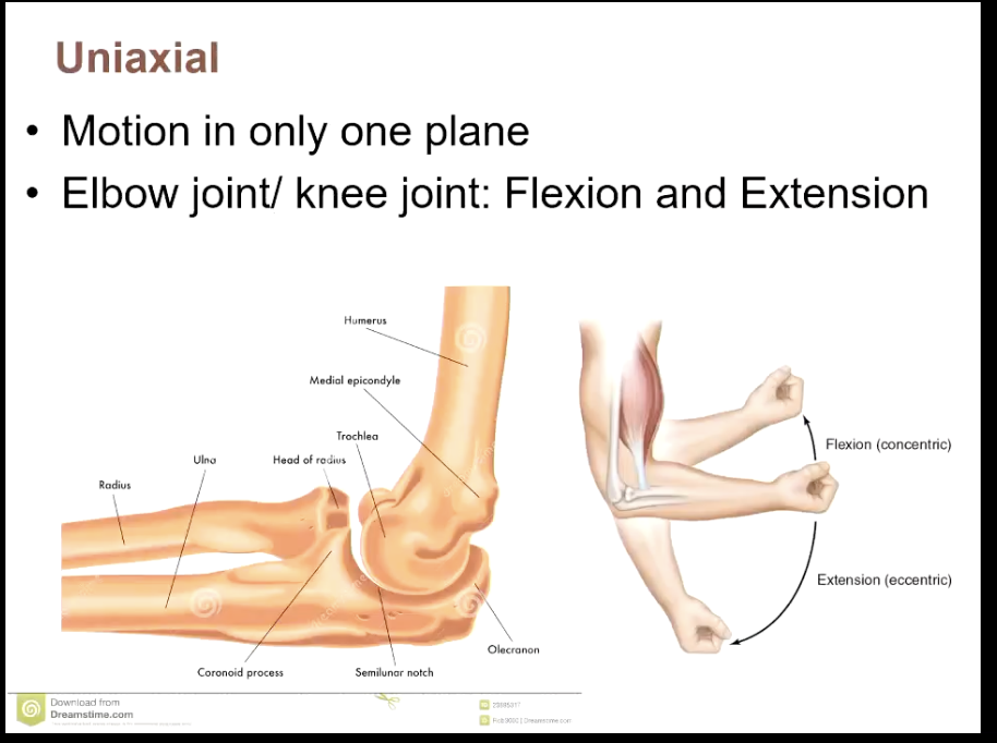
uniaxial
biaxial joint
