Module 4: Skeletal System
 Call Kai
 Learn
 Practice Test
 Spaced Repetition
 Match
 Flashcards
 Knowt Play
1/109

There's no tags or description
Looks like no tags are added yet.
Name | Mastery | Learn | Test | Matching | Spaced | Call with Kai | Chat |
|---|
No analytics yet
Send a link to your students to track their progress
110 Terms
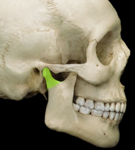
Condylar Process of Mandible
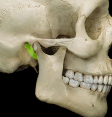
External Acoustic Meatus
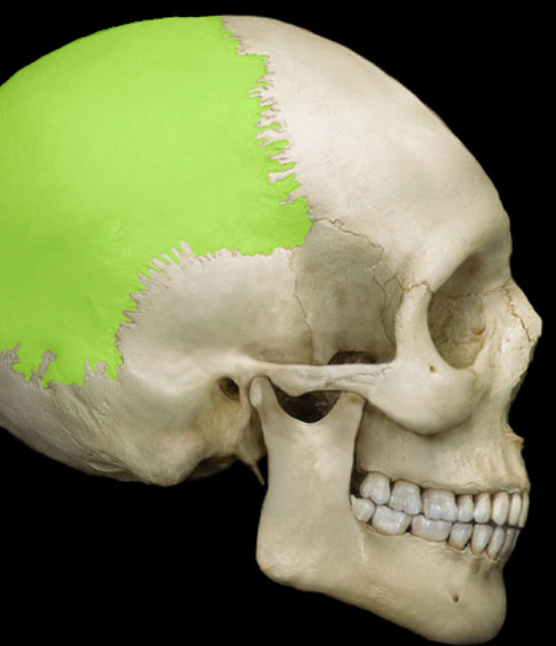
Parietal Bone
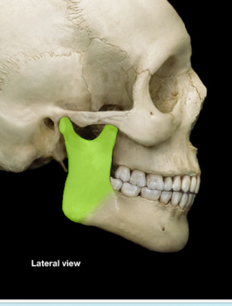
Ramus of Mandible
Temporal Bone
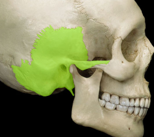
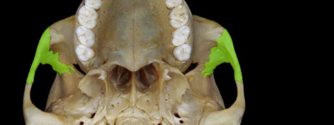
Zygomatic Process of Temporal Bone
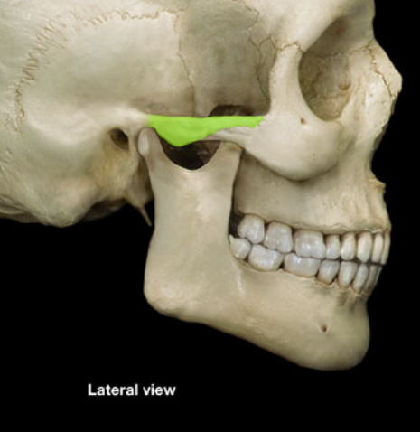
Ethmoid Bone
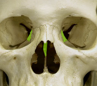
Frontal Bone
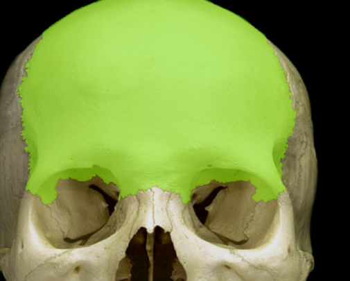
Inferior Nasal Concha
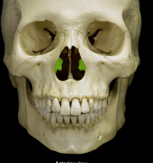
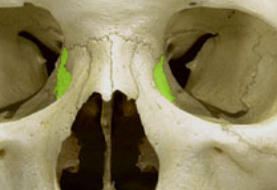
Lacrimal Bone
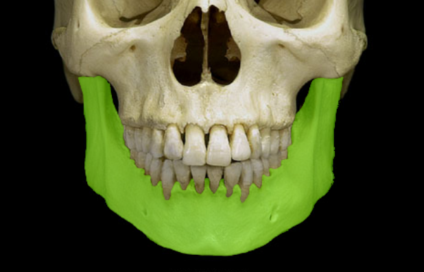
Mandible
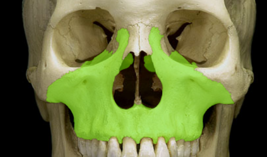
Maxilla
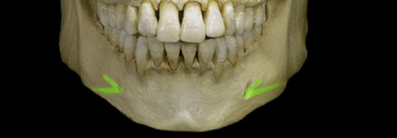
Mental Foramen
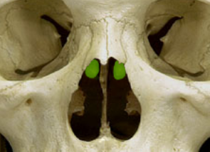
Middle Nasal Concha
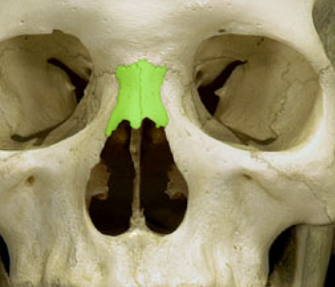
Nasal Bone
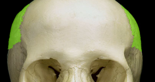
Parietal Bone
Sphenoid Bone
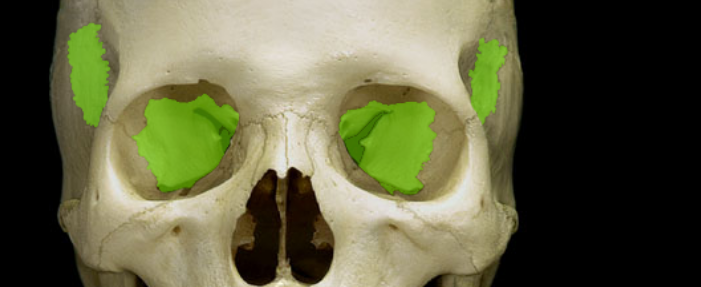
Temporal Bone
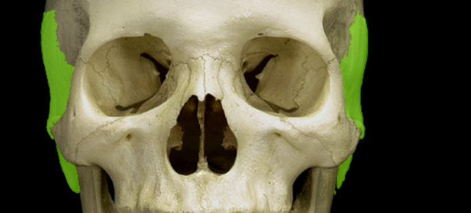
Vomer
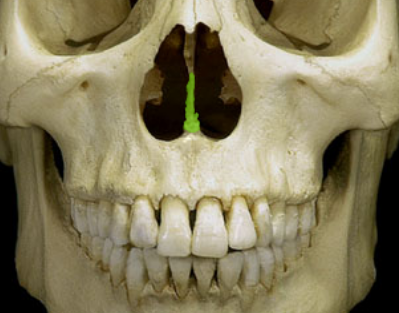
Zygomatic Bone
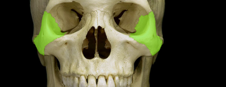
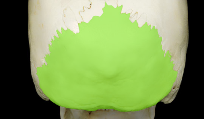
Occipital Bone
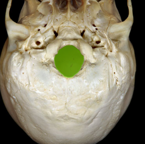
Foramen Magnum
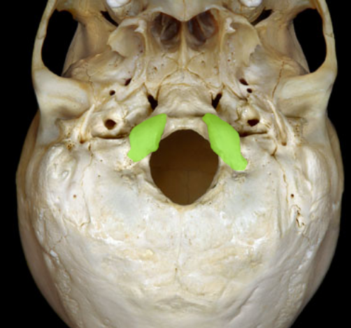
Occipital Condyle
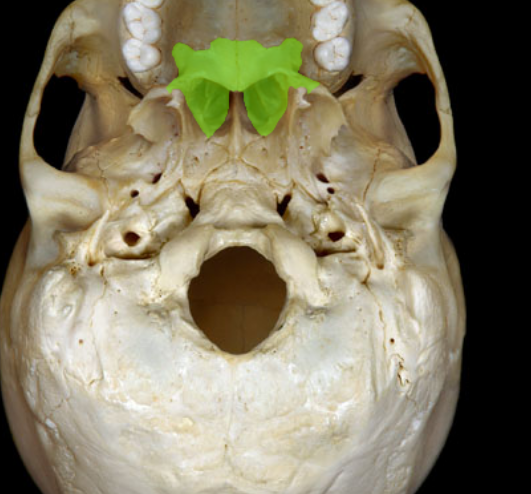
Palatine Bone
Zygomatic Bone
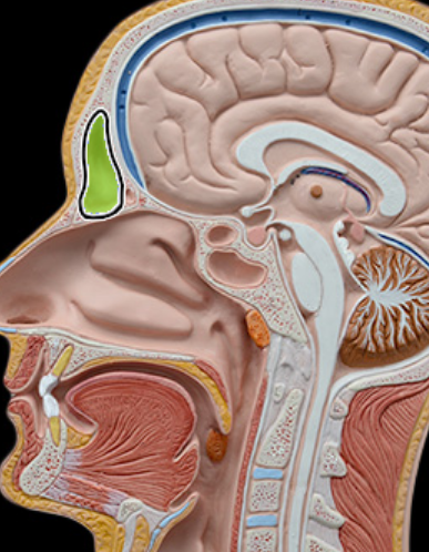
Frontal Sinus
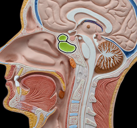
Sphenoid Sinus
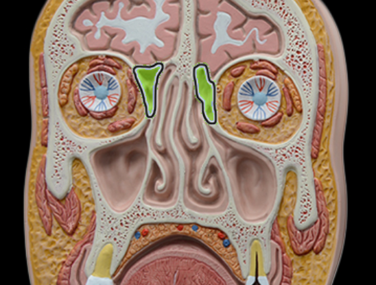
Ethmoidal Cells
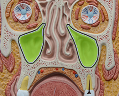
Maxillary Sinus
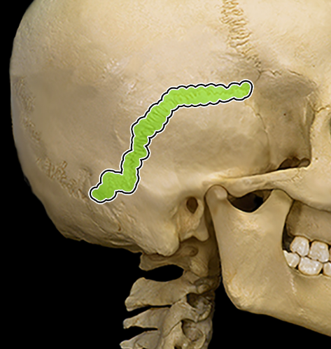
Squamous Suture
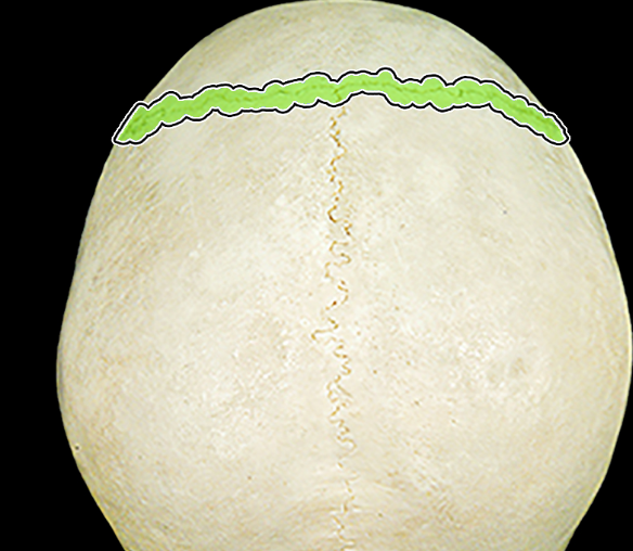
Coronal Suture
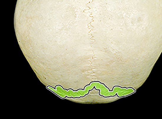
Lambdoid Suture
Sagittal Suture
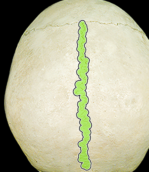
Lacunae
Tiny spaces occupied by osetocytes
Canaliculi
Tiny channels in which the processes of osteocytes exchange nutrients and wastes
Hematopoiesis
Blood formation
Skull, ribs, sternum, clavicles, vertebrae, hip bone
Areas of red marrow in adults
Hydroxyapatite
Calcium Phosphate
Osteogenesis imperfecta
Hereditary collagen defect, in which forming bones are brittle, and fetus may have many bone fractures
Primary Ossification Center
Area in center of diaphysis, where bone tissue first replaces cartilage
Secondary Ossification Center
Area of the epiphyses, where spongy bone forms later in development
Intramembranous Ossification
Mesenchyme differentiates into osteoblasts, build bone, and periosteum forms
Endochondral Ossification
Hyaline cartilage is ossified first by primary centers (diaphysis) and then secondary centers (epiphysis)
Zone of resting cartilage
Layer closest to end of epiphysis; resting cells; anchor epiphyseal plate to epiphysis
Zone of Proliferating Cartilage
Rows of young cells, undergoing mitosis
Zone of Hypertrophic Cartilage
Rows of older cells left behind when new cells appear; thicken epiphyseal plate, lengthening the bone; matrix calcifies, cartilage cells (chondrocytes) die
Zone of Calcified Cartilage
Thin layer of dead cartilage cells and calcified matrix
Vitamin D
Calcium absorption; deficiency causes rickets, osteomalacia
Vitamin A
Osteoblast and osteoclast activity; deficiency retards bone development
Vitamin C
Collagen synthesis; deficiency results in slender, fragile bones
Growth Hormone
Stimulates cartilage cell division; insufficiency in a child can result in pituitary dwarfism; excess causes gigantism in child, acromegaly in adult
Thyroid Hormone
Causes replacement of cartilage with bone in epiphyseal plate, osteoblast activity
Parathyroid Hormone
Stimulates osteoclasts, bone breakdown
Sex Hormone
Promote bone formation; stimulate ossification of epiphyseal plates
Condyle
Rounded process that usually articulates with another bone
Crest
Narrow, ridge-like projection
Epicondyle
Projection above a condyle
Facet
Small, nearly flat surface
Fissure
Cleft or groove
Fontanel
Soft spot in the skull where membranes cover the space between bones
Foramen
Opening through a bone that usually serves as a passageway for blood vessels, nerves, or ligaments
Fossa
Relatively deep pit or depression
Fovea
Tiny pit or depression
Head
Enlargement on the end of a bone
Linea
Narrow ridge
Meatus
Tube-like passageway within a bone
Process
Prominent projection on a bone
Ramus
Branch or similar extension
Sinus
Cavity within a bone
Spine
Thorn-like projection
Sulcus
Furrow or groove
Suture
Interlocking line of union between bones
Trochanter
Relatively large process
Tubercle
Knob-like process
Tuberosity
Knob-like process usually larger than a tubercle
7
Number of cervical vertebrae
12
Number of thoracic vertebrae
5
Number of lumbar vertebrae
5
Number of sacral vertebrae
4
Number of coccyx vertebrae
Body
Drum-shaped, anterior portion
Pedicles
Stalks that project from body, form sides of vertebral foramen
Laminae
Plates that arise from pedicles, fuse to become spinous process
Spinous Process
Posterior projection of spine
Transverse Process
Lateral projection of spine
Vertebral Foramen
Opening for spinal cord
Superior and inferior articular processes
Project upward and downward, to articulate with adjacent vertebrae
Facets
Cartilage-covered articulating surfaces of articular processes
Transverse formina
openings for arteries leading to brain; only found on cervical vertebrae
Vertebral Prominens
C7, spinous process is longer than those of other cervical vertebrae
Atlas
C1, supports head, articulates with occipital condyles
Axis
C2; Atlas pivots around the dens of Axis
Thoracic Vertebrae
Long, pointed spinous process, slopes downward
Lumbar Vertebrae
Thick, short spinous processes, almost horizontal
Median Sacral Crest
Fused spinous processes of vertebrae
Posterior Sacral Foramina
Openings for blood vessels and nerves on sacrum
Sacroiliac joints
transmit weight from spine to legs
Sacral Promontory
Projection at upper margin of sacrum
Sacral Canal
Formed by vertebral foramina on sacrum
Sacral Hiatus
Opening at inferior tip of sacrum