Chapter 12: Nervous system
 Call Kai
 Learn
 Practice Test
 Spaced Repetition
 Match
 Flashcards
 Knowt Play
1/119

There's no tags or description
Looks like no tags are added yet.
120 Terms
nervous system vs endocrine
endocrine uses hormones to communicate while nervous system uses electrical and chemical means
central nervous system
brain and spinal cord
peripheral nervous system
nerves and ganglia
nerve
bundle of nerve fibers (axons) wrapped in fibrous connective tissue
ganglion
location of neuron cell bodies in PNS

peripheral nervous system is divided into BLANK and BLANK, each with BLANK and BLANK subdivisions
Peripheral nervous system is functionally divided into
sensory and motor divisions, each with somatic and
visceral subdivisions
sensory (afferent) division
carries signals from receptors (sense organs) to CNS
somatic sensory division
carries signals from receptors in the skin, muscles, bones, and joints
Visceral sensory division
carries signals from the viscera (heart, lungs, stomach, and urinary bladder)
motor (efferent) division
carries signals from CNS to effectors
somatic motor division
carries signals to skeletal muscles, voluntary
visceral motor division
carries signals to glands, cardiac and smooth muscle, involuntary. further divides into sympathetic, parasympathetic and enteric plexus
sympathetic division of ANS
stimulates and prepares for action
parasympathetic division of ANS
calming
enteric plexus
in digestive tract wall, coordination and communication
3 properties of neurons
excitability, conductivity, and secretion
sensory (afferent) neurons
detect stimuli and transmit information toward the CNS
interneurons
receive signals from other neurons, process it, and make decision. found in CNS
motor (efferent) neurons
send signals out to muscles and gland cells
cell body (neurosoma, soma, or perikaryon)
contains nucleus and organelles. Mitochondria, lysosomes, inclusions, and rough ER. no centrioles
neurites
extensions of the neuron
dendrites
primary site for receiving signals from other neurons

axon (nerve) fiber
rapid conduction of nerve signals

axon hillock
mound on one side of cell body
axoplasm
cytoplasm of axon
axolemma
membrane of axon
axon terminal
forms synapse with next cell

multipolar neuron
one axon and multiple dendrites, most common in body

bipolar neuron
one axon and one dendrite. olfactory cells
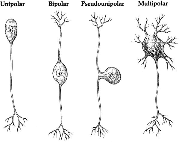
unipolar
single process leading away from cell body. the single branch acts as both the axon and the dendrite

anaxonic neuron
many dendrites but no axon. brain, retina, and adrenal gland

axonal transport
passage of materials along an axon
anterograde transport
movement away from cell body, via kinesin


retrograde transport
movement up the axon toward the cell body, via dynein
fast anterograde transport
organelles, enzymes, synaptic vesicles, small molecules
fast retrograde transport
recycled materials or some pathogens
slow axonal transport
always anterograde. transport of enzymes, cytoskeletal components, new axoplasm
neuroglia
supportive cells of nervous system
oligodendrocytes
form myelin sheaths in CNS
ependymal cells
line internal cavities of brain and secrete CSF (CNS)
microglia
macrophages in CNS
astrocytes
create BBB in CNS, diverse functions
Schwann cells
envelop axons of PNS and form myelin sheath, also help with regeneration of nerve fibers
satellite cells
surround nerve cell bodies in ganglia in PNS and provide insulation, also regulate chemical environment
gliomas
tumors of glial cells
can mature neurons perform mitosis? relate this to tumors
no, so they rarely form tumors. brain tumors usually arise from meninges and metastasis
myelin sheath
layers of insulation around an axon. mostly lipid, 20% protein
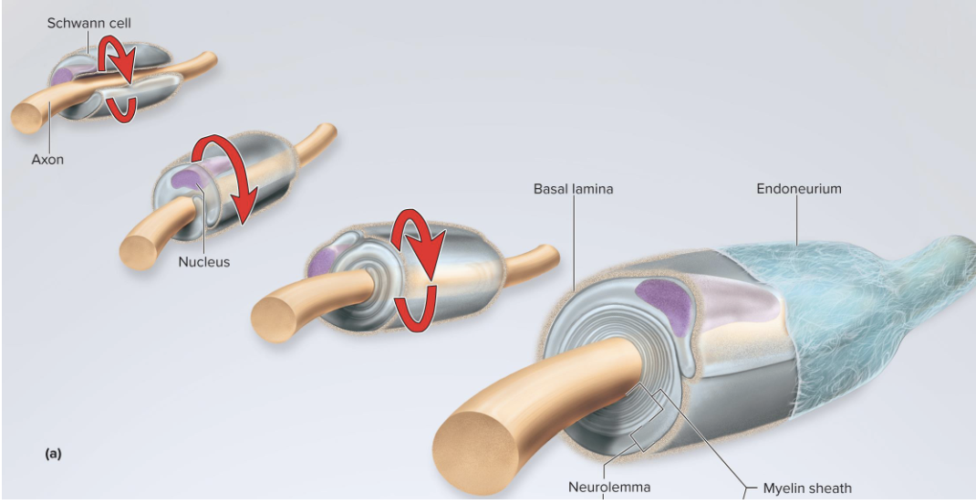
How does myelination happen in PNS?
Schwann cell spirals around a small section of single axon
lays down layers of its membrane which form the myelin sheath
neurilemma is the outermost coil
endoneurium
thin layer of fibrous connective tissue external to the neurilemma

what is external to the neurilemma?
the basal laminate and endoneurium
neurilemma
thick outermost coil of myelin in the PNS. contains the cell nucleus and most of the cytoplasm of the Schwann cell
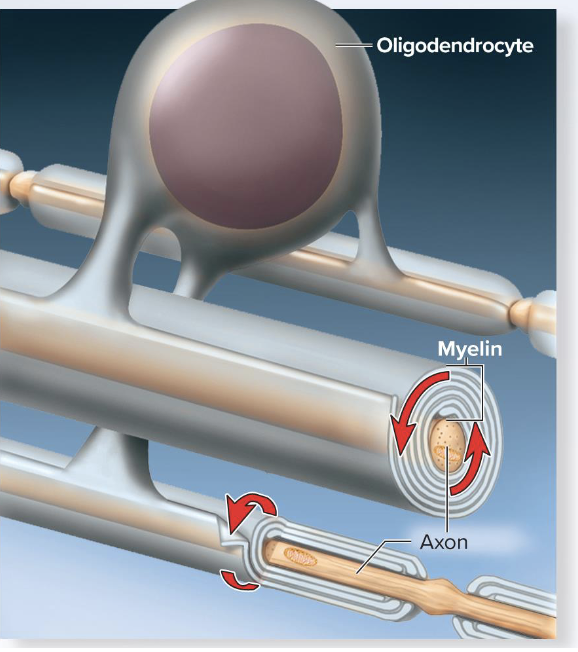
How does myelination occur in the CNS?
oligodendrocyte can wrap around small portions of many axons
myelination spirals inward toward axon
no neurilemma
node of ranvier
gaps between myelin
internal segments
myelin covered segments
initial segment
bare section of axon between the axon hillock and first glial cell
trigger zone
axon hillock and initial segment

multiple sclerosis
oligodendrocytes and myelin sheets in CNS deteriorate.
tay-sachs disease
abnormal accumulation of glycolipid GM2 in the myelin sheath
What affects speed of a neuron?
diameter (large= faster)
myelin (myelinated = faster)
nerve regeneration
Can occur in PNS if cell body remains intact
-macrophages clean up tissue debris
-cell body swells
-axon creates growth processes
-Schwann cell forms regeneration tube
Very slow
Electrical potential
Difference in concentration between charged particle from one point to another
Current
Flow of a charged particle
Resting membrane potential
Charge across membrane. -70mV
Is the inside of the membrane negative or positive?
the ICF is MORE NEGATIVE
Potassium influence of RMP
K+ is more concentrated in the ICF. The membrane is more permeable to K+ so it can leak out and membrane becomes more negative. The electrical charge pulls the positive K back inside
Sodium influence of RMP
Na+ is more concentrated in the ECF and the membrane is less permeable to Na+. Na+ diffuses into the cell down concentration gradient but only cancels a small amount of negative charge
Sodium-potassium pump
Helps establish the RMP and ensure there are more Na+ outside and more K+ inside. Uses one ATP to bring 3 Na+ out of cell and 2 K+ into cell.
Local potentials
Local, temporary change in membrane potential. Occurs at dendrites and soma. Purpose is to process sensory info and determine whether neuron should trigger an action potential.
What are the characteristics of local potentials?
-graded: Varys in magnitude depending on stimulus strength (ex: strong stimuli can open more Na+ channels)
-decremental: weaker as they get further away
-reversible: if stimulus ceases, membrane potential returns to RMP
-can be excitatory: depolarize = more likely to have AP
-can be inhibitory: hyper-polarize = less likely to have AP
Action potential
Rapid up and down change in voltage created by opening and closing of voltage-gated channels. Begins in the trigger zone, where there are enough voltage-gated ion channels

Describe the steps of an action potential
-local potential spreads to axon hillock
-threshold must be met (-55 mV)
-Na+ channels open quickly and K+ voltage gated channels open slowly
-depolarization: Na+ influx into cell and membrane potential reaches +35 mV
-Na+ channels close and inactivate
-Polarization: the K+ channels are now fully open and K+ leaves the cell. Membrane becomes more negative
-hyperpolarization: membrane becomes more negative than RMP
-membrane returns to RMP
Explain depolarization
Influx of Na+ makes the membrane more positive and it peaks at +35 mV.
Explain repolarization
Efflux of K+ out of the cell. Makes the membrane negative again
Do the concentrations of Na+ and K+ change significantly during an AP?
No. Movement of only a few ions has a big effect on the membrane potential
What are the characteristics of an AP?
All or none law: neuron fires go to maximum voltage if threshold meets.
Non decremental: does not get weaker with distance
Irreversible: travels all the way down the axon and cannot be stopped
All or none law
Threshold must be met in order for action potential to proceed. If threshold is met, the action potential proceeds to maximum voltage.
Absolute refractory period
No stimulus of any strength will trigger an action potential. Caused by closing of Na+ voltage-gated channels, which prevents another depolarization
Relative refractory
An unusually strong stimulus can trigger an AP. During hyperpolarization, a larger depolarization (local potential) can trigger an AP
Continuous conduction
Occurs on unmyelinated axons because they have voltage-gated channels down the length of their axon. Action potential begins at the trigger zone and depolarization travels down. voltage-gated channels are opened immediately next to new action potential

Saltatory conduction
Action potentials “jump” from node to node. Electrical signal passes passively between nodes but is strong enough to depolarize membrane to threshold at nodes of ranvier and Na+ channels open, so a full strength action potential occurs
How does myelin increase speed of action potential?
-insulates and prevents ions from leaking
-creates more distance between the intracellular fluid and extracellular fluid so fewer ions are needed to depolarize the membrane
Synapse
Point where an axon terminal meets the next cell
Presynaptic neuron
Nerve cell that sends the signal across the synapse
Postsynaptic neuron
Neuron that receives signals from the presynaptic neuron at the synapse
synaptic cleft
Gap between neurons
Chemical synapse
Release of neurotransmitters
Electrical synapse
gap junctions join cells and electrical signals spread from cell to cell. Very fast but cannot integrate into
Synaptic vesicles
Found in axon terminal of presynaptic neuron and contain the neurotransmitter
Acetylcholine
Neurotransmitter formed from acetic acid and choline
Amino acids
Neurotransmitters glycine, glutamate, aspartate, and GABA
Monoamines
Class of neurotransmitters. Remove the -COOH group from amino acids but amine group remains
purines
Adenosine and ATP
Gases
Nitric acid and carbon monoxide
Neuropeptide
Chains of amino acids that function as hormones or neuromodulators
How do synapses vary in their modes of action?
Depends on type of receptor on post synaptic neuron. Can be excitatory or inhibitory

Excitatory cholinergic synapse
• Cholinergic synapse—acetylcholine (ACh) is theneurotransmitter
• Steps in synaptic transmission:
• Action potential depolarizes the axon terminal, opens voltage-gated Ca channels
• Ca enters, triggers exocytosis of ACh
• ACh diffuses across cleft, binds to postsynaptic receptors
• ACh receptors are ligand-gated ion channels that open and allow Na and K across the membrane
• Entry of Na causes depolarizing postsynaptic potential
• If strong enough, depolarization spreads to the trigger zone, raises the membrane potential up to threshold to trigger an action potential
GABA-nergic synapse
y-aminobutyric acid (GABA) is the neurotransmitter. This is an inhibitory synapse.
AP triggers the release of GABA into the synaptic cleft.
GABA receptors are chloride channels
Cl- entry hypolerpolarizes the postsynaptic membrane
Postsynaptic neuron is inhibited (less likely to fire an action potential)
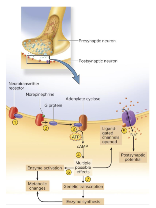
adrenergic synapse
norepinephrine is the neurotransmitter. slower than cholingeric and GABA-ergic synapses. advantage is enzyme amplification
monoamines and neuropeptides bind to G-protein coupled receptors on postsynaptic membrane
activation of second messenger systems such as cAMP
How does the signal cease at the synapse?
presynaptic cell stops releasing neurotransmitters
neurotransmitter already in the synapse is cleared
Explain clearance of a neurotransmitter
neurotransmitter degradation: enzyme in synaptic cleft breaks down neurotransmitter
reuptake: neurotransmitter or its breakdown products reabsorbed into axon terminal
Diffusion: neurotransmitter or its breakdown products diffuse away from synapse into the ECF