Ch 12: Kidneys
 Call Kai
 Learn
 Practice Test
 Spaced Repetition
 Match
 Flashcards
 Knowt Play
1/166

Earn XP
Description and Tags
powerpoint
167 Terms
How much of the nephrons needs to function to ensure survival
1/3 of it
3 important functions of nephron
control blood concentration and volume by removing selected amounts of water and solutes
help regulate pH
remove toxic waste from blood
What is in charge of balancing fluid in the body
Antidiuretic Hormone (ADH)
What does the body do with water when ADH is secreted
body holds onto water
Where does ADH come from
secreted by posterior pituitary gland
Where does ADH increase water permeability and what does this allow
Increases water permeability of collecting tubules which allows water to pass into interstitial spaces
When is ADH secretion increased
in cases of increased water loss (sweating) or reduced blood volume (hemorrhage)
What is diuresis
excessive urine production
Function of Aldosterone Hormone
it is a sodium potassium regulator
What does secretion of Aldosterone do in regards to sodium and what does this cause
it increases the rate of sodium absorption which produces concurrent loss of potassium
What does excess extracellular potassium promote
ADH secretion
What happens when ADH is secreted due to excess cellular potassium
It increases the potassium excretion and sodium retention by kidneys
What is aldosterone secretion also controlled by
renin-angiotensin
What is the relation of renin and sodium and BP
renin increases in response to decreased BP secondary to sodium depletion or to change from supine to upright
How does renin increases BP
renin acts on plasma to produce angiotensin I, which is converted to angiotensin II, which increases systemic blood pressure
Variants and Congenital anomalies of the kidneys
agenesis, supernumerary (multiple), duplicated collecting system, ectopic, horseshoe, extrarenal, hypertrophied column of Bertin, dromedary hump, and junctional parenchyma defect
Extrarenal pelvis
Renal pelvis sits right outside the kidney

Junctional Parenchymal Defect and where its usually seen
Fusion defect usually seen in upper pole of right kidney

Duplicated Collecting System
2 renal sinuses w/ hypoechoic division

Ectopic Kidneys
When kidney isn’t it the normal spot.
left image is RUQ with empty renal fossa
right image is LUQ with empty renal fossa

Where are transplanted kidneys usually located
pelvic kidney

Cross-fused kidney and what can it be confused with
Both kidneys fused together (both on one side of the body). can be confused with horseshoe kidney
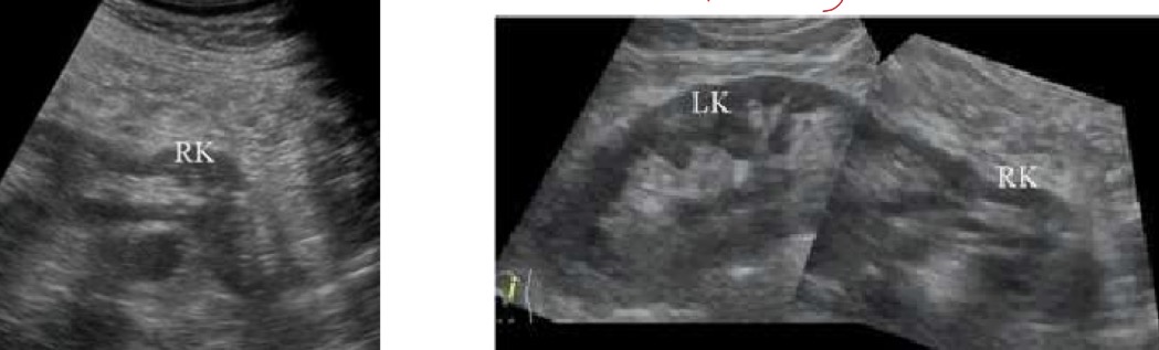
Horseshoe kidney
no clear border at portion where they connect.
midline anterior to aorta

Types of cystic renal disease
acquired, congenital (hereditary), or developmental
How common is cystic renal disease and what may it cause
it is relatively common and may be a cause of renal failure
What is a differential of cystic renal disease and what indicates it
malignancy is a differential diagnosis, and vascularity indicates that the lesion is malignant/not simple cyst
What is Autosomal Dominant Polycystic Kidney Disease (ADPKD)
a common, inherited dominant autosomal trait
Where may you concurrently find cysts with ADPKD
lungs, liver, or pancreas
Complications of ADPKD and which is most serious
Most serious complication is cerebral berry aneurysm (circle of Willis)
others are infections, calculi, or cyst rupture
When does ADPKD usually manifest clinically and what happens with increased age
usually manifests clinically in 4th decade of life, and cysts enlarge/kidney function declines with age
ADPKD clinical symptoms
abdominal/lumbar pain, hematuria (from compression of ureters/rupture of cyst), hypertension, or increased BP
ADPKD Sonographic appearance
bilateral enlarged kidneys, numerous cysts of various size, poorly demarcated (noted/visualized) renal capsule contour, and may distort central echo complex

What is Medullary sponge kidney
Bilateral cystic dilatation of medullary and papillary portions of collecting tubules
What does medullary sponge kidney do to kidney function
Kidneys function normally with this disease
Medullary sponge kidney sonographic appearance (uni/bi, echogencity)
it is bilateral and has increased medullary echogenicity

What is medullary cystic disease
a progressive, familial, or hereditary disorder characterized by presence of variable numbers of cysts in medulla
What is medullary cystic disease associated with
interstitial fibrosis and cortical tubular atrophy
What does damage from fibrosis/atrophy from medullary cystic disease result in
renal insufficiency and progresses to terminal renal failure over 5-10 years
Who is autosomal dominant medullary cystic disease onset in
young adults
Who is autosomal recessive medullary cystic disease onset in
children
Where do simple cysts commonly originate
renal cortex

Simple cyst pathogenesis (origin) and who is it prevalent in
has an unknown pathogenesis and prevalence increases after age 40

Simple cyst symptoms
usually asymptomatic unless there’s an obstruction of collecting system.
often incidental finding

Appearance of complex hemorrhagic cyst
Appearance may change over time
may see calcifications, wall thickening, and septations may form

What should you always do when evaluating a septated/multilocular cyst and why
Use color Doppler because vascular separations can indicate malignancy

Etiology of parapelvic cyst
it is unknown
Where do parapelvic cysts originate from
originate from renal parenchyma and protrude into the renal sinus
Parapelvic cyst sonographic appearance (side and number)
generally solitary and large

Peripelvic cyst etiology
it is unknown
What is a peripelvic considered
a true sinus cyst
Peripelvis cyst sonographic appearance (size, number, borders)
small, multiple, and often irregular outline

What can peripelvic cyst be confused with
hydronephrosis
Most prevalent benign kidney tumor
adenoma
Adenoma symptoms and size
typically measure 1cm or less.
are often found incidentally and are asymptomatic unless they enlarge, and can be cause of painless hematuria
Adenoma sonographic appearance
Pattern similar to renal cell carcinoma, and highly vascular with internal echoes and sound attenuation (shadowing)
hyperechoic-hypoechoic to the normal renal cortex

DDx (differential diagnosis) for renal adenoma
angiomyolipoma and renal cell carcinoma

What is an Oncocytoma and who is it prevalent in
a large vascular benign adenoma and has a male prevalence in later decades of life
Oncocytoma symptoms
usually asymptomatic but may have pain or hematuria depending on the size
What may a Oncocytoma do regarding vascularity and what does this produce
it may outstrip blood supply with concurrent infarction, hemorrhage, and necrosis which produced a central stellate scar
Oncocytoma sonographic appearance (echogenicity, borders, color)
it has a varying appearance and can be difficult to distinguish from renal cell carcinoma on ultrasound
hypo-hyperechoic, homogenous, well defined wall, hypoechoic stellate star, flow in periphery and strip like signals within the mass

What is Angiomyolipoma (AML) aka renal hamartoma
a benign tumor composed of fat cells intermixed with blood vessel and smooth muscle cells
Angiomyolipoma (AML) symptoms and predominance in who
Most AML are asymptomatic and has a female predominance
Angiomyolipoma sonographic appearance (echogenicity, and size)
variable echogenicity but most are hyperechoic, and varying size.
heterogeneous in presence of hemorrhage or necrosis, and the risk of this is when its >4cm
What is Mesoblastic Nephroma aka fetal renal hamartoma
a pediatric benign tumor (counterpart to Wilms tumor)
Mesoblastic nephroma clinical presentation
palpable flank mass and hypertension
Mesoblastic Nephroma sonographic appearance
varying echogenicity, possible areas of cystic degeneration and may show calcifications
What is the most common malignant tumor of the kidney
Renal cell carcinoma (RCC) aka hypernephroma
Who does renal cell carcinoma occur most often in
occurs most often after age 50 and more prevalent in men
renal cell carcinoma symptoms and which is most significant
costovertebral angle pain, palpable mass, or hematuria (most significant symptom)
Size of renal cell carcinoma
size of tumor varies
Where can renal cell carcinoma spread to
perinephric fat and renal vein (more commonly the right)
What is the 5 year survival rate of renal cell carcinoma with and without spread
5 year survival rate is 80-100% if tumor has not spread
5 year survival rate is 0-20% if there’s adjacent invasion of distant metastasis
How does RCC spread and what does that allow
Lymphogeneous and hematogenous spread allows metastasis to develop before patient has local symptoms
What % of patients have metastasis of RCC at time of diagnosis, and what % is to the lungs
30% of patients have metastasis, and 75% of them being to the lung
What can renal cell carcinoma cause/result in
IVC thrombosis (use color/spectral doppler to assess flow/phasicity), or obstruction or lower extremity swelling
Renal cell carcinoma sonographic appearance
Highly vascular with numerous areas of hemorrhage, necrosis, or cystic degeneration; varied appearance from solid to multilocular cyst; calcifications

What sonographic finding indicates that renal cell carcinoma has spread
Masses seen in renal vein, IVC, or contralateral kidney

How is tumor vascularity and echogenicity related in renal cell carcinoma
there is no correlation between the two

What does the presence of hemorrhage or necrosis determine about renal cell carcinoma appearance
the echogenicity

What is the primary malignant epithelial tumor originating in urinary collecting system
Transitional cell carcinoma/TCC (urothelial carcinoma)
When does transitional cell carcinoma (TCC) become clinically apparent
when there is presence of painless hematuria
Where does Transitional cell carcinoma originate
in the medulla?
Transitional cell carcinoma size and what can it cause
they are small and may block urinary outflow, causing palpable hydronephrosis
Transitional cell carcinoma sonographic appearance
iso-hypoechoic mass within collecting system, and can see obliteration of normal renal sinus

What is Wilms tumor (nephroblastoma)
pediatric malignant mixed tumor composed of embryonal elements
Wilms tumor clinical presentation
fever, HTN, hematuria, or palpable flank mass
Wilms tumor sonographic appearance (echogenicity, size)
most commonly homogenous and echogenic, large in size, and calcifications

Is metastasis to the kidneys common or rare
kidneys are a frequent site for metastasis
What is the only malignancy that selectively metastasizes to the opposite kidney
renal cell carcinoma
Main sources of metastasis to the kidneys
lung and breast tumors
What is hydronephrosis
urine dilatation of renal pelvis and calyces

Hydronephrosis symptoms
may be asymptomatic or have abdomen, back, and/or flank pain
Hydronephrosis intrinsic and extrinsic causes
Intrinsic: calculi, hematoma, neoplasm, ureteropelvic obstruction
Extrinsic: neoplasm, trauma, neurogenic bladder, bladder outlet obstruction, pregnancy
What should be elvaluated in a hydronephrosis sonographic exam
ureters for dilatation
prostate for hyperplasia/neoplasm
bladder for calculi or tumors
pelvic structures

3 Grades of hydronephrosis and their appearance
Grade 1: no dilation of ureter/pelvis
Grade 2: pelvis has some dilation
Grade 3: loss of recognition of renal sinus

What normal variants/increased urine flow can stimulate mild hydronephrosis
variants: extrarenal pelvis or full bladder
increased urine flow: overhydration or medications
What inflammatory disease can stimulate hydronephrosis
pyelonephritis
What renal cystic diseases can stimulate hydronephrosis
PCKD, parapelvis cyst, or medullary cystic disease
What is urolithiasis
calculi in the urinary system (can occur anywhere in the system)
Where do majority of urolithiasis develop and what is it called
in the kidney - nephrolithiasis