Chapter 4 Lecture
 Call Kai
 Learn
Learn Practice Test
 Spaced Repetition
 Match
Match Flashcards
 Knowt Play
1/58

Name | Mastery | Learn | Test | Matching | Spaced | Call with Kai | Chat |
|---|
No analytics yet
Send a link to your students to track their progress
59 Terms
4 types of tissues
1. Epithelial
2. Connective
3. Muscle
4. Nervous

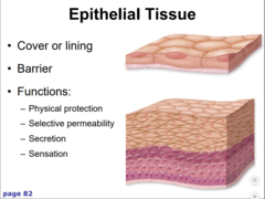
Epithelial tissue functions
- lining/covering
- absorption and secretion
- sensation
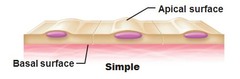
Apical Surface of epithelial tissue
an upper free surface exposed to exterior

Basolateral Surface of epithelial tissue
Areas between two adjacent cells or a cell and the basement membrane
Epithelial tissues are ___________. But they have ___________ abilities supported by division of _____ _______.
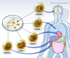
Epithelial tissues are AVASCULAR. But they have REGENERATIVE abilities supported by division of STEM CELLS.
Epithelial cells are bound by what
intercellular junctions

3 types of intercellular junctions
1. Gap
2. Tight
3. Desmosomes

Gap Junction
- allows the passage of materials between cells
- coordinate involuntary muscle contractions

Tight junctions
prevent leakage of extracellular fluid across a layer of epithelial cells

Desmosomes
Anchoring junctions that prevent cells from being pulled apart

2 specialized epithelium types
Transitional
Glandular

Simple Squamous - function
- ease of diffusion & barrier
* Capillaries (endothelium)
* Serous membranes (mesothelium)

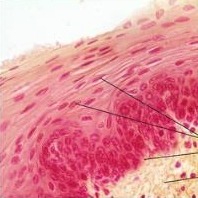
Stratified Squamous - function
- keratinized epidermis
- nonkeratinized orifices, espophagous
simple cuboidal - function
lines renal tubules, thyroid follicles, & more
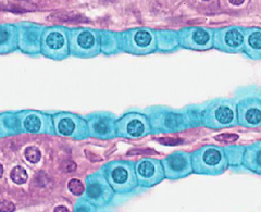
Stratified cuboidal
Lines mammary ducts, sweat, and salivary glands

microvilli
* increase absorption surface area
* may be in simple columnar E.

stratified columnar locations
pharynx epiglottis, exocrine glands

Pseudostratified Columnar location
respiratory tract

glandular epithelium - 2 types
secreting glands
- Endocrine
- Exocrine
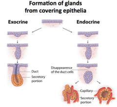
endocrine glands
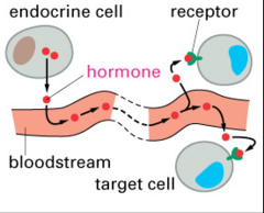
ductless ; secrete hormones into blood stream
Exocrine glands
Secrete onto surfaces via ducts
- sweat, salivary, etc

Classifying Exocrine - 3 catagories
1. structure
2. Mode of secretion
3. Type of secretion
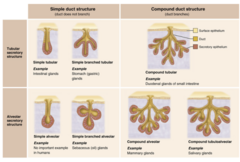
Exocrine step 1 in classifying
- unicellular
- multicellular : structure of duct & shape of secretory
Structure of duct - 3 types (Exocrine gland)
1. simple : straight pipe
2. simple branched : multiple leading to 1 pipe
3. compound : pipe splits into multiple

Shape of secretory portion (Exocrine gland)
1. Alveolar
2. Tubular ; can be coiled
3. Tubuloalveolar : combined

Secretion Modes
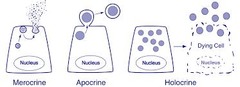
1. Merocrine
2. Apocrine
3. Holocrine

Merocrine secretion mode
A strategy of secreting hormones via exocytosis
- salivary and most exocrine glands
Apocrine secretion mode
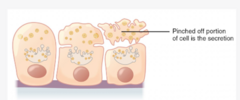
involves the loss of cytoplasm as well as the secretory product
- mammary glands
Halocrine secretion mode
cells accumulate a product until they disintegrate
- sebaceous glands

Serous secretion type
Thin, watery fluid
*tears

Mucous secretion type
- mucin lubricant
* intestinal submucosal glands
mixed exocrine glands
both serous and mucous
*submandibular salivary glands
CT 3 types
1. CT proper
2. Fluid CT
3. Supporting CT
Fibroblasts
In CT, cells that secrete the proteins of the fibers.

Macrophage
Large white blood cell that removes bacteria, foreign particles, and dead cells
Mast Cells
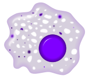
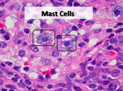
Cells that release chemicals (such as histamine) that promote inflammation.
Adipocytes
store triglycerides

Melanocytes
cells that produce melanin

Collagen Fibers
provides flexibility and strength

Reticular fibers
Fibers found near blood vessels that add strength and support

Elastic fibers
Flexible and "stretchy" fibers that add elasticity to tissue

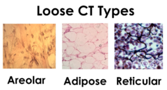
3 types of loose CT
1. Areolar
2. Adipose
3. Reticular
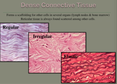
3 types of dense
regular, irregular, elastic
3 types of Fasciae layers
1. superficial fascia
2. deep fascia
3. subserous fascia
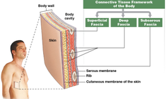
superficial fasciae
A layer of loose connective tissue that lies just beneath the skin, providing insulation and cushioning for underlying structures. It also allows for the movement of skin over tissues.
deep fasciae
dense, fibrous layer of connective tissue that lies beneath the superficial fascia and wraps around muscles, bones, nerves, and blood vessels
subserous fascia
layer of connective tissue that suspends, wraps, and supports internal organs within body cavities
Tissue membranes 4 types
1. Mucous membranes
2. Serous membranes
3. Cutaneous membrane
4. Synovial membranes
Line or cover body surfaces
Typically consist of epithelium supported by connective tissue
Mucosal membranes
Line body openings
-Produce mucin and release antimicrobial substances
-Underlying areolar tissue (Lamina Propria)

Goblet cells
secrete mucus

Serous Membranes
Thin epithelium (mesothelium)
- Underlying areolar tissue

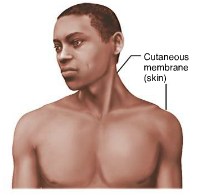
Cutaneous Membranes
covers body, skin
Synovial membranes
Loose, connective tissue that lines the joint cavity

Types of Fluid CT
blood & lymph

Cartilage - ( _________ cells occupying __________)
Chondrocytes occupy lacunae within gel-lie ECM

Perichondrium
Dense connective tissue surrounding cartilage.

Endothelium
Capillaries
Mesothelium
serous membranes
underlying areolar layer at base of mucosal membrane
Lamina Propria