Nervous System (Pt. 2)
 Call Kai
 Learn
 Practice Test
 Spaced Repetition
 Match
 Flashcards
 Knowt Play
1/61

There's no tags or description
Looks like no tags are added yet.
62 Terms
2 major functions of Nerves
1. Irritability
2. Conductivity
Irritability
Response to a stimulus
Conductivity
Transmit impulse to other nerves, muscles or glands
Resting neuron electrical condition
Polarized
Polarized condition of Neuron
more negative ions inside the neuron than outside
Synapse transmission
Neurotransmitter released to cross the synapse to excite adjacent nerve

Synapse transmission step 1
Action potential (AP) reaches axon terminal and electrical charge opens Ca channel
Synapse transmission step 2
Ca cause vesicles containing neurotransmitters to fuse with axon terminal membrane
Synapse transmission step 3
Vesicle opens causing release of neurotransmitter into synaptic cleft
Synapse transmission step 4
Neurotransmitter binds to neurotransmitter receptors on adjacent neuron, initiating previous cycle of Na entry into neuron
Synapse transmission step 5
Neurotransmitter effect is brief as it is removed by diffusion, reuptake into the axon or broken down by enzyme activity
Reflexes
Rapid responses programmed in the body, Predictable and involuntary, Brought by stimulus
Reflex arc involves
CNS and PNS

2 Types of reflexes
1. Somatic (ex. pull hand out of fire)
2. Autonomic (ex. regulation of body functions)

5 Different elements of reflex arc
1. Sensory receptor
2. Sensory neuron
3. Interneuron
4. Motor neuron
5. Effector organ

Sensory receptor
Reacts to stimulus

Sensory neuron
Transmits signal to CNS

Interneuron
Communication center between Afferent and Efferent pathways

Motor neuron
nerve transmitting signal from CNS

Effector organ
muscle/ organ stimulated

Anatomy of brain
1. Divided into two main cerebral hemispheres
2. Hemispheres collectively known as cerebrum
Gyri
Elevated portions of brain
Sulci
1. Shallow grooves of brain
2. Separate gyri
Fissures
1. Deep grooves of brain
2. Separate brain into different lobes
3 Features of brain hemispheres
1. Superficial cortex of grey matter
2. Internal white matter
3. Basal nuclei
Cerebral cortex
Contains areas controlling...
1. Speech
2. Memory
3. Logical and emotional responses
4. consciousness
5. Sensation
6. Voluntary movements

2 Areas of cerebral cortex
1. Primary somatic sensory area
2. Primary motor area

Primary somatic sensory area location
1. Parietal lobe
2. Posterior to central sulcus

Primary motor area location
1. Frontal lobe
2. Anterior to central sulcus

Primary somatic sensory area
1. Interprets all sensory input (excludes special senses)
2. Interprets signals involving threat, temperature, pressure and light touch
3. Sensory signals are crossed (left body corresponds to right brain and vice versa)

Primary motor area
1. Allows person to move body voluntarily
2. Major axons from this region descend to form corticospinal tract
3. Sensory signals are crossed (left body corresponds to right brain and vice versa)

4 Specialized areas of the cortex
1. Broca's area
2. Anterior association area
3. Posterior association area
4. Wernicke's area

Broca's area
1. Found at base of precentral gyrus
2. Anterior to central sulcus
3. Only in left hemisphere
4. Controls speech

Anterior association area
1. Anterior part of frontal lobe
2. Involved with higher intellectual activity and socially acceptable behavior

Posterior association area
1. Encompasses posterior cortex
2. Recognition of patterns and faces

Wernicke's area
1. Posterior cortex
2. Language comprehension

Location of cell body for specialized areas in the cortex
Outermost grey matter

Cerebral white matter
1. Makes up majority of remaining tissue of cortex
2. Impulses are transmitted to, from and within the cortex
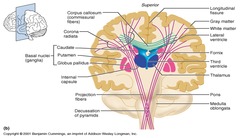
3. Large, central fiber tract is the corpus callosum
4. Association fiber tracts
5. Projection fiber tracts

Association fiber tracts
Connect areas within hemispheres
Projection fiber tracts
connect cerebrum with lower CNS

Fiber tract
connects brain hemispheres
Basal nuclei
1. AKA basal ganglia
2. Grey matter deep within white matter
3. Help regulate voluntary motor activities. y modifying instructions
4. Internal capsule passes from basal ganglia to thalamus

Internal capsule
tight band of fibers
Diencephalon location
Top of brain stem
3 Parts of Diencephalon
1. Thalamus
2. Hypothalamus
3. Epithalamus
Thalamus
Relay center for sensory impulses passing into sensory cortex

Hypothalamus
1. Below thalamus
2. Responsible for regulation of temperature, water balance and metabolism
3. Important for emotional drive (appetite, sex, pain, etc.)
4. Member of limbic system
5. Control pituitary gland

2 Hypothalamus hormones
1. Anti-diuretic
2. Oxytocin

Epithalamus
Forms floor of third ventricle

2 Parts of epithalamus
1. Pineal gland
2. Choroid plexus

Pineal gland
1. Part of endocrine system
2. Melatonin
3. Dark/ light cycle

Choroid plexus
Forms CSF

3 Parts of brain stem
1. Mid brain
2. Pons
3. Medulla oblongata

Midbrain
transmit ascending and descending impulses
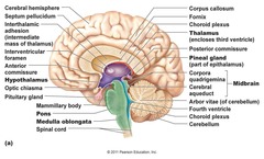
corpora quadrigemina
1. Rounded protrusions
2. reflex centers for vision and hearing

cerebral peduncles
Transmit ascending and descending impulses
Cerebral aqueduct
Canal connecting 3rd and 4th ventricles

Pon
1. Below mid brain
2. Made of fiber tracts
3. Contains nuclei involved in breathing

Medulla oblongata
Contain pyramidal or corticospinal motor tracts, this is the area where they cross over to the opposite site
Also contains fibers that control visceral activity, HR, BP, breathing, swallowing and vomitting

Reticular formation
1. Extends length of brain stem
2. Made of grey matter
3. Involved in motor control of visceral organs
4. Reticular activation system (RAS) play role in consciousness/ sleep and awake cycle
5. Filters sensory input
6. Damage can result in coma
Cerebellum location
Under occipital lobe of cortex
Cerebellum
Provides timing for muscle activity, balance, and equilibrium, Allows for smooth, fluid and coordinated movement