IB 131 Unit 1: Lecture 1-14
 Call Kai
 Learn
Learn Practice Test
 Spaced Repetition
 Match
Match Flashcards
 Knowt Play
1/245

There's no tags or description
Looks like no tags are added yet.
Name | Mastery | Learn | Test | Matching | Spaced | Call with Kai | Chat |
|---|
No analytics yet
Send a link to your students to track their progress
246 Terms
In Anatomical Position, what is the difference between 'axial' and 'appendicular'?
Axial = along the midline; Appendicular = attached to the axial skeleton
What plane divides the body into left and right parts?
Sagittal plane
Which plane is horizontal and divides the body into superior and inferior parts?
transverse plane
Which vertical plane divides the body into anterior and posterior parts?
Coronal/Frontal plane
What structure separates the thoracic cavity from the abdominal cavity?
Diaphragm
What is the structure of serous membranes?
2 layers: parietal and visceral with serous fluid filling in between them
Parietal layer: permanent outer layer
Visceral layer: inner layer, lines the organs
What are mesenteries?
Continuous double folds of the parietal peritoneum that attaches the organ to the body wall
How does light microscopy work?
Illuminates tissue with a beam of light, lower magnification
How does electron microscopy work?
Uses beams of electrons for higher magnification
How does scanning electron microscopy microscopy work?
Provides 3D images of unsectioned surfaces
What does the Right Upper Abdominopelvic Quadrant contain?
Right lobe of liver, gallbladder, right kidney, parts of stomach, small/large intestines
What does the Left Upper Abdominopelvic Quadrant contain?
Left lobe of liver, stomach, pancreas, left kidney, spleen, parts of large intestines
What does the Right Lower Abdominopelvic Quadrant contain?
Cecum, appendix, parts of small intestines, reproductive organs, right ureter
What does the Left Lower Abdominopelvic Quadrant contain?
Most of small intestines, parts of large intestine, left ureter, reproductive organs
Name all of the Abdominopelvic regions

What movements does the pectoral girdle allow?
Moving shoulders up/down, protraction and retraction
Which bone attaches the upper limb to the axial skeleton?
Clavicle
The clavicle has two ends: lateral/acromial and medial/sternal. What does each end attach to?
Lateral/acromial end → scapula
Medial/sternal end → sternum

What are the bony projections in the clavicle?
Conoid tubercle: attachment point for muscles
Costal tuberosity: attached ribs
Which part of the scapula attaches to the acromial end of the clavicle and points posteriorly?
Acromion

Where on the scapula does the head of the humerus attach?
Glenoid cavity
What is the function of the subscapular fossa?
Allows muscle attachments
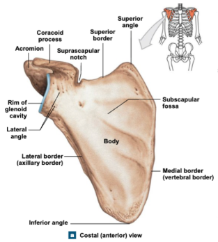
Which direction does the coracoid process of the scapula point?
Anteriorly

What is the largest bone of the upper limb?
Humerus, head is proximal and points medially

The humerus attaches to the ulna at what landmarks?
The trochlea (always posterior) of the humerus attaches to the trochlear notch of the ulna
What are condyles and their functions in the humerus?
Condyle articulates bones to one another
Trochlea: attaches to ulna at the trochlear notch
Capitulum (anterior condyle): articulates with radius
What is the intertubercular sulcus?
A groove on the anterior side of the humerus, provides attachments for muscles
Regardless of supination or pronation of the forearm, which bone is always positioned medially?
Ulna
What structures form the PROXIMAL radioulnar joint?
Head of radius + radial notch of ulna
What structures form the DISTAL radioulnar joint?
Head of ulna + ulnar notch of radius
What is the name of the membrane located between the radius and ulna?
Interosseous membrane
What are the PROXIMAL bones of the carpal?
Scaphoid, Lunate: articulate with radius
Triquetrum, Pisiform

What are the DISTAL bones of the carpal?
Trapezium, Trapezoid, Capitate, Hamate

What are metacarpals and their structure?
5 metacarpals make up the palm of the hand: long bones, each with a base, shaft and head
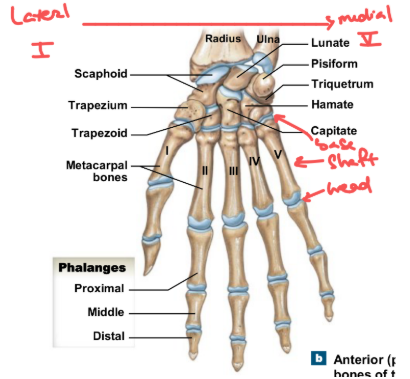
What are phalanges and how many phalanx does each one have?
Phalanges are long bones in the fingers:
Pollex (first phalanx) has 2 phallanges
Digits 2-5 each have 3 phalanges

Joints are generally classified based on what two general criteria?
Structure: histological structure of the joint
Function: range of motion
What is a synarthrosis joint (functionally), and what structural types fall into this category?
Immovable - Fibrous and Cartilaginous joint
What is an amphiarthrosis joint (functionally), and what structural types fall into this category?
Slightly moveable - Fibrous, Cartilaginous joint
What is a diarthrosis joint (functionally), and what structural types fall into this category?
Freely moveable - Synovial joints
What are two examples of fibrous synarthrosis joints?
Skull sutures, gomphosis
What is an example of a fibrous amphiarthrosis (slightly moveable fibrous) joint?
Interosseous membrane
What is an example of a cartilaginous synarthrosis joint?
Epiphysis of long bones
What is an example of an amphiarthrosis cartilaginous joint?
Cartilaginous joint of the ribs
What functional category do all synovial joints fall into?
Diarthrosis joints
What are the structures of a synovial joint?
Articular capsule: fibrous exterior
Synovial membrane: thickenings that make the capsule ligament
Articular cartilage: articular surfaces covered in hyaline cartilage
Joint cavity: contains synovial fluid for lubrication and protects bones

What type of joint movement occurs in the sagittal plane?
Flexion/extension
What type of joint movement occurs in the coronal plane?
Abduction/adduction
What type of joint movement occurs in the transverse plane?
Internal and External rotation OR medial and lateral rotation
What are the joints of the pectoral girdle and their properties?
Acromioclavicular joint: synovial gliding joint
Sternoclavicular joint: fibrocartilaginous saddle joint, amphiarthrosis
Where is the origin and insertion of all chest muscles?
All chest muscles start the axial skeleton and insert on the limbs/pectoral girdle
What movements does the Glenohumeral joint allow?
Most freely moveable joint: Flexion/extension, IROT/EROT, abduction/adductionW
What muscles at the Glenohumeral joint allow for stability?
4 rotator cuff muscles
What is the function of the Glenoid labrum?
Increases the depth of the glenoid cavity => keeps the humerus secured in place
What type of joint is the Elbow/Humeroulnar joint, and what bony structures are involved?
Hinge joint between the trochlea, coronoid process, olecranon, trochlear notch
What is the Humeroradial joint and its components?
Pivot joint consisting of the Capitulum and head of radius
What type of joint is the Radiocarpal joint and what movements does it allow?
The Radiocarpal joint is a condyloid synovial joint, allowing flexion/extension, radial and ulnar deviation
Describe the curvature of the vertebral column
Convex curvatures = Secondary/Lordotic curvatures develop later in life
Concave curvatures = Primary/Kyphotic curvatures that develop first

Which regions of the vertebral column allow flexion/extension?
Cervical and lumbar
Which regions of the vertebral column allow rotation?
Cervical and Thoracic
Which regions of the vertebral column allow lateral bending?
Mostly cervical, some lumbar
What are vertebrae processes and their functions?
Processes are bony projections that provide attachments for muscles
Transverse processes: have unique structures in different regions = allows regional identification

What are facets and their functions in vertebrae?
Facets are smooth surfaces that provide stability and guide movements

What is the pedicle and its functions in vertebrae?
The pedicle connects the vertebral body to the transverse process of the next vertebra

How to distinguish if a vertebra is cervical, thoracic or lumbar?
Cervical vertebrae: have transverse foramen that contains vertebral artery
Thoracic vertebrae: have transverse costal facets
Lumbar vertebrae: no transverse costal facets and transverse foramen

What does the Sacrum consist and what does it articulate with?
The Sacrum is made of 5 fused vertebrae, articulating with L5 superiorly and the Coccyx inferiorly
Which structure of the sacrum is formed by the fused vertebral bodies?
Transverse ridges
Which structure of the sacrum is formed by the processes being fused together?
Sacral crest:
Medial sacral crest: fused spinous processes
Lateral sacral crest: fused transverse processes
Sacral foramina: contain sacral nerves
What is the structure where the sacrum articulates with the hip bone?
Sacral ala
Describes the Vertebral development
Vertebral development begins with a cartilage model then bone develops at the ossification centers

What is Spina bifida and its cause?
A neural tube defects caused by the lack of folic acid/folate that impacts neural arches fusion
What are the components of the intervertebral discs?
Hyaline cartilage: remnant of embryonic skeleton
Annulus fibrosus: fibrocartilage ring for stability
Nucleus pulposus: gel-like, absorbs compressive forces

What happens in a herniated disc?
The nucleus pulposus broke through the annulus fibrosus
What is the function of the thoracic cage?
Protects vital organs in the thoracic cavity, supports pectoral girdle and upper limbs
How do we classify the ribs?
True ribs: 1-7, directly articulates with the sternum via cartilaginous synarthrosis joints
False ribs: 8-12, do not directly articulate with the sternum
8-10: Vertebrochondral ribs
11-12: Floating ribs

What are the 3 sections of the Sternum?
Manubrium, body, xiphoid process

What are some of the anatomical landmarks of the Sternum and their articulation?
Jugular notch: horizontally in plane with T2/3
Sternal angle: articulate with T4/5
Xiphisternal joint: articulate with T9

What does the head of the rib articulate with?
Vertebral body
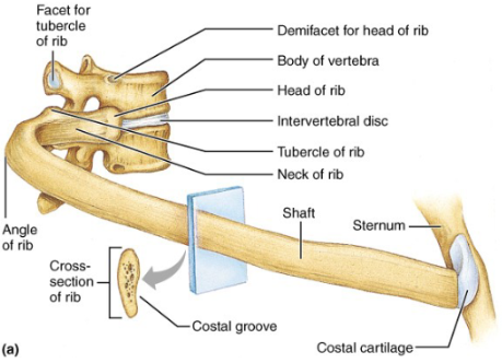
What does the tubercle of the rib articulate with?
Transverse costal facets

What does the costotransverse joint connect?
Rib and Inferior vertebra

What does the costovertebral joint mark?
Where the head of the rib articulate with vertebral body

What are skeletal muscles and what are their components?
Skeletal muscle: striated, highly metabolic tissues, 40% of BW
Skeletal muscle fibers contain myofibrils made of thick (myosin) and thin (actin) filaments

What is a Sarcomere and where are the bands within it?
Sarcomere: a working unit of skeletal muscle
I-band: contains thin filaments anchored at Z-disks
H-zone: contains thick filaments anchored at middle of H-zone
A-band: overlap zone

What happens during muscle contraction?
Myosin binds actin, pull along Z-disks towards the middle => Cross-bridge cycling
What is the difference between muscle’s origin and insertion
Origin: point of muscle attachment that remains stationary
Insertion: point of muscle attachment that is moveable

What is a muscle action?
Function of the muscle upon contraction: always pull the insertion towards origin
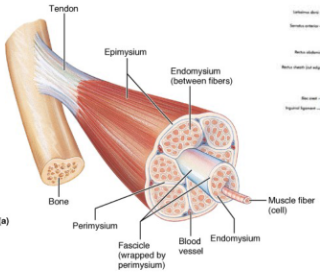
What are tendons and their categories?
Tendons are connective tissues that anchors muscles to bones
Endomysium: surrounds single muscle cells
Fascicle: bundle of muscle fibers
Perimysium: surrounds fascicles
Epimysium: surrounds entire muscles

What is the Periosteum?
Outer layer of bone that tendon attaches to
What are the different fiber orientations and how is force produced?
Force is produced along the line of contraction:
Parallel fibers: Strap, Fusiform
Angled fibers: Unipennate, Bipennate, Multipennate

What are the muscular components of every movement?
All movements require more than 1 muscle:
Antagonists: oppose action of prime movers
Agonists/prime movers/synergists: facilitate primary movement
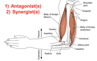
What are the factors that impact muscle performance?
Muscle force generation is optimized at a certain resting sarcomere length
Greater muscle size/cross-sectional area = more sarcomeres = more force
Length of muscle allows for greater ROM = greater torque arm
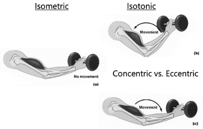
What is Isometric contraction?
Contraction without changing muscle length

What is Isotonic contraction?
Consists of Concentric and Eccentric contractions:
Concentric: shortening contraction
Eccentric: allows smooth/controlled movements
What is a Motor unit and how can we increase force production?
Motor unit: a single motor neuron and the fibers it innervates
Recruitment = adding more motor units = more force

Where do the scapular muscles originate and insert?
Originate in axial skeleton, insert on scapula
What movements does the Trapezius allow?
Elevation, depression, retract scapula
Upward rotation of scapula: often comes with arm movement
Cervical extension, lateral flexion of the head/neck

What movements does the Rhomboids allow?
Opposite functions as trapezius: downward rotation of scapula, retraction

What movements does the Serratus Anterior allow?
Starts at the ribs, attaches to the side of scapula: allows protraction and upward rotation of the scapula

What movements does the Levator Scapulae allow?
Elevation and downward rotation of the scapula

What movements does the Pectoralis Minor allow?
Originate in the ribs and insert on coracoid process: scapular protraction

Where do the Glenohumeral muscles originate and insert?
Originate on axial skeleton, insert on intertubercular sulcus