Skin, Hair, & Nails + Musculoskeletal System
 Call Kai
 Learn
 Practice Test
 Spaced Repetition
 Match
 Flashcards
 Knowt Play
1/54

Earn XP
Description and Tags
Health Assess Exam 2
55 Terms
Wheal
Elevated, irregular-shaped area of subcutaneous edema.
Nodule
Elevated, firm, circumscribed lesion; deeper in the dermis than a papule. It is 1-2 cm in diameter, such as a lipoma, hemangioma, or neurofibroma.
Macule
Small, flat, circumscribed areas that are a change in the color of the skin and are less than 1 cm in diameter; freckle or flat mole
Papule
Elevated, firm, circumscribed area less than 1 cm in diameter, such as a wart, elevated mole, or skin tag.
Patch
Flat, nonpalpable, irregular-shaped macules greater than 1 cm in diameter.
Vesicle
Elevated, circumscribed, superficial (do not extend into dermis), filled with serous fluid, and less than 1 cm in diameter.
Keloid
A hypertrophic scar.
Pustule
Elevated, superficial lesion similar to a vesicle but filled with purulent fluid (example: acne).
Bullae
Large vesicles greater than 1 cm in diameter.
Fissure
A linear crack with abrupt edges that extends into the dermis; it can be dry or moist.
Grade & (Lovett Scale) for Muscle Strength
0 (Zero) = No evidence of contractility
1 (Trace) = Evidence of slight contractility
2 (Poor) = Complete ROM w/ gravity eliminated
3 (Fair) = Complete ROM w/ gravity
4 (Good) = Complete ROM against gravity w/ some resistance
5 (Normal) = Complete ROM against gravity w/ full resistance
Abnormal findings of Muscle Strength
- Paralysis - a lack of voluntary movement that is spastic or flaccid:
Spastic paralysis (exaggerated muscle tone) - the involuntary contraction of muscles that occurs after a spinal cord injury or Cerebrovascular Accident (Stroke)
Flaccid paralysis is a lack of muscle tone or deep tendon reflexes after damage to lower motor neurons
Assessment for Evaluating Muscle Strength
Bicep: pt. flexes the arm while the RN pulls to extend the arm
Triceps: pt. extends the arm while the RN pushes to flex the arm
Ankle & Foot Muscles: pt. performs dorsiflexion while the RN pushes the foot to plantar; pt. performs plantar flexion while the RN pushes the foot to dorsiflexion
Range of Motion (ROM)
Active ROM - movement done by client & should be smooth and painless through its complete range
Passive ROM - examiner moves the relaxed joint through the limits of movement; stop if encounter pain or resistance
Flexion
bending (decrease in angle)

Extension
straightening limb to increase angle


Hyperextension
bend as far as possible

Abduction
away from midline

Adduction
toward midline

Internal Rotation
turning the part/joint toward midline

External Rotation
turning joint away from midline

Circumduction
circular movement

Supination
turning palm up
Pronation
turning palm down

Opposition
touching thumb to each finger on same hand
Inversion
turn inward


Eversion
turn outward
Dorsiflexion
move foot so that toes are pointed upward
Plantar flexion
move foot so that toes are pointed downward
ROM Cervical Spine Head & Neck
Flexion, extension, hyperextension, lateral flexion, & rotation
Jaw: open & close; side to side (smooth & w/o pain); protrude & retract
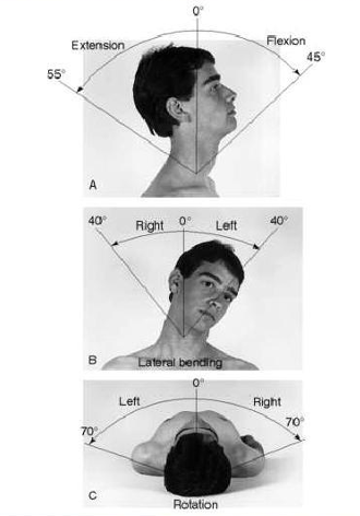
ROM of Shoulders
Forward flexion, extension, hyperextension, abduction, adduction, internal rotation, & external rotation

ROM of Thoracic & Lumbar Spine
Flexion, extension, hyperextension, lateral bending, & rotation of upper trunk

Normal Spine Curvature
Cervical - concave
Thoracic - convex
Lumbar - concave
* No matter which way you approach the patient
Scoliosis
Structural Scoliosis - lateral curvature of thoracic or lumbar vertebrae; most often in adolescence and females. Treatment includes a brace to prevent further progression of curvature
Functional Scoliosis - (temporary change) due to unequal leg length, muscle spasms, poor posture; correct underlying problem to eliminate spinal curvature
- assess by having pt. do forward flexion at the waist & rise slowly: shoulders & iliac crests are unequal

Kyphosis
- excessive convexity of the thoracic spine
causes: aging, poor posture, Scheuermann’s disease, arthritis, osteoporosis, disk degeneration, fractures, or cancer/cancer treatments
Lordosis
- excessive concavity of lumbar spine
causes: obesity, pregnancy, osteoporosis, benign (juvenile), achondroplasia (associated w/ short stature in dwarfism), kyphosis
ROM of Elbows
flexion, extension, supination, pronation

ROM of Hands & Wrists
- knuckle joints: metacarpophalangeal flexion & extension, metacarpophalangeal hyperextension
- thumb: thumb flexion & extension
- fingers: finger flexion (make a fist) & extension, abduction & adduction, opposition
- wrist: flexion, extension, hyperextension, radial deviation, ulnar deviation

ROM of Hips
hip flexion (knee straight - 90 degrees), hip flexion (knee bent & to chest - 120 degrees), hip extension (back to straight leg in standing position/supine), abduction, adduction, hyperextension, internal rotation (turn knee toward inside), external rotation (turn knee toward outside)

ROM of Knees
flexion & extension
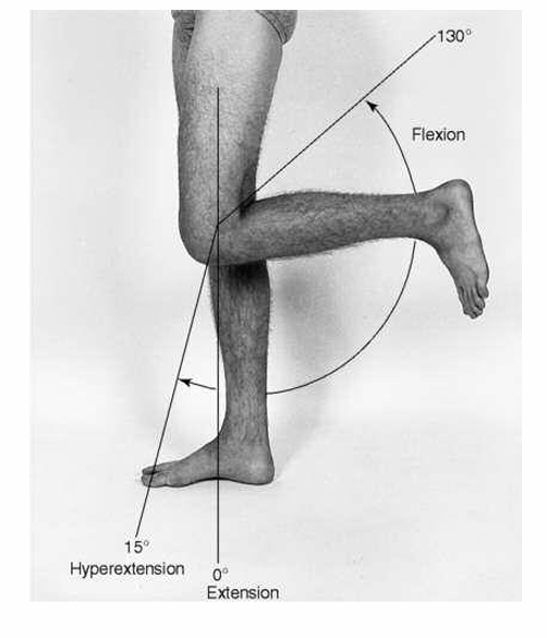
ROM of Ankles
dorsiflexion, plantar flexion, inversion, eversion, abduction, & adduction
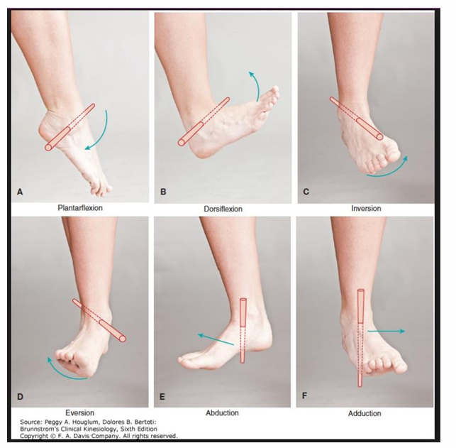
Carpal Tunnel Syndrome
caused by pressure on the median nerve as it travels through the carpal tunnel
Tinel’s Sign
- test for carpal tunnel syndrome
- tap on median nerve where it passes through Carpel Tunnel (at wrist); no pain or tingling is a negative test

Phalen’s Sign
- test for carpal tunnel syndrome
- flex both wrists and press dorsal aspects against each other for 1 minute; if pain, numbness, or tingling: positive for Carpal Tunnel

Rheumatoid Arthritis (RA)
- chronic inflammatory, autoimmune disease (cause unknown)
- causes destruction of connective tissue
- synovial joints become inflamed causing swelling, which leads to deterioration of cartilage and erosion of bone surface
- own body will attack joints bilaterally
- risk factors: female (usual onset in 30s & 40s), viral infection trigger, small frames & genetic predisposition
Rheumatoid Arthritis Onset, Symptoms, & Deformities
- onset: usually begins insidiously over several weeks to months, but can be sudden (24-48 hours); progressive, with exacerbations & remissions
- symptoms: joint involvement is usually bilateral; pain, edema, stiffness: fingers, wrists, ankles, feet, & knees; morning stiffness usually lasts more than 30 minutes
- deformities: ulnar deviation, Swan-Neck, & Boutonniere
Osteoporosis
- bones become porous due to a loss in bone mass/density (osteopenia) and decrease in bone formation
- leads to increased risk of fractures: 1 in 2 women and up to 1 in 4 men age 50+ will break a bone due to osteoporosis
- known as the “silent disease” because bone loss occurs w/o signs or symptoms
- thin, frail, & athletic affected as well
Osteoporosis Risk Factors
- uncontrollable risk factors: gender, age, body size, ethnicity, bone fractures
- modifiable risk factors: lack of exercise, low calcium, anorexia nervosa, low estrogen levels, smoking, caffeine, alcohol, medication intake
- risk reduction: increase physical activity, increase calcium & Vitamin D intake; avoid excessive caffeine, alcohol, steroids, or smoking; estrogen replacement therapy & prevent falls
Osteoarthritis - Degenerative Joint Disease (DJD)
- progressive breakdown & loss of cartilage in one or more joints
- caused by wear & tear, trauma
- obesity, frequent pregnancies
- affects: vertebrae, hips, knees, & ankles; can also be noted in fingers
- symptoms: joint edema, aching; diffuse pain w/ movement
morning stiffness lasts less than 30 minutes & decreases w/ movement
deformities such as Heberden nodes & Bouchard nodes
Gout
- form of arthritis that develops from an increase in serum uric acid causing crystals of uric acid to deposit in one or more joints
- great toe is most common place affected, but can occur in wrists, knees, & ankles
- patients report sudden & progressively severe onset of redness, pain, & edema in affected joint
- causes: diet, increase in animal organs/red meat, increase in alcohol consumption, medications; more common in men
Special Considerations for Infants
- palpate clavicle for fractures (fracture - pain, bruising, loss of function)
- inspect back & spine for alignment, tufts of hair, bulges or obvious malformation
- assess hips
Barlow maneuver (up to age 3 months) & Ortolani maneuver - should be no clicking of hips = misalignment
Allis sign (Galeazzi Test)
Infant Hip Assessment
Hip dysplasia - medial term for a hip socket that doesn’t fully cover the ball portion of the upper thighbone; allows the hip joint to become partially or completely dislocated
- most people w/ hip dysplasia are born w/ it
Special Considerations for a Child
- compare data to tables of normal age & sequence of motor development
- observe gait for steadiness
- examine back - shoulders, scapula, iliac crest for symmetry
- wide stance & gait until 24-36 months
- outward rotation of the lower extremities can be normal in children ages 2-3.5 years
- inward rotation requires further evaluation
- balance problems or increases in falls should be evaluated
- lordosis (more common in African American children) can be present up to age 6
Special Considerations for Adolescents
- screen for scoliosis, kyphosis, & lordosis
- injuries due to sports or trauma
Special Considerations for Older Adults
- decreased bone and muscle mass
- intervertebral disk space narrows (result: decrease in height by ~1-4 in)
- increased risk for falls & fractures
- decreased agility & endurance for exercise
- gait is slower