Movement, skeletal system and bone
 Call Kai
 Learn
Learn Practice Test
 Spaced Repetition
 Match
Match Flashcards
 Knowt Play
1/44

There's no tags or description
Looks like no tags are added yet.
Name | Mastery | Learn | Test | Matching | Spaced | Call with Kai | Chat |
|---|
No analytics yet
Send a link to your students to track their progress
45 Terms
Frontal/ coronal plane
divides body into anterior and posterior parts
medial (midsagittal)
lies on middle of body, splits into left and right
sagittal plane
splits body into two uneven halves
transverse (horizontal) plane
divides body into superior and anterior parts
anatomical position
universal reference point for anatomical description. standing with palms facing forward
flexion
reducing angle of the lamp (bending elbow)
extension
increasing angle of the joint
abduction
movement laterally away from the body
adduction
movement towards the body
rotation
movement along axis of a bone (bone is spinning)
circumduction
distal end of limb draws a circle
inversion
turning foot medially (sole in)
eversion
turning sole of foot laterally (out)
5 functions of the skeleton
1 support and attachment
2 movement
3 protection
4 storage (calcium in bones, fats in yellow bone marrow)
5 haematopoiesis
haematopoiesis
blood cell formation in red bone marrow
axial skeleton
longitudunal axis of the body, contains the skull, verebral column and thoracic cage
appendicular skeleton
upper limbs and attatching pectoral and pelvic girdle
what is a bone
a form of supporting connective tissue
why is a bone an organ
it has 2 tissue types and blood vessels
what forms a bone
calcium phosphate crystallised around protein fibres made by osteoblasts
osteoblast
bone making cells
long bone
longer than they are wide. Found in the limbs and act as
levers for movement. eg humerus
short bone
Cube shaped found in the wrist and ankles and provide strength
and stability. eg talus
flat bone
thin and flattened, protects organs and serves as muscle attatchment eg sternum
irregular bones
vertebrae of the spine
sesamoid bone
bone embedded into tendon that resembles a sesame seed.
function of sesamoid bone
acts as a pulley to increase tendon’s mechanical advantage and protects from stress eg patella

closed fracture

open fracture

greenstick

impacted fracture
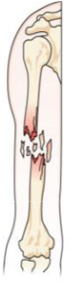
comminuted

spiral fracture

transverse
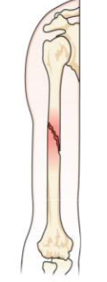
oblique fracture
compact bone
dense tube like structures (osteons) running in one direction. Very strong in longitudinal directions
spongy (cancellous bone)
looser meshworks of struts (trabeculae) giving strength in all directions
osteon
structural and functional unit of compact bone. Consists of concentric layers of bone matrix (lamellae)
function of osteons
structural support
nutrient supply (vascular network within canal ensures bone cells receive nutrients)
bone remodeling (formed and reabsorbed during continuous bone remodeling to adapt to mechanical loads
first few days after fracture
fracture haematoma (blood clot) formation and phagocytosis (breakdown) of damaged material
few weeks after fracture
periosteum cells lay external soft callus made of cartilage and spongy bone across break to stabilise broken ends
6-12 weeks post fracture
cartilage tissue in callus is replaced with spongy bone (withstands gentle stresses- cast can be removed)
several months post fracture
compact bone replaces spongy bone where appropriate
what affects healing process of a fracture
nutritional status, age, general health, nature and extent of the break
why are some bones more difficult to heal?
some bones have poor circulation eg scaphoid, neck of femur and shaft of tibia