9/11 and 9/14 (after - unit 1 (post quiz))
 Call Kai
 Learn
Learn Practice Test
 Spaced Repetition
 Match
Match Flashcards
 Knowt Play
1/51

There's no tags or description
Looks like no tags are added yet.
Name | Mastery | Learn | Test | Matching | Spaced | Call with Kai | Chat |
|---|
No analytics yet
Send a link to your students to track their progress
52 Terms
what drives a post-synaptic potential?
channel opening
Excitatory postsynaptic potential (EPSP)
raises Vm closer to AP threshold
Inhibitory postsynaptic potential (IPSP)
lowers Vm further from AP threshold
ionotropic receptor vs metabotropic receptor
ionotropic receptor
ion channel
neurotransmitter binds → channel immediately opens or closes → ions move down their concentration gradient → membrane potential changes
fast and direct
metabotropic receptor
NOT an ion channel
usually a G-protein coupled receptor (GPCR)
when neurotransmittor binds it starts a chain of events inside the cell that affects ion channels or other proteins
slow (tens of ms to sec) but effects last longer
provides amplification: many 2nd messenger molecules generate
metabotropic receptor pathway
neurotransmittor binds to the metobotropic receptor
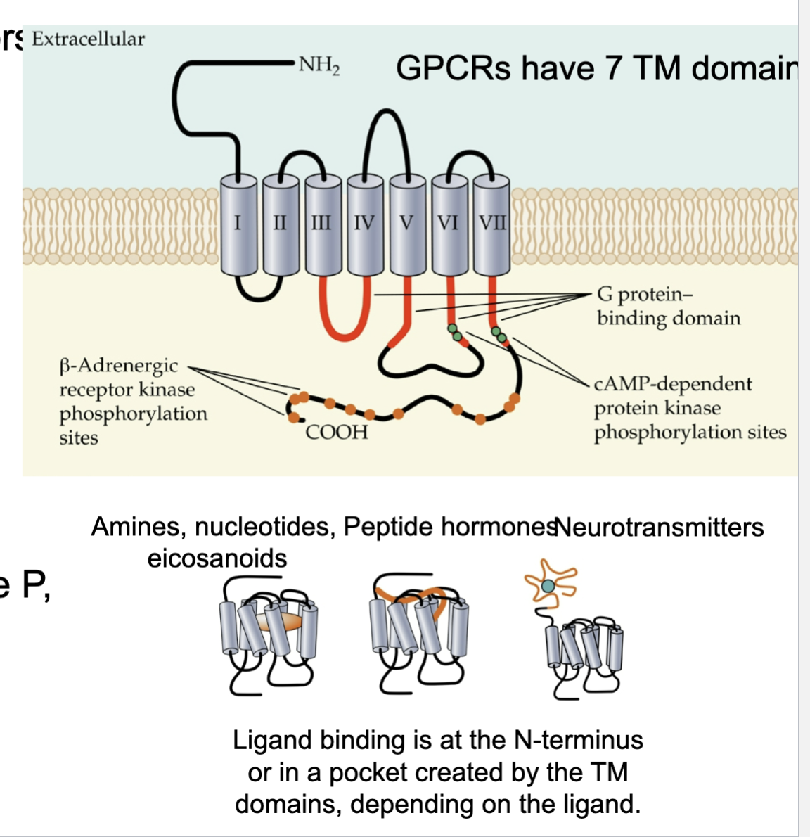
receptor has 7 transmembrane segments
receotor activates a G protein
G protein is made up of three subunits: alpha, beta, and gamma
alpha separates and can interact with another protein
G protein activates an enzyme
in this example it activates adenyl cyclase
Adenyl cyclase produces a second messanger
converts ATP → cAMP
first messenger = neurotransmittor outside cell
second messenger = cAMP (molecule produced inside cell that carries signal forward)
cAMP activates another enzyme (protein kinase A)
PKA → phosphorylkates proteins
PKA phosphorylates the K+ channel
phosphorylation causes K+ channel to close
K+ channel closes
less K+ leaves the cell
in this example causing depolarization but in other examples can also hyperpolarize

how can metabotropic receptors affect the cell?
depolarization or hypoerpolarization
GPCR structure
7 transmembrane domains that cross the membrane each time (so 7 total)
N-terminus on outside (NH2)
C-terminus on inside (COOH)
ligand binds outside → receptor changes shape → G protein inside is affected
what is a motor neuron, motor nerve, motor unit, muscle
motor neuron carries signals from the CNS to the skeletal muscle
one motor neuron can control multiple muscle fibers
motor nerve contains multiple motor neuron axons
motor unit - one motor neuron + ALL the muscle fibers that it innervates
one muscle contains many motor units

neuromuscular junction
specialized synapse between the presynaptic cell = motor neuron and the postsynaptic cell = skeletal muscle fiber
neuron→ muscle synapse

pathway of how the nervous system gets a signal to the muscle
motor neuron fires an action potential, and it arrives at the motor neuron terminal depolarizing the presynaptic membrane
depolarization opens voltage gated Ca2+ channels
Ca2+ moves from outside to inside
Ca2+ is the signal that triggers synaptic vesicle fusion
vesicles release ACh by exocytosis into the synaptic cleft
ACh binds to nicotine ACh receptors(ionotropic) located on the motor end plate
the channel opens allowing Na+ to flow into the muscle(greater influx) and K+ out of the muscle (smaller eflux)
causing local depolarization (end-plate potential)
the depolarization spreads
positive current spreads and deploarizes nearby membrane containing voltage gated Na+ channels
muscle AP is generated
if meets threshold*
now made transition from motor neuron AP to muscle AP (not the same thing)
ACh has to be broken down

nicotine ACh receptor
ionotropic receptor - fast and direct
ligand-gated ion channel
pentamer (5 subunits, each with 4 transmembrane domains) crosses membrane 4 times; forms central pore ion flows thru
non-selective cation channel
Na+ can enter
K+ can leave

for NMJ mechanism why does the membrane depolarize after AcH binds to nAChR if Na+ is going in and K+ is going out?
the inward pull (driving force) on sodium (\(\text{Na}^{+}\)) is much larger than the outward push on potassium (\(\text{K}^{+}\)), making the net movement of positive charge enter the cell
What happens to postsynaptic Vm when ACh binds to the channels at rest?
ACh opens nAChRs, which are non selective cation channels. Na+ enters and K+ exits, but at resting Vm there is a net inward positive current, causing depolarization (EPSP) and driving Vm towards the nACHR reversal potential (around 0 mV)
there is a net inward positive current because ENa (+60 mV) is far from Vm(-70 mV) causing a strong driving force inward and EK (-90 mV) is closer to Vm making it a smaller driving force
what are the three types of muscles?
skeletal muscle
cardiac muscle
smooth muscle
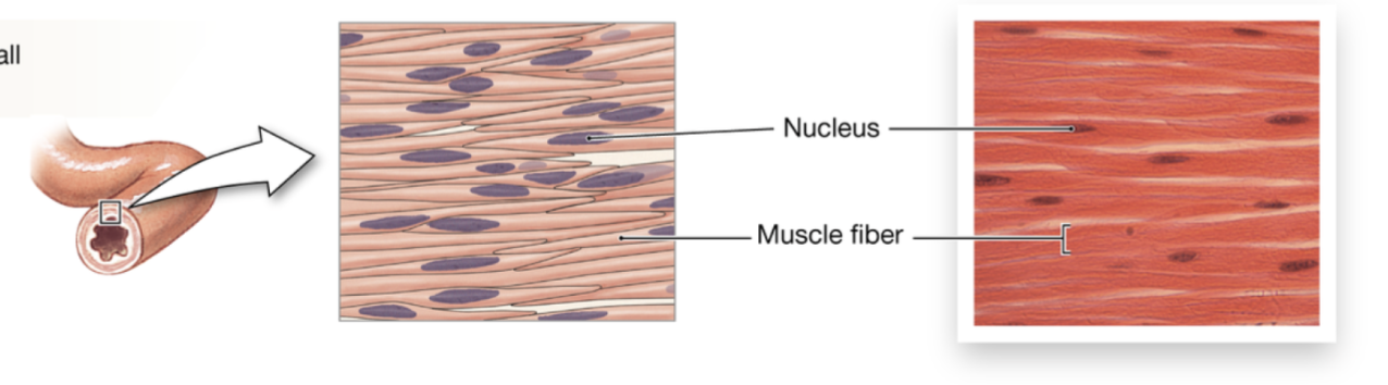
skeletal muscle fibers
large, long, striated, and multinucleated
one skeletal muscle fiber = one muscle cell
type involved in NMJ

cardiac muscle fibers
striated, smaller, branched, and uninucleated
intercalated disks are type of junction that joins cardiac cells

smooth muscle fibers
small and lack striations
muscle fiber structure hierarchy
one muscle fiber = one muscle cell = one myocyte
sarcolemma - plasma membrane of muscle cell
each fiber contains myofibrils (long cylindrical structures)
each myofibril is made up of repeating units called sarcomeres
each sarcomere contains thin (actin) and thick (myosin) filaments
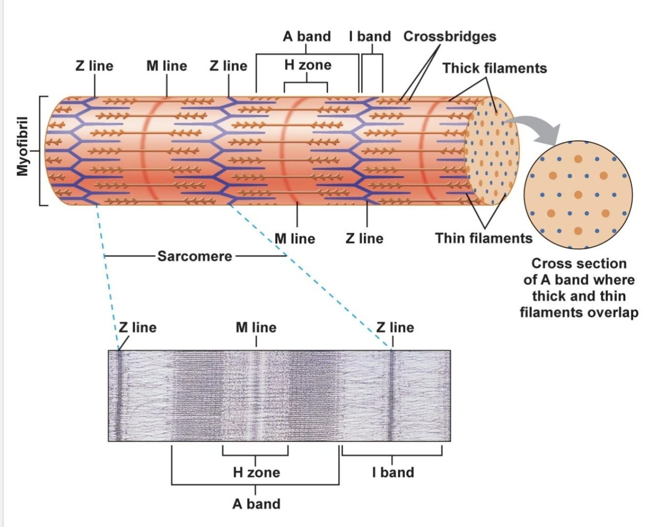
sarcomere - contractile machinery + structure
sarcomere is the fundamental unit of striated muscle
thick (myosin) and thin (actin) filaments make up the contractile machinery
move relative to one another to contract muscle fiber and driven by depolarization and Ca2+ signalling
zline → zline = one whole sarcomere
zline - actin - myosin - actin - zline

A band - sarcomere
corresponds to the entire length of the thick/myosin filaments
regions with myosin only + regions where myosin + actin overlap
I band
contains only thin flaments
actin only

H zone
center portion of A band where there is thick/myosin filament only

crossbridges
when myosin heads attach to actin they form crossbridges

how do myosin and actin interact with each other in the sarcomere?
thick filaments (myosin) pulls on thin filaments (actin) [not other way around]
myosin heads attach to the actin forming a crossbridge
the myosin head will pivot and pull actin toward the middle of the sarcomere
![<ul><li><p>thick filaments (myosin) pulls on thin filaments (actin) [not other way around]</p><ul><li><p>myosin heads attach to the actin forming a crossbridge</p></li><li><p>the myosin head will pivot and pull actin toward the middle of the sarcomere</p></li></ul></li></ul><p></p>](https://assets.knowt.com/user-attachments/91f2c783-d761-4985-80a6-cad996c251aa.png)
thin filament made of process?
thin filaments are made of actin
individual actin proteins are called G actin and each one contains a myosin-binding site where the head will attach
G-actin molecules polymerize together gotming F-actin
two F-actin strands twist together to form double-helical actin strands
regulatory proteins (tropomyosin and troponin) modulate binding of motor of myosin to binding sites

thick filament made of process?
thick filaments are made of myosin which is an ATPase (catalyze ATP and use released energy)
single myosin molecule contains:
tail + 2 heads(molecular motors waiting to grab actin)
head contains: actin-binding site and ATPase sites
two myosin molecules bound at theur tail ends
several hundreds of myosin molecules assemble together to form one thick filament
Ca2+ importance in crossbridge cycling?
In the absence of Ca2+:
relaxed muscle
muscle does not want myosin grabbing actin constantly so tropomyosin covers the myosin-binding sites on actin
myosin cannot bind to actin → no cross-bridge cycling → no contraction
In the prescence of Ca2+:
contracting muscle
Ca2+ binds to troponin and causes troponin to change shape, which causes tropomyosin to move away from the myosin-binding sites on actin exposing them
binding sites exposed → myosin can bind to actin
The crossbridge cycle process
requires ATP and Ca2+ to occur
1. A new ATP molecule binds to the myosin head causing myosin to release actin
2. myosin (ATPase) hydrolyzes ATP providing the energy to “cock” the myosin head
low energy myosin → ATP hydrolysis → high-energy/cocked myosin with ADP +Pi still attached to myosin head (ready to bind to actin form)
relaxed state bc myosin no longer bound to actin
3. Ca2+ must be present for this: myosin binds to actin and a coss-bridge has formed
Ca2+ binds to troponin → tropomyosin moves → actin binding sites exposed so the cocked myosin head can attach to the actin
4. high energy phosphate is released to provide energy for the power stroke
power stroke - as the myosin head pivots: myosin pulls actin toward the middle/M line of the sarcomere
5. ADP is released causing it to be in rigor state (myosin is in low-energy form)
myosin is still bound to actin therefore maintining tension hence rigor state
myosin can not detach from actin until another ATP binds
(only 3-4 nm per stroke!)

What is needed for cross-bridge cycling and why?
ATP
allows myosin to detach → cock → cycle again
detaches and energixes myosin
Ca2+
determines whether myosin has access to the myosin binding sites on actin
exposes actin
why does rigor mortis happen?
after death, ATP production stops:
without ATP
myosin cannot detach from actin so cross bridges become stuck and muscles become stiff causing rigor mortis
where does the Ca2+ needed for cross bridge cycling come from?
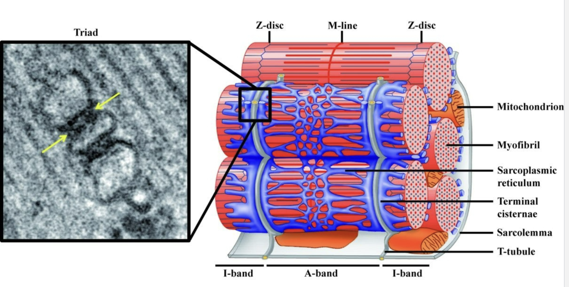
for skeletal muscle the major source is the sarcoplasmic reticulum
basically muscle cell’s specialized smooth ER and its main job is to store Ca2+
T-tubules bring the action potential inside the muscle fiber for calcium release
invagination of sarcolemma allowing for deep activation
AP travels along sarcolemma → enters T-tubules → electrical signal travels deep into the muscle fiber → eventually triggering Ca2+ release from the neighborinhg SR

sarcoplasmic reticulum and T tubules
excitation-contraction coupling
how does the muscle action potential cause the muscle to contract?
big pic sequence
ACh released at NMJ
released fom axon terminal of a motor neuron and binds to receptors in the motor end plate
ACh binds nAChRs
end-plate potential
muscle AP
AP travels along sarcolemma
AP travels down T-tubules
SR releases Ca2+
Ca2+ binds troponin
tropomyosin moves
cross-bridge cycling
contraction

DHP vs RyR1 and their roles and how they work
DHP receptor
Ca2+ channel protein located in the T-tubular membrane
functions as a voltage sensor for skeletal muscle excitation-contraction coupling
so when muscle AP depolarizes the T-tubule membrane; DHP detects the change in membrane voltage
RyR1
located in the SR membrane
it functions as the Ca2+ release channel
mechanically coupled to each other

Sr Ca2+ release pathway
action potential depolarizes the T-tubule
DHP changes conformation
because DHP and RyR1 are mechanically coupled; RyR1 opens
Ca2+ leaves SR

Where does Ca2+ move when RyR1 opens and why?
SR has a very high Ca2+ concentration compared with the cytosol
So when RyR1 opens: Ca2+: SR → cytosol
Ca2+ moves down its electrochemical/concentration gradient through RyR1
SERCA vs RyR1 vs Calsequestrin
SERCA
pump (ATPase) in the SR membrane
Ca2+ moves against its gradient, requiring ATP (cytosol → SR)
RyR1
Ca2+ moves down its gradient through a channel (SR → cytosol)
Calsequestrin
is inside SR and binds Ca2+
helps SR store a large amount of Ca2+ and calsequestrin helps to do that by binding Ca2+ inside the SR
reversal process - contraction stopping (relaxation)
Ca2+ stays bound to troponin and cross-bridge cycling continues for as long as cystolic Ca2+ remains high
as cystolic Ca2+ decreases:
SERCA uses ATP (pumps Ca2+ cytosol → SR) → Ca2+ dissociates from troponin → troponin returns towards its resting conformation → tropomyosin moves back over the myosin-binding sites → new cross bridges can no longer form (cross-bridge cycling stopped) → muscle relaxes

ATP involvement - contraction vs relaxation?
ATP is used for:
cross-bridge cycling: ATP binding allows myosin to detach, and ATP hydrolysis energizes/cocks the head
relaxation: SERCA hydrolyzes ATP to pump Ca2+ back into the SR
why is the AP highly probable?
a motor neuron can form multiple axon-terminal contacts with a muscle fiber
when motor neuron fires terminals release ACh onto the motor end plate
sliding filament model - what happens?
during contraction the filaments don’t shorten, the whole sarcomere does
action and myosin DO NOT get shorter
actin slides past myosin
thin actin filaments are attached to the Z lines so during the power stroke, myosin pulls actin toward the M line, which id the center if the sarcomere
actin pulled inward → z lines move closer together → sarcomere shortens
sliding filament model - what happens to the bands?
sarcomere - shortens during contraction
I band - shortens during contraction
H zone - shortens during contractio
A band - stays the same during contraction
Thick filament - stays same length during contraction
Thin filament - stays same length during contraction
actin-myosin overlaps - increases during contraction

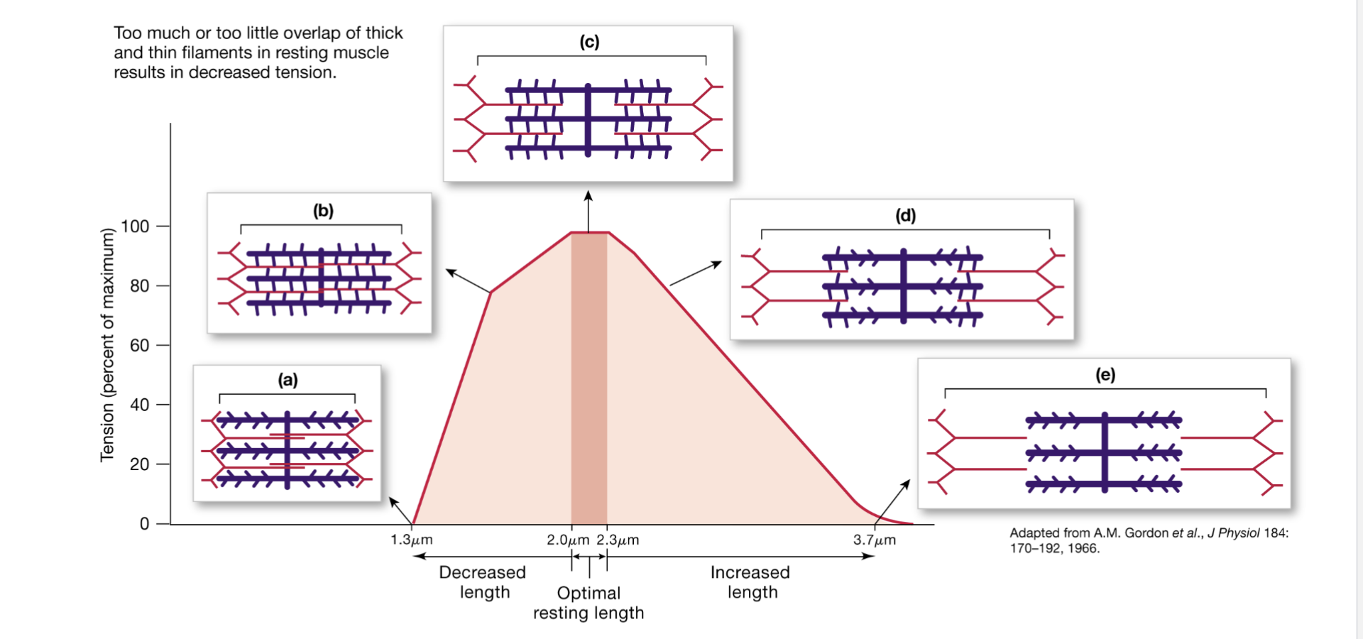
force-length relationship
a muscle produces different amounts of force depending on how stretched/shortened it is
the amount of force depends on the starting length of the sarcomere, because starting length determines how much actin and myosin overlap
actin - myosin overlap → determines how many cross-bridges can form → determines force
general relationship: appropriate overlap → many cross-bridges → high force
can be too little or too much overlap of thick and thin filaments in resting muscles which results in decreased tension
graph - descriptions
a and b
sarcomere is too short (increased overlap)
filaments begin interfering with one another and the thick filament approaches/compresses against the z discs; thin filaments from opposite sides also overlap excessively
interferes with effective cross-bridge production so force falls
c - (2-2.3 um)
actin and myosin have optimal amount of overlap
many myosin heads can interact with actin → lots of cross bridges → maximum active tension
d and e
sarcomere is too stretched (less overlap)
fewer myosin heads can reach actin→ fewer cross bridges → less force
Immediately following an action potential at the NMJ that causes a muscle twitch, what three things have to happen to stop excitation-contraction coupling?
neurotransmitter clearance
the ACh the motor neuron released has to be cleared or else it wuld continue activating receptors
acetylcholinesterase (AChE) - breaks doen ACh in the synaptic cleft
myocyte repolarization
muscle membrane was depolarized during its AP so it has to return toward resting membrane potential
voltage gated K+ channels open → K+ leaves the muscle cell → membrane repolarizes
Ca2+ clearance
SERCA uses ATP to pump Ca2+ from the cytosol back into the SR (once Ca2+ back inside SR → calquestrin helps bind/store it there)
as cytosolic Ca2+ decreases: Ca2+ comes off troponin → troponin returns toward resting conformation → tropomyosin moves back over the myosin-binding sites → myosin can no longer continue forming productive cross bridges → contraction stops/muscle relaxes
what determines the amount of force produced in a contraction?
actin-myosin cross bridge number is proportional to force
force-length relationship

where is force generated during sarcomere contraction?
myosin power stroke
myosin binds actin → power stroke → actin pulled → force generated
elastic structures
elastic elements within the sarcomere → titin
elastic elements at muscle attachment points → tendon recoil
If myosin pulls on actin inside the cell, how does that force get transmitted to structures outside the muscle fiber?
dystophin helps connect the internal contractile machinery to the sarcolemma and extracellular matrix, allowing force to be transmitted while stabilizing the muscle fiber

where does muscles need energy (which processes in excitation-contraction coupling)?
Muscles need energy to:
maintain concentration gradients (Na+/K+ ATPase)
drive myosin power stroke (ATPase)
pump calcium into the SR (SERCA ATPase)
During active cross-bridge cycling is the entire muscle getting shorter or longer?
the muscle could be getting longer or shorter or staying the same
does contraction always generate movement?
contraction generates force, but not necessarily movement
isotonic contraction
muscle contracts, shortens, and create enough force to move this load
muscle contracts → fore exceeds required load → muscle shortens → load moves

isometric contraction
muscle contracts but does not shorten. force cannot move this load.
muscle generates force but it doesnt generate enough force to move the load
so cross bridges are active and force is being generated but muscle length does not shorten
