Found 2.2 CM
 Call Kai
 Learn
 Practice Test
 Spaced Repetition
 Match
 Flashcards
 Knowt Play
1/42

There's no tags or description
Looks like no tags are added yet.
43 Terms
Passive 3 vs Active Transport 1 Summary
speed is inverse to size
Passive Transport
Diffusion = net movement down its gradient
simple diffusion: S & M
facilitated diffusion: L
every transported substance moves down its own gradient
Active is the only one that has at least one substance going against it’s concentration gradient (CG)
CG vs EG 2 vs PG
concentration gradient (CG) truly the number of molecules
Electrochemical Gradient (EG)
Electrical charge gradient created by the difference in charge across the lipid bilayer
ions can move based on charge difference across membrane
Originally the concentration gradient was equal, but now it is unequal & wants to achieve electoral neutrality
Nernst Potential (equilibrium potential)
Basically just describes how electrical & chemical gradients oppose each other
Pressure Gradient (PG): unilateral increase in pressure can force solutes across caused by greater numbers of molecules striking the membrane per second on one side than on the other side
CG, EG, PG Visual and recap
motion = ?
direction vs net
osmosis
All molecules/ions in body fluids (including dissolved substances) are in constant motion that we call "heat"
Greater motion = more heat
Motion never ceases completely except in absolute zero
Molecules move in all directions, but here we are concerned with the NET movement of those molecules. So it’s not that molecules move ONLY from high concentration to low concentration areas. Rather, they move in both directions, but the NET movement is from higher concentration to lower concentration until equilibrium is attained.
Osmosis is similar to diffusion, except it’s the movement of water the solvent rather than solute

Diffusion: Lipid- vs. Water-Soluble thru CM?
Lipid-Soluble Substances
Readily traverse through phospholipid bilayer
Ex: oxygen, nitrogen, carbon dioxide, alcohols, VA etc.
Water-Soluble Substances
Can readily traverse membrane through selectively permeable and gating membranous protein channels even if charged
Ex: Water is highly insoluble through membrane bilayer, but readily passes through protein channels (aquaporins) that penetrate through membrane
Voltage-Gated Na+ Ion Channels - 2
When the inside of the cell loses its negative charge (b/c more positive), VG-Na+ channel opens to allow Na+ to enter the cell as it flows down its concentration gradient (basic mechanism for depolarization and eliciting action potentials in nerves)
Selectivity filter lined with strong negative charges, pulling small dehydrated Na+ ions through channel
Na+ makes inside of the cell more positive

Voltage-Gated K+ Ion Channels - 2
3 step process
When the inside of cell is (+), K+ channels open to allow K+ to leave cell as it flows down its concentration gradient (basic mechanism for repolarization of cell)
Permit K+ ions passage through cell membrane ~ 1000x more readily than Na+ ions despite its larger size d/t inter-carbonyl oxygen distance on the inside of the protein.
K+ makes inside of the cell more negative
Hydrated K⁺ in body fluid is surrounded by water molecules—a hydration shell
Carbonyl oxygens temporarily replace the surrounding water molecules as it passes through the narrow filter.
Dehydrated K⁺ becomes surrounded by water again.

Ligand-Gated Channels - nAchR
Nicotinic Acetylcholine Receptor (nAchR)
Involved in skeletal muscle contraction
Ach binds nAchR, opens the gate, and allows Na+ ions to flow into the cell
This influx of Na+ makes the inside of the cell more positive, bringing the membrane potential to threshold potential, and elicits an action potential (see action potential section for more detail)
Ligand gated open → developed EPP → TP met → VG channels open
Other Types of Gated Ion Channels - 2

Carrier-Mediated: Facilitated Diffusion - ex
saturation kinetics 2
Water-soluble molecules cross cell membrane via carrier proteins
Example: glucose, fructose, galactose
Saturation kinetics
Once at max rate (Vmax), protein cannot increase transport rate, despite concentration of solute (it can't move solutes across any faster)
For a good part until Vmax, facilitated diffusion is faster than simple diffusion

Carrier-Mediated: Active Transport
Primary vs Secondary
Uses energy to move at least one substance against its CG (uphill)
Requires ATP (energy) either directly or indirectly
Primary Active Transport
Energy derived directly from ATP hydrolysis (or some other high-energy PO4)
Na+/K+ pump, Ca+ pump, H+ pump
Secondary Active Transport
Energy derived indirectly from ATP hydrolysis involved in an active transport process that formed an ionic concentration difference across membrane
Basically, it piggybacks off an active transport process
Na-K-ATPase - define 3
secondary fx
Uses energy (ATP) to pump both Na+ & K+ against their concentration gradients
Moves 2 K+ into cell & 3 Na+ out of cell
Creates & maintains ionic gradient (Na+ & K+)
Establishes resting net (-) charge inside cell
Also a secondary fx is to maintain the osmotic balance
Controls cells volume and prevents bursting by pumping Na+ out of the cell (water follows Na+)

Na-K-ATPase Visual
5 steps
Phosphorylation –addition of a phosphate group following the hydrolysis of ATP which leads to a conformational change in the target protein.
3 Na+ intracellularly binds first
Phosphorylation by ATP causes conformational change
3 Na+ released extracellularly as 2 K+ binds to other site
Dephosphorylation favors original form
2 K+ released intracellularly

Active Transport: Calcium Ions - 1 ex 2
Ca++ in ICF is normally very low (10,000x less than ECF)
Achieved by 2 active transport calcium pumps
PMCA: In the cell membrane to pump Ca2+ out of cell
SERS: Pumps Ca2+ into organelles such as the sarcoplasmic reticulum
Active Transport: Hydrogen Ions - ex 2
Gastric glands (hydrochloric acid)
At the secretory ends of the gastric gland, H+ ion concentration is increased up to 1 million-fold along with Cl- ions to form hydrochloric acid
Renal tubules (pH buffer)
Large amounts of H+ ions are secreted against its gradient (by 900-fold) from blood into renal tubular fluid for elimination
Na/H exchanger is a 2nd active transport
Secondary Active Transport:
relies on which ion gradient?
Two Forms
Energy in the form of an ionic gradient created by a primary active transport process
Usually involves Na+ ion gradient created by Na/K pump
Co-Transport: When solute moves down its gradient (created by active transport process), it can go with another substance with it
Counter-Transport: When solute moves down its gradient (created by active transport process), it can be exchanged for another substance
Secondary Active Transport: Co-Transport
sodium-x Co-T where? 2
Others? ntk
Sodium-Glucose Co-Transporter
Renal and intestinal epithelial cells
Glucose transported into cell with Na+ once both molecules bind to receptor
Glucose moves against gradient
Na+ moves with gradient
No ATP used!
More Ex
with Na+: Amino Acids, Phosphate, iodine
with others: K+, Cl-, HCO3-, Fe, urate

Secondary Active Transport: Counter-Transport - aka
Na-X Con-T where?
Na-X Con-T where? weaker than?
aka exchanger
Sodium-Calcium Counter-Transport (NCX)
Ca2+ is moving against its gradient
Na+ is moving with its gradient
cardiac > smooth muscles, neurons
Sodium-Hydrogen Counter-Transport (NHX)
proximal tubule (PT) of kidneys (Na+ reabsorbed)
Weaker vs ATP H+ transport in distal tubule (DT)

Action Potential: Outline
Membrane potentials
Diffusion potential
Equilibrium potential
Action potential
Refractory period
Anatomy of the neuron
Conduction types
Potential Difference
Membrane Potential
Resting Membrane Potential (RMP)
inside vs outside mV?
what else is highly negative inside the cell?
Potential Difference: Voltage difference b/w two points (Ohms Law)
Membrane Potential: Voltage difference across the plasma membrane (inside relative to outside)
Resting Membrane Potential (RMP): Voltage difference across the plasma membrane when cell is at rest
Intracellular: -90 mV
Extracellular: 0 mV
Charge inside of cell at (RMP) is negative
Leaky K+ channels allow K+ to leave the cell down gradient
High intracellular protein (protein is more negatively charged) concentration relative to extracellular concentration
Equilibrium Potential 2
Law of Electroneutrality 1
neutral vs balanced
Equilibrium Potential: The membrane potential that counters the chemical forces (concentration gradient) that would move an ion across the cell membrane, thus putting the ion in equilibrium
Charge at which a specific ion will not move across membrane
Equilibrium potential is not automatically 0 mV. Each ion has its own equilibrium potential based on its concentration gradient.
Law of Electroneutrality: the # of (+) & (-) charges must be equal across membrane
Desire for the charge to be neutral across membrane
This is more about ensuring that the charges are balanced more so than being perfectly neutral
Ex: in acidosis H+ goes in and to keep balance, K+ must leave the cell
↑ pH but hyperkalemia risk increases
Membrane Potential: Pump-Leak Model - 2
2 passive
1 active
Na/K ATPase = ↑ Na+ outside & ↑ K+ inside
Leaky Na+ & K+ channels allows each to flow down gradient
K+ diffuses faster than Na+ = ionic asymmetry = more (+) charges leaving cell than entering cell = (-) membrane potential is created
Resting Membrane Potential = -90 to -70 mV
Closest to Equilibrium Potential for most permeable ion (K+)
Equilibrium Potential (Nernst Potential) - define 1
Nernst equation
Membrane potential (voltage difference across cell membrane) at which a specific ion would be in "electrochemical equilibrium" - electrical & chemical forces are equal in force but opposite in direction
resulting in No net movement of that specific ion in either direction across membrane
Calculated by Nernst equation, which considers the concentration gradient of the ion across membrane
EMF (mV) = ±61 log (Ci / Co)
Considers the concentration gradient of the ion across the membrane
Equilibrium Potential of:
Na+
Cl-
K+
Ca2+
Knowing this helps you see which direction the ions want to go

Action Potential
Resting Membrane Potential:
Depolarization:
Threshold Potential:
Action Potential:
Repolarization:
Hyperpolarizaton:
Resting Membrane Potential: voltage of cell at "rest"
Depolarization: cell b/c more positive compared to the RMP
Threshold Potential: voltage threshold a depolarization must reach to initiate an action potential
Action Potential: transient electrical impulse in which the membrane potential rapidly rises to a much more positive level than RMP
Repolarization: cell b/c more negative, returning to RMP
Hyperpolarizaton: cell b/c more negative, below RMP
NaKATPase will fix this
Resting Membrane Potential - 2
maintained by?
Resting stage b/f action potential begins
Different for specific cell types (usually around –70 mV)
Membrane is said to be "polarized"
Established by Na+/K+ pump
Na+/K+ leak channels allow Na+/K+ to leak out of cell, even at rest
Depolarization - define
influx vs efflux
overshoot?
Membrane now permeable to Na+
Na+ & K+ channels open (K+ is very slow to peak)
Na+ influx occurs down its concentration gradient, bringing a positive charge inside axon
Influx = movement into the cell
Polarized state is neutralized (depolarized)
Can "overshoot" above zero (positive charge)
Repolarization - 3
Na+ channels close & K+ channels takes over (peak)
Rapid K+ efflux down its concentration gradient, bringing a positive charge outside axon
Efflux = movement out of the cell
Polarized state is restored
Voltage-Gated Sodium Channel
activation 2
inactivation 2 why?
Activation
As MP rises (depolarizes), it reaches a threshold (-55 mV) that induces a conformational change
Activation gate opens, allowing Na+ influx
Na+ permeability increases 500- to 5000-fold
Inactivation
Once +35 mV is reached the inactivation gate closes
Na+ influx stops
Inactivation gate remains closed until at or near RMP
Nerve can't depolarize again until repolarization has happened
Protective mechanism that prevents Na+ channels from reopening too quickly, and ensures that AP moves in one direction
Voltage-Gated Sodium Channel Visual
how many gates? aka?
gate movement as AP occurs 3
Activation Gate aka m gate (movement)
Inactivation Gate aka h gate (halt)

Voltage-Gated Potassium Channel
gates open when? why?
During Resting State
Closed during resting state, preventing K+ ions from moving down their concentration gradient
During Action Potential
When TP is met conformational change opens gate slowly and is fully open (peak) at about the same time VG-Na+ channels close at peak AP
When EPP is met, BOTH channels are open!
K+ is slower than Na+
Na+ has an inactivation gate which K+ does not
Voltage-Gated Potassium Channel Visual
Activation Gate aka n gate (not yet)

Na+/K+ Conductance - define
Conductance means how easily ions can cross the membrane because their channels are open.
Na+ conductance increases several thousand–fold during the early stages of the action potential
K+ conductance increases only about 30-fold during the latter stages of the action potential and for a short period thereafter.

AP Summary - 2
Each AP = a sudden change from (-) membrane potential to a (+) potential and ends with (-) potential
Once MP reaches TP, an AP is created (all-or-none fashion) - can’t be stopped
Are rapid changes in membrane potential that spread rapidly along the nerve fiber membrane (like a domino effect)
To conduct nerve signal, the AP moves along nerve fiber until nerve terminal

Threshold Potential
too little vs too much stimulus?
A weak electrical stimulus may not be able to excite a fiber (subthreshold potential)
However, when the voltage of the stimulus is increased, there comes a point at which enough excitation occurs to elicit an action potential
This point is called a threshold potential (about –55 mV)
Note: The action potential does not get larger with a larger stimulus
Refractory Period - define
2 zones - exactly when it starts and end
what’s a greater stimulus?
Period during which the cell is incapable of eliciting a normal action potential d/t inactivated Na+ channels being closed
Absolute
From initiation of action potential until membrane potential falls back below threshold potential
Na+ channel h gate is closed until open = start of next zone
No matter how great the stimulus, it is impossible to elicit another AP
Relative
Begins just after absolute refractory period, when membrane potential falls below TP
Lasts until membrane potential is reset to RMP
some h gates reopened, but K⁺ conductance remains high → greater stimulus required to reach TP
Doesn’t change TP of -55 mV, you just need more ligands, more nGpcr, more ions compared to resting state
Re-Establishing Na+ & K+ Gradients
1 active vs 2 passive
what ions are missing? where?
Na+ and K+ concentration differences are re-established by the Na+/K+ pump, essentially "recharging" the nerve for another AP
When one gradient gets too much, then that is when the Na+/K+ leak channels start to send overflow back to the other side
Ca+ and Cl- are more in cardiac cells
The Neuron - define
direction of AP
NT made? transported where? how?
Neurons are excitable cells
Once AP gets to Axon it goes ONLY in one direction
Dendrites receive graded signals; the axon initial segment generates action potentials.
Neurotransmitters synthesized in the Soma (body) then packaged & transported ⇆ axon terminal via microtubules
Neuron Make up visual
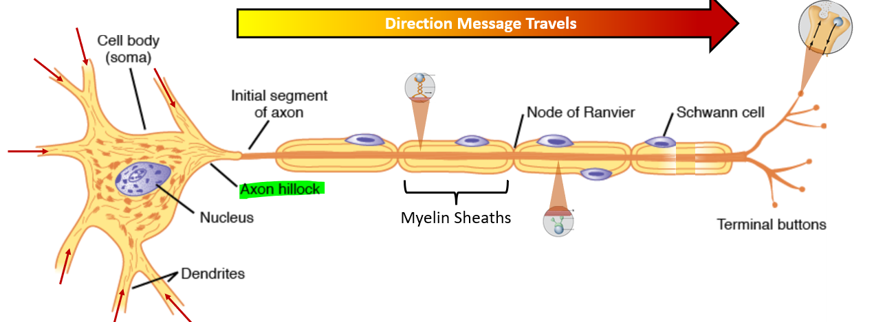
Myelin Sheath - b/t? measurement?
PNS vs CNS ex names
Myelination = ↑ insulation = ↑ speed
More myelination = more speed
Sheath interrupted every 1-3 mm at Node of Ranvier
Peripheral Nervous System
Schwann Cell
Central Nervous System
Oligodendrocyte

Propagation of AP:
Continuous Conduction - 2
Continuous conduction: An AP spreads along the entire unmyelinated membrane by depolarizing each adjacent section in a ripple fashion.
normally moves forward because the membrane behind it is in refractory.
Action Potential:
Saltatory Conduction
where?
benefits? 2
Action potential only occurs at nodes of Ranvier (Myelinated nerve fibers)
Increased velocity
Energy conservation
Action Potential: Manipulation using K+ - 2
RMP directly correlates with serum K+ levels
↑ K+ = ↑ RMP (more positive)
In hyperkalemia, it pushes RMP closer to threshold, too much it can cause your AP to get stuck in the absolute refractory period
h gate stays closed and won’t open
h gate does not open at threshold potential. It reopens when the membrane becomes sufficiently negative during repolarization

Action Potential: Manipulation using Ca2+ - 2
TP directly correlates with serum Ca2+ levels
↑ Ca2+ = ↑ TP (more positive)
Giving Ca chloride IVP can help increase the distance between the RMP and TP
where ↑ K+ decreases the distance
↓ Ca2+ = ↓ TP (more negative)
