RT 110+111 Unit 3: Vertebral Column Anatomy and Positioning
 Call Kai
 Learn
 Practice Test
 Spaced Repetition
 Match
 Flashcards
 Knowt Play
1/204

Earn XP
Description and Tags
Overview of anatomy of vertebral column, positioning, and evaluation criteria and scenarios for spine examinations
205 Terms
How many sections does the vertebral column contain?
5: cervical, thoracic, lumbar, sacral, and coccygeal.

What purpose does the vertebral column serve?
Protects the spinal cord
Provides flexible support for the trunk and head
Acts as attachment sites for deep muscles of the back and ribs
The openings within each vertebrae create a tube-like opening for the spinal cord called the ___.
spinal canal
Where does the spinal cord begin? Where does it end?
Begins at the medulla oblongata
Ends at the lower border of the 1st lumbar vertebrae
Describe the structural components of the vertebral column
The vertebral column is composed of singular segments called vertebrae held together by ligaments.
What is located between each vertebrae? What purpose do they serve?
Fibrocartilaginous intervertebral disks
Allows for movement and flexibility of the vertebral column
T/F: The average adult vertebral column is composed of 33 separate bones
False
How many vertebrae are in each division of the vertebral column?
Cervical - 7
Thoracic - 12
Lumber - 5
Sacrum - 1 (3-5 fused bones)
Coccyx - 1 (3-5 fused bones)
The largest and strongest weightbearing vertebrae is the ___.
lumbar vertebrae
What terms are used to describe the curvature of the vertebral column?
Concave = rounded inward or depressed surface
Convex = rounded outward or elevated surface

Which sections of the vertebral column curve inward?
Cervical and lumbar

Which sections of the vertebral column curve outward?
Thoracic and sacral

An abnormal or exaggerated lumbar curvature or increased concavity is called ___.
lordosis

The abnormal increased convexity of the thoracic spine curvature creating a “humpback” appearance is called ____.
kyphosis

An abnormal or exaggerated lateral curvature of the spine is called ___.
scoliosis

An abnormal or exaggerated lateral curvature to the right side is called ___.
dextroscoliosis
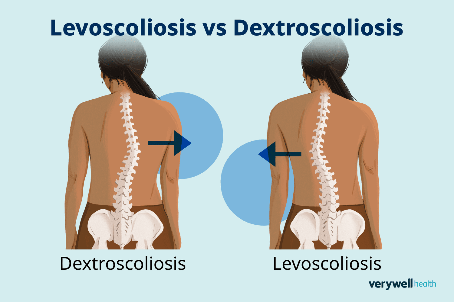
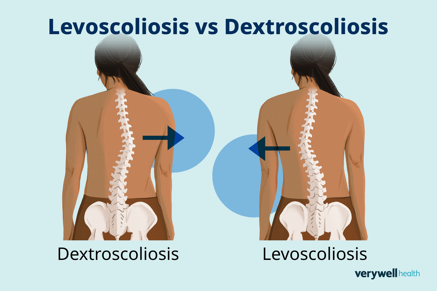
An abnormal or exaggerated lateral curvature to the left side is called ___.
levoscoliosis
The most superior portion of the vertebral column is the ____.
cervical spine


The small horns extending laterally from the pedicle and body are the ___.
transverse processes (F)


The bridge extending from each side of the vertebral body that forms part of the transverse foramen is the ___.
pedicle (C)


Openings on each side of the vertebrae that allow arteries, veins, and nerves to pass through are called ___.
transverse foramina (E)


Posterior bony structures extending from the lamina that form bifid tips on C3-C6 are called ___.
spinous process (H)
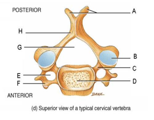

Circular opening between the posterior surface of the body and vertebral arch that allows the spinal cord to pass through is known as ____.
vertebral foramen (G)

What characteristics do cervical vertebrae possess that no other vertebrae have?
Bifid spinous processes on C3-C6
Articular pillars
3 foramina
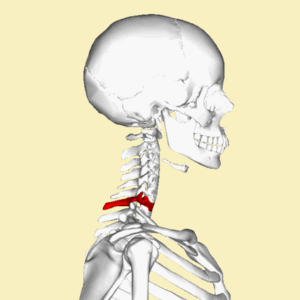
Another name for C7 spinous process is ___.
vertebra prominens
The superior thoracic vertebrae (T1-T4) are smaller and share features of ____ vertebrae. The inferior thoracic vertebrae (T9-T12) are larger and share features of ____.
cervical; lumbar
All 12 thoracic vertebrae possess surfaces covered in fibrocartilage located on the posterolateral margin of the body that help with articulation of the heads of the ribs called ___.
costal facets (demifacets)
Concavities on the inferior surface of the pedicle that form the thoracic intervertebral foramina are called ___.
vertebral notches
Thoracic intervertebral foramina are best visualized in a ____.
lateral view
Thoracic zygapophyseal joints are best visualized in an ____.
oblique view
The column of bone between the superior and inferior articular processes is known as ___.
articular pillar/lateral mass (C1)
The superior and inferior articular processes come together to form a joint between them that allows movement between vertebrae known as ___.
zygapophyseal joints
Zygapophyseal joints of the second through seventh cervical vertebrae can only be visualized in ____.
lateral projection
Zygapophyseal joints of the first and second cervical vertebrae (atlantoaxial) can only be visualized in ___.
AP open mouth projection

The cervical intervertebral foramina furthest from the IR can only be visualized radiographically ___.
45* posterior oblique with a 15-20* cephalic angle
The cervical intervertebral foramina closest to the IR can only be visualized radiographically ___.
45* anterior oblique with a 15*-20* caudal angle
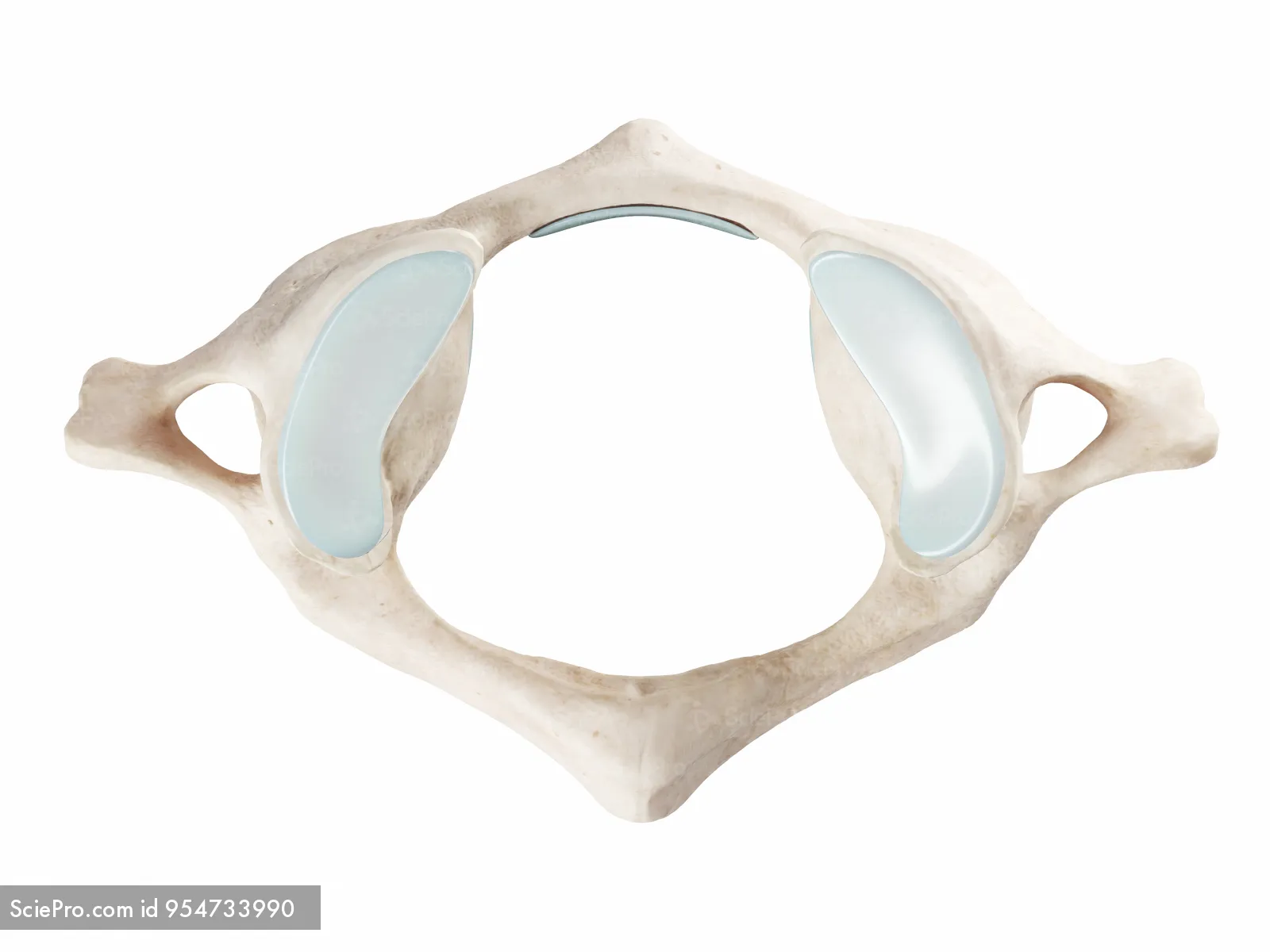
What makes the first cervical vertebrae different from the rest?
No anterior body, posterior laminae, or spinous process
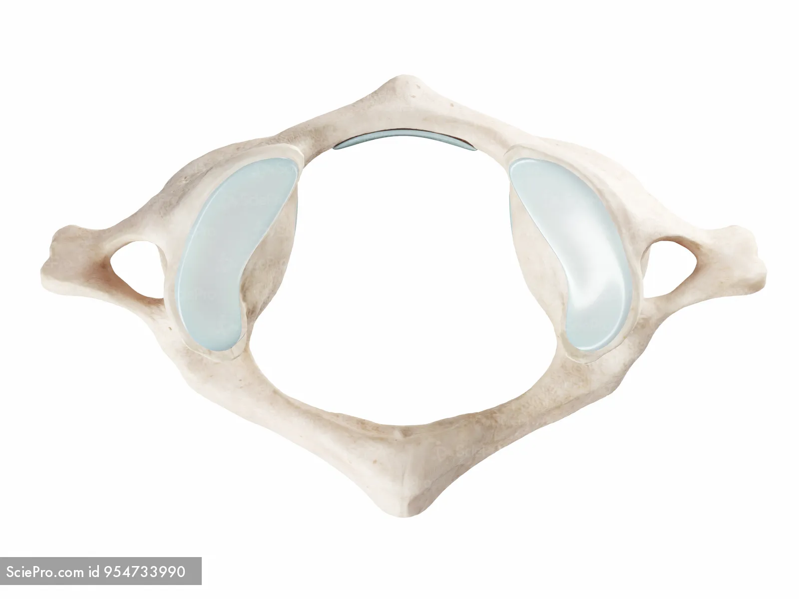
The first cervical vertebrae (atlas) is composed of a thick arch of bone called ___, which includes a smaller tubercle.
anterior arch
The first cervical vertebrae (atlas) contains a thick posterior arch of bone called ___, which also includes a smaller tubercle instead of a spinous process.
posterior arch

Large depressed surfaces on each side of C1 that articulate with the occipital condyles are called ___.
superior articular facets
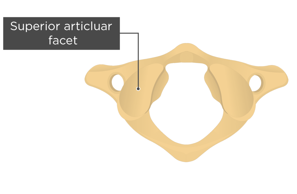

The large pillars between the superior and inferior articular processes that support the weight of the head and assist in rotation of the head are called ___.
lateral masses

The most distinctive feature of the second cervical vertebra (axis) is a conical process projecting up from the superior surface of the body called ___.
odontoid process/dens

T/F: Forced flexion-hyperextension “whiplash” injuries may fracture the dens
True
The ___ for articulation with C3 lies inferior to the lamina.
inferior articular process
T/F: The transverse process and foramina of the second cervical vertebrae lie inferior and lateral to the superior articular process
True
Which projection is used to demonstrate pathology and fractures in the mid and lower cervical spine?
AP axial cervical spine
What technical factors are used to visualize an optimal AP axial projection of the cervical spine?
40” SID, 14×17” IR portrait, collimation on both sides of jaw and top of ear, 70-85 kVp, grid, center photocell, 15-20* cephalic angle, suspend respiration
How is the patient and part positioned for an AP axial projection of the cervical spine?
Patient is erect or supine with MSP aligned to CR and midline of the table
Adjust head so that a line from the lower margin of upper incisors to mastoid processes is perpendicular to table and/or IR
Line from tip of mandible to base of skull is parallel to angled CR
Where is the CR centered for an AP axial cervical spine projection?
CR is angled 15*-20* cephalad and directed to the level of the upper margin of thyroid cartilage to pass through C4
To properly demonstrate open intervertebral disks spaces between cervical vertebrae, what must happen to the CR?
Angle 15*-20* cephalic
If a patient has a smaller lordotic curvature in their neck, the CR should be angled ___.
15* cephalic
If a patient has a greater lordotic curvature in their neck, the CR should be angled ___.
20* or more cephalic
What anatomy should be demonstrated in an AP axial projection of the cervical spine?
C3-T2 vertebral bodies, space between pedicles, open intervertebral disk spaces seen
If the mandible amd base of the skull superimpose the first two cervical vertebrae on an AP axial projection, what error occurred?
No error occurred. It fits with the evaluation criteria
How is no rotation indicated in an AP axial projection of the cervical spine?
Spinous processes and sternoclavicular joints (if visible) are equidistant from the spinal column lateral borders
Which projection best demonstrates the odontoid process and C1 and C2 structures?
AP open mouth
What technical factors are used on an AP open mouth projection?
40” SID, small collimation from cheek to cheek, 14×17” IR portrait, grid, 70-85 kVp, no AEC, suspend respiration
How is the patient and part positioned for an AP open mouth?
Patient is erect or spine with their MSP aligned to CR and midline of table
Adjust head so that, with mouth open, a line from the lower margin of upper incisors to the base of the skull is perpendicular to the table and/or IR
No rotation of the head
Ensure the mouth is open wide, only have the lower jaw drop
Where is the CR centered for an AP open mouth?
CR is directed through the center of open mouth
What anatomy should be demonstrated in an AP open mouth?
Odontoid process (dens) and vertebral body of C2, lateral masses and transverse processes of C1, and atlantoaxial joints
If the teeth are superimposed on the upper dens, what positioning error occurred?
Insufficient extension of the neck
Fix: slightly tilt head back (extend) or angle CR slightly cephalic (up)
An AP open mouth projection was taken and the image shows the odontoid process being superimposed by the base of the skull. What positioning error occurred? If an error occurred, how should it be fixed?
Insufficient flexion of the neck
Fix: Tilt the head down or angle slightly caudal
How is no rotation indicated in an AP open mouth?
Equal distances from lateral masses and/or transverse processes of C1 to condyles of mandible
Center alignment of spinous processes of C2
If the upper odontoid process cannot be demonstrated with an AP open mouth, what other projections are used?
Fuchs or Judd methods
What technical factors are used for Fuchs (AP) and Judd (PA) method?
40” SID, small collimation from chin to middle of throat or midoccipital lobe, grid, 70-85 kVp, no AEC, suspend respiration
How is a patient and part positioned for an AP projection of the dens Fuchs method?
Patient is erect or supine
Chin is elevated as needed to bring the MML near perpendicular to tabletop
No rotation of the head
Where is the CR centered for an AP projection of the dens Fuchs method?
CR is parallel to MML, directed 1” below mastoid tips and mandible angles
How is a patient and part positioned for a PA projection of the dens Judd method?
Patient is erect or supine
Chin is resting on Bucky or tabletop and extended to bring MML near perpendicular to IR
No rotation of the head
Where is the CR centered for a PA projection of the dens Judd method?
CR is parallel to MML through midoccipital bone, about 1” below mastoid tips and mandible angles
What anatomy should be demonstrated in AP/PA projections of the dens Fuchs/Judd methods?
Dens is centered within the foramen magnum
Dens is free of superimposition from the mandible
How is no rotation indicated in an AP/PA projection of the dens (Fuchs/Judd methods)?
Symmetric appearance of the mandible arched over foramen magnum
Why is AEC not recommended for AP open mouth, Fuchs, and Judds?
The odontoid has a smaller field of view
If trauma is present, should the Fuchs or Judd method projections be performed? Why or why not?
No because it could further injure the patient's neck
Which projection demonstrates the zygapophyseal joints in the cervical neck?
Lateral cervical spine
What technical factors are used for a lateral projection of the cervical spine?
72” SID, 10”×12” collimation from top of shoulder to EAM and both sides of the neck, 14×17” IR portrait, grid, center photocell, suspend respiration on full expiration, small focal spot, 70-85 kVp
How is the patient and part positioned for a non-trauma lateral projection of the cervical spine?
Patient’s left side is against Bucky with MCP aligned to the middle
Relax shoulders and “drop” them as far as possible
Elevate chin to place AML parallel with the floor
Extend chin out to prevent the mandible superimposing upper vertebrae
Where is the CR centered for a non-trauma lateral projection of the cervical spine?
Top of IR is about 1-2” above EAM
CR is directed at C4, which is at the level of upper margin of thyroid cartilage)
Why is a 72” SID necessary for a lateral projection of the cervical spine?
To reduce OID
What anatomy should be demonstrated in a lateral projection of the cervical spine?
C1-T1, open intervertebral joint spaces, right and left articular pillars superimposed for each vertebrae, spinous processes, and open zygapophyseal joints
If the shoulders superimpose C7-T1 in a lateral projection, what other projection is used to visualize C7-T1?
Cervicothoracic lateral (Swimmers)
If trauma is present in the cervical spine, what projection is used to visualize fractures or subluxations?
Cross-table lateral c-spine
What technical factors are used for a cross-table lateral cervical spine trauma?
60-72” SID if possible, 14×17” IR along the side of the body, with or without grid, 70-85 kVp with grid, 8-10 mAs, suspend respiration on full expiration, no photocell, collimation to area
How is a patient and part positioned for a cross-table lateral cervical spine?
Patient is supine with C-collar around neck, no pillow
IR is placed against patient’s left shoulder, top of IR is placed 1-2” above EAM
Shoulders are relaxed and “dropped” as far as they can
Where is the CR centered for a cross-table lateral cervical spine?
CR is horizontal and directed at C4 (upper margin of thyroid cartilage)
T/F: A cervical collar cannot be removed unless authorized by a physician who evaluated the image
True
What technical factors are used for a Swimmers view?
40” SID for table, 72” SID for upright Bucky, 75-95 kVp, 14×17” IR portrait, suspend respiration on full expiration or orthostatic breathing technique, 200 mA, 2 density, and 3-5 seconds exposure time for breathing technique, center photocell, grid
Why should you use an orthostatic breathing technique for lateral projections of the cervical and thoracic spine?
To blur out details of the lungs, which helps improve image quality
How is a patient and part positioned for a Swimmers lateral?
Patient’s left side is laying on the table or upright against Bucky, with MCP aligned with the midline of the IR
Arm closest to the IR is placed above patient’s head
Arm furthest away from IR is relaxed and “dropped”
No rotation in the head or chest
Where is the CR centered for a Swimmers?
Directed at T1, which is about 1” above jugular notch or at level with C7
If a patient has broad shoulders or cannot relax shoulders, what can be done to the CR to separate the shoulders?
Angle CR 3*-5* caudal
T/F: A high mA and short exposure time is used for an orthostatic breathing technique to blur out overlaying lung structures
False
What anatomy should be demonstrated in a cervicothoracic lateral (Swimmers)?
C5-T3 vertebral bodies and intervertebral disk spaces, humeral head farthest from IR is magnified and appears below T4/T5, superimposed zygapophyseal joints and articular pillars and posterior ribs
Which positions are used in functional studies to evaluate whiplash injuries or assess stability following spinal fusion surgery?
Lateral hyperflexion and hyperextension
What technical factors are used for flexion and extension lateral projections of the cervical spine?
60-72” SID, 14×17” IR portrait, grid or nongrid, center photocell, 70-85 kVp, suspend respiration on full expiration
How is the part positioned for a flexion lateral of the cervical spine?
Chin is tucked to chest cavity
How is the part positioned for an extension lateral of the cervical spine?
Chin is raised and head is tilted back
Where is the CR centered for flexion and extension lateral projections of the cervical spine?
Directed at C4 (upper thyroid cartilage), with top of light slightly above EAM and bottom of light slightly above shoulder
If the cervical spinous processes are well separated, then the patient is ___.
hyperflexing
If the cervical spinous processes are close in proximity, then the patient is ___.
hyperextending
Which position best visualizes the cervical intervertebral foramina?
Anterior and posterior obliques