Test 1
 Call Kai
 Learn
 Practice Test
 Spaced Repetition
 Match
 Flashcards
 Knowt Play
1/133

Earn XP
Description and Tags
134 Terms
Amputation Disparities Due to Age and Race
Non-whites approximately 42% of limb loss
African Americans with diabetes 2x higher amputation rate in 2008
Age: increased risk with increase in age
Greatest Causes of Amputation
Amputation Secondary to Peripheral Vascular Disease (PVD) & Peripheral Arterial Disease (PAD)
Secondary to Trauma
Secondary to Cancer
Most common cause of limb loss
Amputation Secondary to Peripheral Vascular Disease (PVD)
Common Peripheral Arterial Disease (PAD) Types
Diabetes Mellitus
Arteriosclerosis
Buerger’s disease
Diabetes Mellitus causing amputation
Leading cause of amputation = 154,000 / year

_ of diabetes-related amputations are preceded by a foot ulcer
85%
5 Year Mortality Percentage of Diabetic Foot Complications
Worst mortality outcome than breast cancer and all cancer

PVD/PAD: Arteriosclerosis
“hardening of the arteries”
Interrupted blood flow
Ischemic tissue
Amputation becomes necessary when ischemic tissue cannot be revascularized
PVD: Buerger’s Disease
AKA: thromboangiitis obliterans
First described - Buerger (1908)
Defined: acute inflammation and thrombosis (clotting) of arteries and veins
Affects the hands and feet Incidence: 20-40 y.o. male cigarette smokers
Buerger’s Disease Treatment
Complete cessation of smoking
Amputation: Secondary to Trauma
Second most common cause; 2% of amputations
45% of amputations due to injury including motor vehicle collisions, workplace accidents, sports injures, and military wounds
Severe fractures, non-union/malunion
Injury/insult to blood supply
Burns
Electrical
Thermal
Frostbite
Amputation Secondary to Cancer
Third most common cause; 2% of amputations
Congenital Limb Deficiency Prevalance
~ 1 in every 1,900 babies is born with a limb reduction defect in the United States.
Congenital Limb Deficiency Categories
Failure of formation of parts
Failure of differentiation
Duplication
Overgrowth
Constricting band syndrome
Skeletal abnormalities
Congenital Deficiency due to Thalidomide
Drug designed as sedative
Used to treat morning sickness in pregnant women (1950’s-1960’s)
Causes limb defects
Resurgence in Brazil (2005-2008)
Congenital Limb Deficiency: ISO/ISPO Classification
Know the concept I don’t think we need to name
Terms such as hemimelia, etc., are avoided due to their lack of precision and translation.
Anatomic and radiologic bases only
Name the bones affected in a proximal-distal sequence by using the name as a noun
State whether each affected bone is totally or partially absent
Transverse
Longitudinal
Any bone not named is present and of normal form
Less common causes of amputation
Cellulitis and Infections
Neurological Conditions
Spider/Insect Bite
Future Projections of Amputation
we have job security
3.6 million people living with an amputation by year 2050.
Numbers increase as the population ages and dysvascular disease increases.
Casting Technique for PTB traditional
2 stage
Anterior splint
Circumferential wrap
Casting Technique for PTB SC or PTB SCSP
3 stage
Anterior splint
Circumferential wrap
Proximal splint
Casting Technique for Hybrid PTB/TSB
2 stage (1-stage optional)
Anterior splint
Circumferential wrap
or
Circumferential wrap only
TSB Casting Techniques
1 stage (2-stage optional)
circumferential wrap
or
Anterior splint
Circumferential wrap
Casting Steps
Gather Casting Supplies
Circumferential Measurements
if using gel liner, do them over liner
Applying the Casting Interface
plastic barrier
optional casting sock
Measure Prosthetics Side
Measure Non-Prosthetics side
Identify and Mark Landmarks
Take Impression (Cast)
What do you measure on prosthetic side when casting?
Residual Limb Length
Bony Length
ML
AP
Proximal ML (PML) for Supracondylar Suspension
What do you measure on non-prosthetic side when casting?
MTP to Floor Measurement
Anatomical Foot Measurement
When marking bony prominence have patient in _ degrees of flexion
20
to accentuate bony prominences
List of landmarks to identify and mark
Patella
mid patella ligament and both sides of the patellar ligament
Tibial crest
Anterior distal tibia
Tibial tubercle
Medial flare of the tibia
Medial shaft of the tibia where it transitions to soft tissue
Lateral tibial flare and lateral aspect of the crest of the tibia
Widest point of the medial and lateral condyles of the knee
Medial condyle of the knee and adductor tubercle if pronounced
Fibular head
Shaft of the fibula
Cut end of the fibula
From the patellar tendon level “0”, make a mark at one-inch intervals as far distal as possible (or transfer these one-inch interval marks from the tape underneath)
Iliotibial band (for supracondylar cast)
Adductor tubercle (for supracondylar cast)
Mark one inch proximal to the proximal border of the patella (supracondylar suprapatellar cast)
Landmark markings: Patella
Circle drawn around patella
A in picture

Landmark markings: mid patella ligament and both sides of the patellar ligament
Mark as an “H” for us
b in picture

Landmark markings: Tibial crest
A straight vertical line drawn directly down the sharpest edge of the shin bone
e in picture

Landmark markings: Anterior distal tibia
where tibia ends
some draw a circle
g in picture

Landmark markings: Tibial tubercle
prominence where patellar tendon inserts into the bony bump on the front of the shin
some people drawn upside down triangle or an oval directly
c in picture

Landmark markings: Medial flare of the tibia
broad, flat, triangular shelf of bone on the upper, medial side of the shin
can trace the curve of your hand for it
h in picture

Landmark markings: Medial shaft of the tibia where it transitions to soft tissue
line where your feel the edge of the tibia on the medial aspect
i in picture
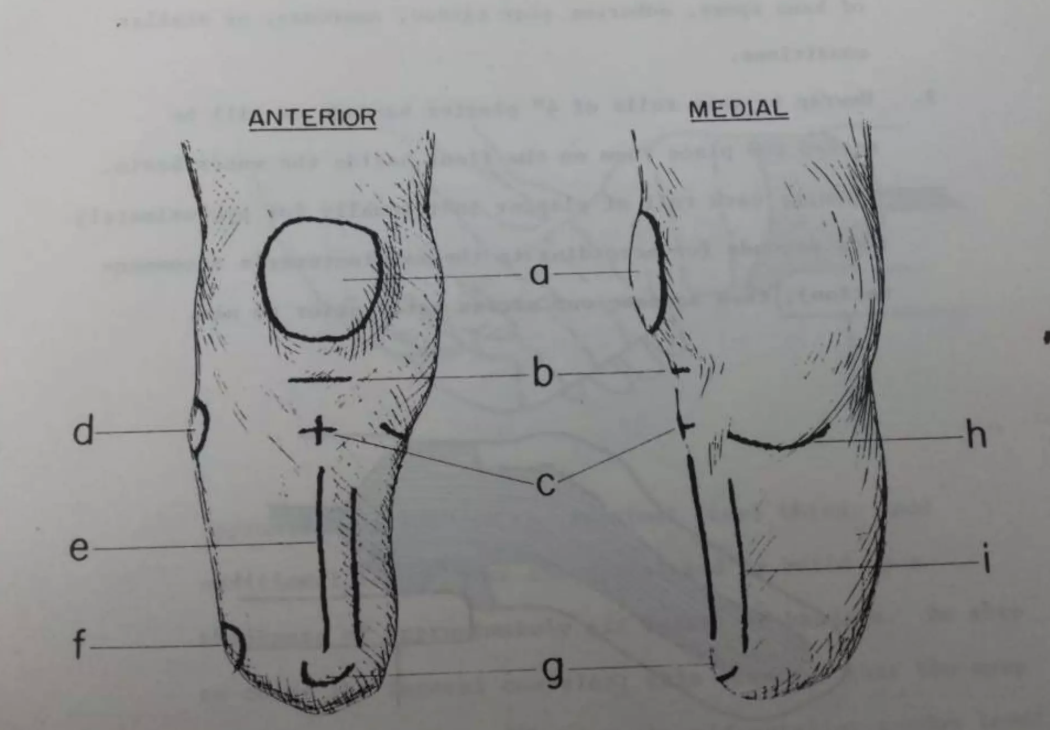
Landmark markings: Lateral tibial flare and lateral aspect of the crest of the tibia
in between tibia and fibula

Landmark markings: Widest point of the medial and lateral condyles of the knee
Feel the sides of the knee joint for the widest bony flares of the femur.
These points are wear you measure the ML

Landmark markings: Medial condyle of the knee and adductor tubercle if pronounced
Palpate the inner side of the knee joint to find the flat medial femoral condyle. Slide your fingers slightly upward and backward along the femur to feel for the small bony projection of the adductor tubercle. Circle it if it is pronounced.
Landmark markings: Fibular head
Circle around the prominent, round bony knob on the upper, outer side of the leg (below the joint line)
d in picture
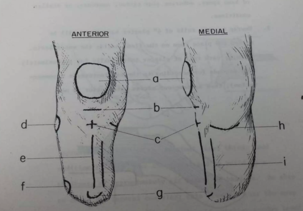
Landmark markings: Shaft of the fibula
Line on the outer bone ridge downward from the fibular head toward the bottom of the limb
Landmark markings: Cut end of the fibula
Circle very bottom tip of the outer bone
Because the fibula is cut shorter than the tibia during amputation, locate its exact terminal end and draw a small circle over it for pressure relief
f in picture

Landmark markings: one-inch intervals
patellar tendon level is “0”
move distally
this is where you measure circumferences

Landmark markings: Iliotibial band
only for supracondylar cast
palpate the thick, fibrous band of tissue running vertically down the outside of the thigh, inserting just below the knee. Mark its borders to ensure the high socket trim line accommodates its movement
Landmark markings: Adductor tubercle
only for supracondylar cast
Landmark markings: one inch proximal to the proximal border of the patella
only for supracondylar suprapatellar cast
another 1in line just proximal this time
K0 Description
This patient does not have the ability or potential to ambulate or transfer safely with or without assistance and a prosthesis does not enhance their quality of life or mobility.
K1 Description
This patient has the ability or potential to use a prosthesis for transfers or ambulation on level surfaces at fixed cadence - a typical limited or unlimited household ambulator.
K2 Description
This patient has the ability or potential for ambulation with the ability to traverse low-level environmental barriers such as curbs, stairs, or uneven surfaces - a typical limited community ambulator
K3 Description
The patient has the ability or potential for ambulation with variable cadence - a typical community ambulator with the ability to traverse most environmental barriers and may have vocational, therapeutic, or exercise activity that demands prosthetic use beyond simple locomotion
K4 Description
The patient has the ability or potential for prosthetic ambulation that exceeds basic ambulation skills, exhibiting high impact, stress, or energy levels - typical of the prosthetic demands of the child, active adult, or athlete
K0 Foot/Ankle Assemblies and Knee Unit
Not eligible for prosthesis
K1 Foot/Ankle Assemblies and Knee Unit
Foot: External keel, SACH feet or single axis ankle/feet
Knee: Single-axis, constant friction knee
K2 Foot/Ankle Assemblies and Knee Unit
Foot: Flexible-keel feet and multi-axial ankle/feet
Knee: Single-axis, constant friction knee
K3 Foot/Ankle Assemblies and Knee Unit
Foot: Flex foot and flex-walk systems, energy storing feet, multi-axial ankle/feet, or dynamic response feet
Knee: Fluid and pneumatic control knees
K4 Foot/Ankle Assemblies and Knee Unit
Foot: Any ankle foot system appropriate
Knee: Any ankle knee system appropriate
Prosthetic Foot Basics Categories
Length
Heel height
Build height
Keel
Color, Toe style
Functional Level
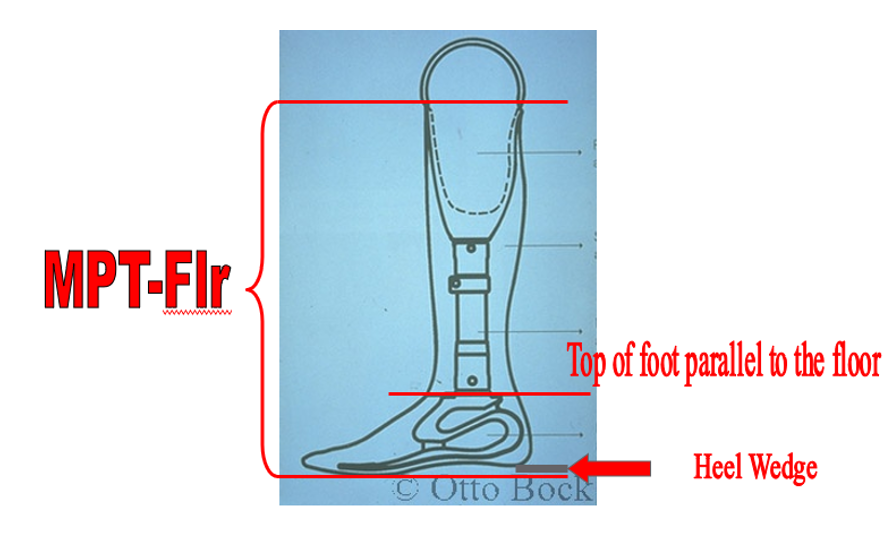
Proper Foot Orientation
top of the foot is parallel to the floor
plantar surface is not usually parallel to the floor
heel wedge must be inserted below the heel of the foot in order to maintain the top of the foot in the transverse plane
thickness of the wedge represents the heel rise of the foot
the mid-patellar tendon to floor measurement is taken from the floor at the ball of the foot or at the heel under the heel wedge
Prosthetic Feet Types
SACH
Single Axis
Flexible Keel (K2)
Dynamic Response (K3)
Specialty
Prosthetic Feet Additional Features
Multiaxial
Torsion
Vertical Shock Absorbing
Integrated Vacuum for suspension
Heel Height adjustability
Hydraulic Ankle
Microprocessor Ankle
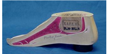
SACH Feet
Solid-Ankle Cushioned-Heel
most basic prosthetic foot available
a rigid, non-deforming keel, but the heel is compressible
K1

SACH Feet Types
Internal keeled
External keeled (discontinued)
Syme (discontinued)
Post Operative (discontinued)

SACH Feet Advantages and Disadvantages
Advantages
Stable
Lightweight
Durable
Disadvantages
No energy return
SACH: Post-Operative
Used on post-operative prosthesis only
Very soft foam covering, rigid keel
No shoe to be worn with this foot
Single-Axis
Articulated in sagittal plane to provide plantar and dorsiflexion
K1
available in both an endoskeletal and an exoskeletal version
It incorporates a single-axis hinge at the ankle.
Single-Axis Bumpers
available with differing firmness, making resistance adjustable
Posterior Bumper (Plantarflexion Bumper):
Gait Phase: Initial contact → Loading response
Simulates: Eccentric dorsiflexors (tibialis anterior).
Function: Compresses to absorb heel strike shock and control the rate of plantarflexion to achieve rapid foot-flat, maximizing knee stability
Anterior Bumper (Dorsiflexion Bumper):
Gait Phase: Midstance → Terminal stance / Toe-off
Simulates: Eccentric plantarflexors (gastrocnemius-soleus complex)
Function: Compresses to resist forward movement of the tibia (dorsiflexion), maintaining a stable toe lever for smooth progression and rollover
Single Axis feet Advantages and Disadvantages
Advantages
Adjustable resistance
Increased knee stability at loading response (good for week quads)
Disadvantages
Maintenance
Potential for noise (fix with garage door lube)
May be more difficult to cover
Flexible Keel Feet
K2
Simulated joint motion
more ground compliant than those feet with rigid keels

Flexible Keel Foot Advantages and Disadvantages
Advantages
Durable
More compliant than SACH
Easy “rollover” (progression from midstance to late stance)
Disadvantages
No energy return
Dynamic Response Foot
K3
has a deformable spring-type keel that provides a lively, responsive feel while the patient is walking
keel deforms under loading, it absorbs shock and then quickly returns to its original position once the load is removed

Advantages of Energy Return
Increased self-selected walking velocity
Increased stride length
Decreased sound side weight acceptance force
Increased prosthetic side propulsive force

Fiberglass vs Carbon
both experience delamination and deterioration
fiberglass feet do not experience the same type of catastrophic failure as carbon fiber
durability of fiberglass feet is an advantage
offer a different feeling of energy return that some patients report as “softer” or not as “bouncy” as carbon feet
Seattle Lightfoot Dynamic Response
These feet have Delrin or similar composite keels
They provide energy return, but minimal compared to other dynamic response feet
K3
Split Keel Feature
Provides Inversion Eversion motion to accommodate slightly uneven ground.
Not a billable feature.
Dynamic Response vs. Flexible Keel
Dynamic Response
Spring
Energy Storing / Energy Returning
Flexible Keel
Sponge
Energy Absorbing
Additional Features
Multi-Axial
Vertical shock absorption
Heel height adjustability
Hydraulic
Microprocessor Control
Powered
Feature: Vertical Shock Absorption
K3+
allows the prosthesis to dampen the axial loading of the prosthesis and thereby decrease the shock transmitted to the residual limb
Feature: Multi-Axial Rotation
K2 or K3
can increase the base of support by accommodating uneven terrain
Prosthetic joint simulation can be accomplished by true articulation of the prosthetic foot, as well as compression of the foot itself
Feature: Multi-Axial Rotation Advantages and Disadvantages
Advantages
Accommodate variable terrain
Energy return
Disadvantages
Weight
Noise
maintenance
Feature: Heel Height Adjustability
adjust the foot’s dorsi/plantar flexion position providing them the freedom to wear shoes of varying heel heights
Only on dynamic response feet, k-3 and k-4
Insurance doesn’t want to pay, can get on workers comp
Feature: Hydraulic Ankle
K2 or K3
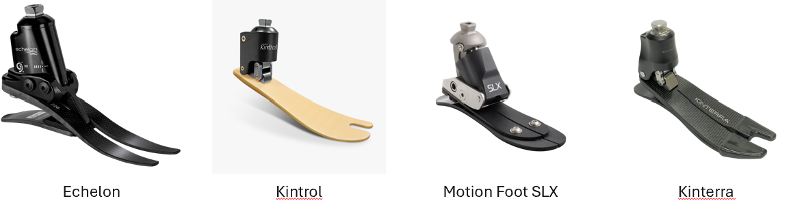
Microprocessor Ankle
K3
microprocessor-controlled and motor-powered dorsiflexion and plantar flexion during the swing phase
make navigation of inclines, stair ascent and descent, and sitting onto or rising from a chair easier
Ankle Units
that can be coupled with the prosthetic foot to provide the multi-axial feature in a foot that does not otherwise provide this motion
Activity Specific Feet
Swimming foot
skiing foot
running feet
sprinting
jogging
stompers
rock climbing
SACH Foot Mechanic at Initial Contact
soft heel cushion compresses under load
compression simulates plantarflexion
musculature: simulates an eccentric contraction of the dorsiflexors
SACH Foot Mechanic in Stance phase
keel does not deform, providing firm support for push-off
simulates an isometric contraction of the dorsiflexors
Toe dorsiflexion is simulated by the rubber toe section of the foot distal to the keel
Patellar Tendon Bearing “PTB” Socket
Construction: Laminated or molded plastic socket
Intimate, total contact fit over entire residual limb
Anterior wall extends to encapsulate distal 1/3 of patella
Socket has a “bar” at the patellar tendon as a major weight-bearing surface
PTB Socket Design
Medial and lateral walls extend proximally to adductor tubercle of femur to control mediolateral forces applied to residual limb
Posterior wall is designed to apply anteriorly directed force to maintain the patellar tendon on the bar
Distal end pad (DEP) and/or soft liner may be used when indicated for added protection and comfort
PTB Indications
Virtually all transtibial amputees
PTB Contraindications
Extremely short residual limbs
may require the “bent-knee” prosthetic design
Severe flexion contractures (beyond 45 degrees)
may require bent-knee prosthetic design
PTB Hard Socket Indications
Mature residual limb with good soft tissue coverage and no sharp bony prominences
PTB Hard Socket Contraindications
Skin coverage that is thin, scarred, grafted, or otherwise subject to breakdown
Diabetes/Peripheral vascular disease (PVD)
Sharp bony prominences
A recent amputee
PTB Hard Socket Advantages and Disadvantages
Advantages
Perspiration does not corrode the socket.
Less bulky at the knee than with an insert
Easy to keep clean
Contours within the socket do not compress or pack down with use.
Reliefs or modifications can be located with exactness
Disadvantages
Requires precision in casting and modification
More difficult to fit to bony or sensitive residual limbs
Not as easily modified as a socket with a liner
PTB Socket with Soft Liner Indications
PVD, thin, sensitive, or scarred skin
Sharp bony prominences, peripheral neuropathy
Noncompliant or unreliable patients, bilateral transtibial patients
Highly active patients, blind patients
PTB Socket with Soft Liner Contraindications
Patient rejection
PTB Socket with Soft Liner Advantages and Disadvantages
Advantages
Provides a soft, protective socket interface
Is appropriate for the majority of residual limbs
Rebound in the liner may aid circulation by providing a "pumping action" and by providing intermittent pressure over bony prominences.
Easily modified
Disadvantages
Not as hygienic as a hard socket because liner tends to absorb perspiration.
Increases bulk around the knee (the entire socket must be enlarged by approximately 1/4 inch)
May increase the weight of the prosthesis
The liner may compress and pack down over time resulting in loss of definition of the original modifications
PTB Design Variants
PTB
proximal brim of the socket does not extend above the adductor tubercle
PTB-SC
a self-contained suspension system requiring no additional straps, etc
socket extends above the condyles of the knee
shaded area marked out above the condyles is a supracondylar wedge
PTB-SCSP
is self-suspending with the supracondylar wedge
Socket extends above the condyles medially and laterally and also extends above the patella
Just superior to the proximal border of the patella, the socket is modified with a slight indentation known as the quadriceps bar

Supracondylar Socket (PTB-SC) Indications
Short residual limb
Mild mediolateral instability
Cannot tolerate cuff
due to scars
pinching during knee flexion
restricted circulation
Supracondylar Socket (PTB-SC) Contraindications
Kneeling
Obese or very muscular patients
Cosmesis
Moderate to severe ML instability
Supracondylar Suprapatellar Socket (PTB-SCSP) Indications
Short residual limb
Mild mediolateral instability or genurecurvatum
Cannot tolerate cuff
due to scars
pinching during knee flexion
restricted circulation
Supracondylar Suprapatellar Socket (PTB-SCSP) Contraindications
Kneeling
Obese or very muscular patients
Cosmesis
Moderate to severe ML instability or genurecurvatum
Total Surface Bearing Designs (TSB)
Disburses pressure evenly over entire residuum
Uses gel insert (locking or non-locking)
Sleeve-type suspension
Locking pin suspension
Suction suspension
Vacuum-Assisted Suspension