histology nervous tissue
 Call Kai
 Learn
Learn Practice Test
 Spaced Repetition
 Match
Match Flashcards
 Knowt Play
1/63

There's no tags or description
Looks like no tags are added yet.
Name | Mastery | Learn | Test | Matching | Spaced | Call with Kai | Chat |
|---|
No analytics yet
Send a link to your students to track their progress
64 Terms

what are the arrows pointing to?
cell bodies of neurons

label the parts of the neuron
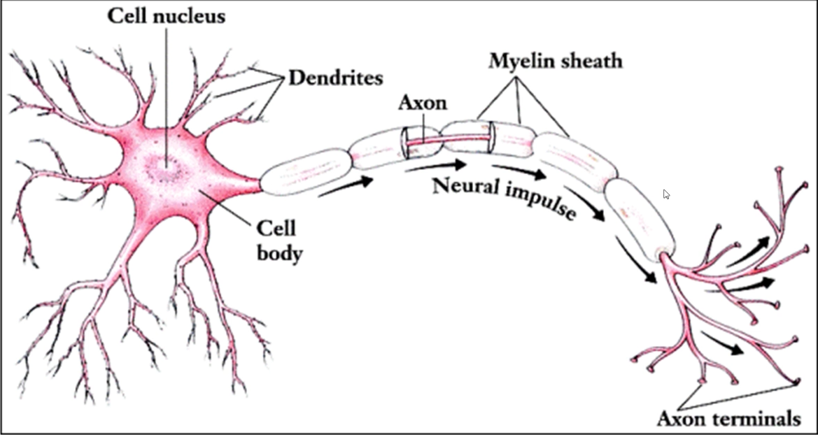

what’s being shown in this picture?
A - Nissel’s Granules
B - Nucleus

what’s being shown in this picture?
dendrites

what are these pictures showing?
dendrites

what are the black arrows pointing to?
myelinated axons

What is each letter labeling?
A - dendritic spines
B - axon
C - cell body
D - dendrites

what is this?
myelinated axon
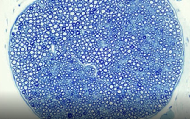
what is this
unmyelinated axon

what’s the blue arrow pointing to?
Node of Ranvier
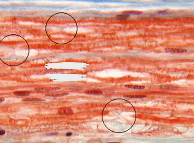
what’s circled in this picture?
Nodes of Ranvier

what’s each letter pointing to?
A - Schmidt-Lanterman Cleft
B - Node of Ranvier
C - Myelin

what are these pictures showing?
Microglia

what’s being shown here?
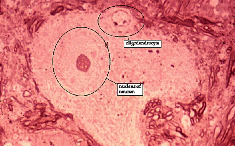
oligodendrocyte
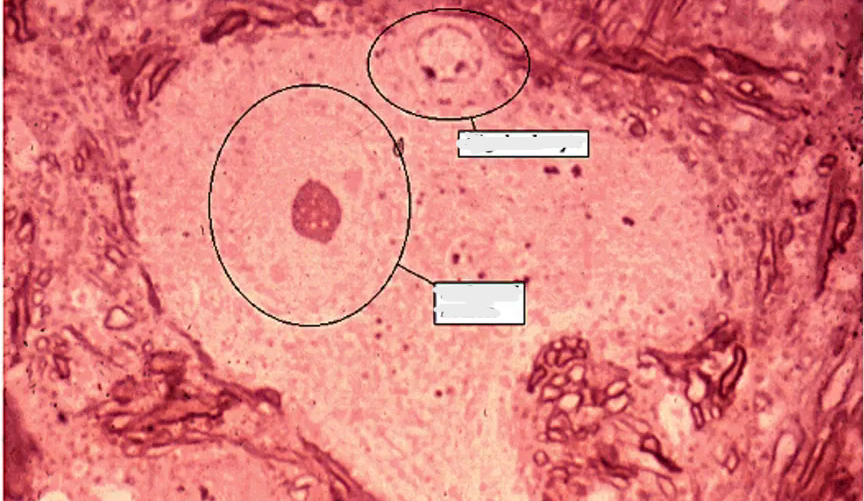
label the two circles
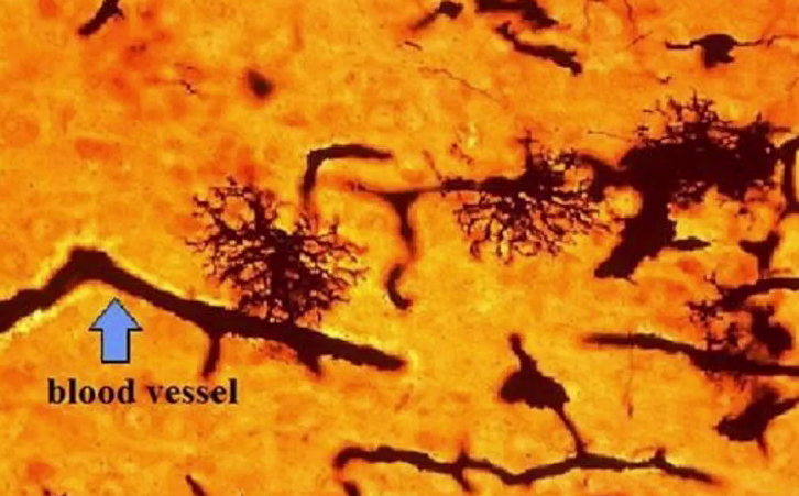
what’s this?
a protoplasmic astrocyte (grey matter)

what’s this?
fibrous astrocyte (white matter)
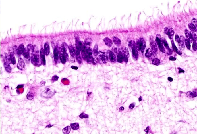
what’s this?
ependymal layer

label the boxes


what’s being pointed to here?
ependymal cells

what are the blue arrows pointing to?
pyramidal cells
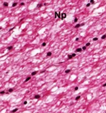
what’s this showing?
white matter with neuropil and glial cells
what are the two types of cells in nervous tissue and what are their functions
neuron: specialized in perception of sensory stimuli and processing/receiving info, and transmitting it further to other neurons via nerve impulses
neuroglia: support, nourish, and protect neurons
what cell components are and aren’t in neurons?
not really a lot of mitochondria but do have neurofibrils to connect dendrites through cell body and axon
dendrite function
conduct impulses TOWARDS body
axon function
conduct impulses away from body, usually one at a time
dendritic spines
small projections off of dendrite that act as a point of contact where they’re contacting other neurons
synapses
contact points for dendritic spines, can either be between two neurons or between a neuron and a muscle fiber (via neuromuscular junction)
function of myelin in an axon
important for insulating axon and helps speed up impulse
axon hillock
point where cell body merges into axon
Schwann Cell placement in myelinated vs unmyelinated axon
myelinated: sheath wraps around and have Schwann cell at the end all bunched up
unmyelinated: Schwann cell cytoplasm
relationship between diameter of axon and action potential
larger axon diameter = faster action potential
importance of Node of Ranvier
important for nerve impulses and salt conduction by exposing small part of membrane to external environment
how are Schmidt-Lanterman Clefts formed and where are they found?
formed from myelin clefts of cytoplasm that was left behind during Schwann cell myelinating process
found in myelinated axons of the PNS
synapse importance with neurotransmitters
action potential comes down and synaptic vesicles release neurotransmitters, which can vary depending on purpose of that neuron and location within body (very specific!)
neuroplasticity
making new synapses to fix or accommodate for injured/damaged synapses; limited ability to produce new neurons but neuroplasticity allows for regrowth and regenerative capacity
3 main phases of neuroplasticity and what happens in each phase
injury: injury to neuron occurs, fibers distal to injury (away from cell body) die but Schwann cells above injury remain intact
Degeneration (Wallerian degeneration): macrophages clean up tissue debris, soma swells, ER breaks up, nucleus moves off center due to lack of nerve growth factor
Regeneration: axon stump sprouts multiple growth processes and regrows through Schwann cells that will create new myelin sheath
Nervous system divisions
CNS: brain/spinal cord, gray/white matter
PNS: nerves/ganglia
Glial Cells
supporting cells of CNS, filling inner spaces of nervous tissue, outnumber nerve cells (10:1) and much smaller than nerve cells
macroglia and microglia cells in CNS
types of macroglia in CNS
Ependymal cells, astrocytes, oligodendrocytes
microglia in the CNS
equivalent to macrophages but reside in brain, dispose of debris, dark stain and dense, found equally in gray and white matter; CEREBRAL CORTEX
oligodendrocytes
not as many processes, end in sheet of myelin, wrap around segment of axon (like Schwann cells but these are only in CNS and these can attach to multiple axons), responsible for normal propagation of action potentials in CNS; MYELINATED WHITE MATTER
astrocytes
most abundant glial cell in the brain, has GFAP (glial fibrillary acidic protein) which is found in the cytoskeleton of astrocytes, star shaped but shape can be varied depending on if it’s in white or gray matter; CAPILLARIES
protoplasmic astrocytes
found in GRAY matter, thicker branches and cell body, more fluffy looking, foot wraps around lots of synapses
fibrous astrocytes
found in WHITE matter, thinner branches, more twig looking, foot process wraps around blood vessels and Nodes of Ranvier
ependymal cells
form epithelial lining of ventricles in the brain and central canal of spinal cord, neuroectoderm, resembles cuboidal or columnar epithelium, dense chromatin, small nucleus and cilia
Wiegart stain
used to highlight different regions of brain and good to differentiate between white and gray matter (gray matter appears lighter and white matter appears darker), usually stains myelin black
Grey Matter
regions with cell bodies together with dendrites and axon terminals (contains lots of other stuff besides myelinated axons), found in surface area of cortex in the brain and deeper in nucleus masses of the brain
white matter
axons gathered into bundles, called white (and usually appears whiter) because of all the myelinated axons (myelin is mostly adipose and adipose appears white), parallel to act as finer tracks/pathways, some distance from cell bodies
betz cells
large pyramidal cells of motor cortex that help in movement; associated with the need to sustain extremely long axons
neuropils
unmyelinated cell processes that look like fibers of CT
what are the 6 layers of the cortex and what is found in each layer
Plexiform/Molecular Layer: sparse neurons/glia
outer granule layer: smaller pyramidal and stellate neurons
outer pyramidal layer: moderate sized pyramidal neurons
inner granule layer: packed with stellate neurons, very dense
inner pyramidal layer: large pyramidal neurons
multiform cellular layer: mix of pyramidal and stellate neurons
*All 6 layers are GREY MATTER, white matter is below these 6 layers*
Cerebellum
gray matter, referred to as “cortex”, has 3 layers
3 layers of cerebellum and whats in each one
outer molecular layer: synaptic, lots of axons/dendrites/granule cells, least dense, very light
Purkinje Cell layer: very thin, single layer of large pear shaped Purkinje cells, ONLY the cell bodies make up these layers (dendrites part of molecular layer and axons part of granular layer)
Inner Granular layer: very dark stained, densely packed granule/golgi cells with small neurons in brain, round/oval, mossy fiber
where are white and gray matter found in Spinal Cord
gray matter: “gray H”, butterfly shape on interior part of spinal cord
white matter: white commissure/area of white matter that’s just interior to central canal
layers of meninges and characteristics of each layer
Dura Mater: outermost layer of very dense and fibrous CT that’s tough/impermeable and has arachnoid grannulation/vila
Pia Mater: inner most layer, light layer of collagen and fibroblast cells that adhere to outermost layer of proper nervous tissue, very delicate
Arachnoid: in between dura and pia, very open/loose, loose CT with some CSF instead of GS, spiderweb-like collagen; subarachnoid space- fluid filled spaces of arachnoid, still within arachnoid layer though
choroid plexus
ventricular system of the brain liked by simple cuboidal epithelium, formed by wrinkles/folds of ependyma, important in forming blood-brain barrier, CSF leaked by these appendable cells at steady state and continues even if drainage points blocked
importance of blood vessels in CNS
small blood vessels are able to penetrate into gray and white matter while large blood vessels remain on surface of brain or spinal cord
2 supporting cells of PNS
Schwann cells and satellite cells
Schwann cells
form myelin around peripheral nerves (only in PNS), can envelope unmyelinated axons but without dense membrane wrapping normally thought of with myelin
satellite cells
round/circular cells found around cell bodies of unipolar axons (Schwann cells are around axons not cell body), sensory ganglia and sensory around unipolar cell bodies
3 layers of CT surrounding bundles of axons/dendrites
Epineurium: continuous with dura, in between fassicles
Perineurium: bundles of nerves, perineurial epithelium made of squamous perineurial cells and form continuous layer to isolate axon from surrounding CT
Endoneurium: each individual nerve fiber, biner network of collagen fibers

what are these showing?
satellite cells

what’s each letter pointing to?
A - Schwann Cell
B - Axon
C - Myelin sheath