A&P 1 - Q5 - Integument & Osteon quiz
 Call Kai
 Learn
 Practice Test
 Spaced Repetition
 Match
 Flashcards
 Knowt Play
1/29

There's no tags or description
Looks like no tags are added yet.
30 Terms
Epidermal layer directly superficial to the stratum lucidum
Stratum corneum
Part of the dermis containing numerous collagen and elastic fibers
Reticular layer
Body response if temperature rises above normal
Blood circulation to the reticular and papillary dermis increases (promoting heat loss)
Destination of melanin after production by melanocytes
Keratinocytes (transported via melanosomes)
Integumentary cancer beginning in the stratum spinosum or superficial strata keratinocytes
Squamous cell carcinoma
Hormone linked to sunlight, vitamin D, and the integumentary system
Calcitriol
Protein found in nails, hair, and the stratum corneum
Keratin
Injection administered directly deep to the deepest strata of the epidermis
Intradermal injection

Most prominent portion of the papillary dermis?
#3

Which number is pointing to the stratum corneum?
#1
Bone term: a narrow crack or slit in a bone
Fissure
Example of an irregular bone
Vertebrae
Hormone that aids calcium absorption from the digestive system via Vitamin D
Calcitriol
Medical condition NOT caused by wear and tear on articular cartilage
Epiphyseal fracture
Structure surrounding the most superficial portion of a bone
Periosteum
Structure NOT present in the central canals of an osteon
Periosteum
Three main sources of calcium in the body
Digestive system/diet, bones, and blood plasma
Large, multinucleate bone cell that secretes acids and enzymes to dissolve matrix
Osteoclast
Biological process that is NOT a function of the skeletal system
ATP generation
Layer of the periosteum containing osteogenic cells, osteoblasts, and osteoclasts
Cellular layer (inner layer)
Bone possessing a coronoid process
Mandible
Medical condition resulting from wear and tear on articular cartilage at the epiphyses
Osteoarthritis
The name for a 'soft spot' in a newborn's skull
Fontanel
Bone tissue layer lining the medullary cavity and covering spongy bone trabeculae
Endosteum
Bone cells most affected by the activity of PTH and Calcitonin
Osteoclasts

What type of ossification will this bone experience?
Intramembranous ossification

Structure hidden or not visible in a standard 3D osteon wedge diagram
Perforating canal (Volkmann's canal)

The microscopic bony spaces where osteocytes reside
Lacunae (pointed to by #2)

Bone diagram showing overactive osteoclasts and under-active osteoblasts
Diagram #2 (representing bone loss/osteoporosis)
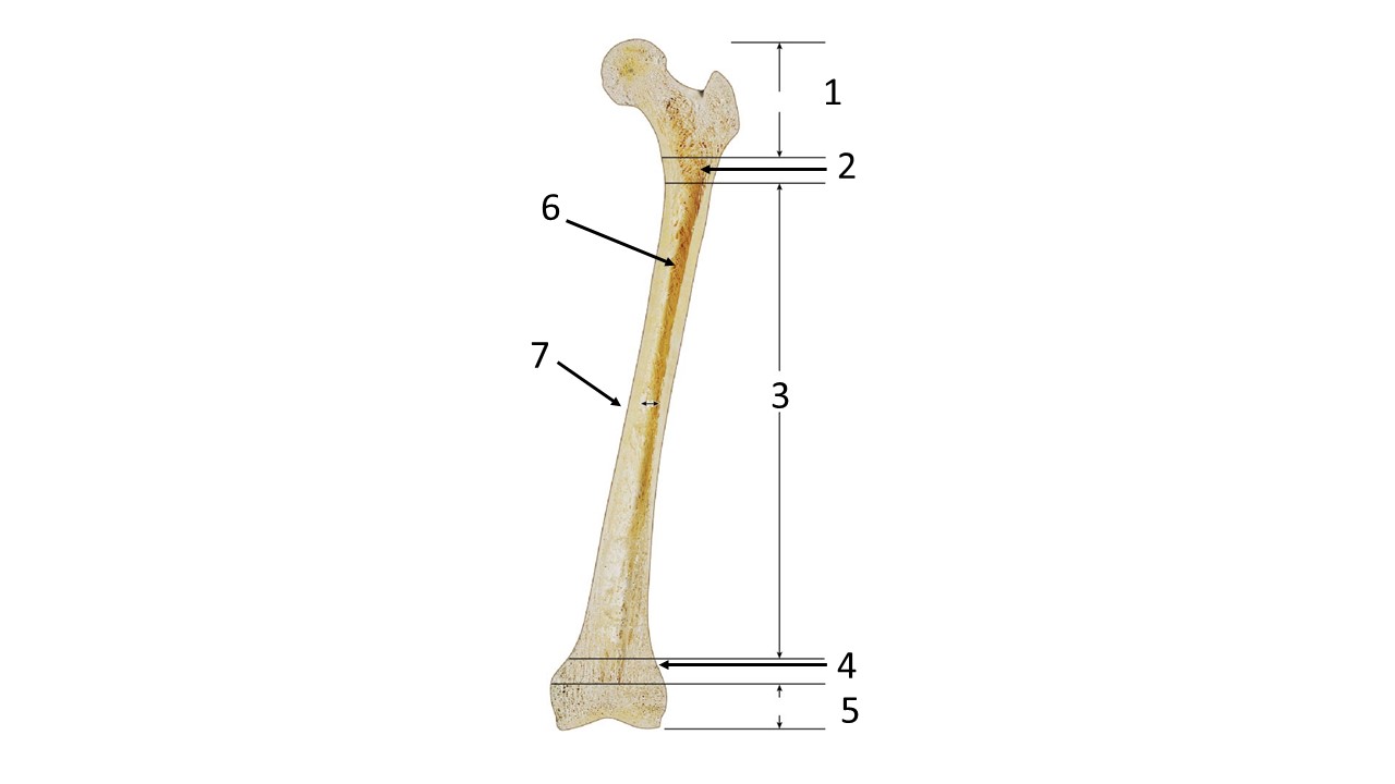
The number pointing to the distal epiphyseal plate (or line) on a long bone diagram
4