Respiratory System AMP II Pratical
 Call Kai
 Learn
 Practice Test
 Spaced Repetition
 Match
 Flashcards
 Knowt Play
1/59

There's no tags or description
Looks like no tags are added yet.
60 Terms
What is pulmonary respiration?
the physical movement of air into and out of the lungs, commonly known as breathing
What is in inhalation(Inspiration)?
The diaphragm and external intercostal (rib) muscles contract. This expands the chest cavity, lowers the pressure inside the lungs below atmospheric pressure, and draws air inward.
What is Exhalation (Expiration)?
The muscles relax, the chest cavity shrinks, and the pressure inside the lungs rises above atmospheric pressure, pushing air outward. In quiet breathing, this is mostly a passive process.
What is external respiration?
is the exchange of oxygen and carbon dioxide between the air in the lungs and the blood in the pulmonary capillaries
What is internal respiration?
the exchange of gases between the blood in systemic capillaries and the body's tissues
What is aerobic cellular respiration?
the metabolic process in eukaryotic cells that converts glucose and oxygen into carbon dioxide, water, and ATP (adenosine triphosphate), the primary energy currency of the cell.
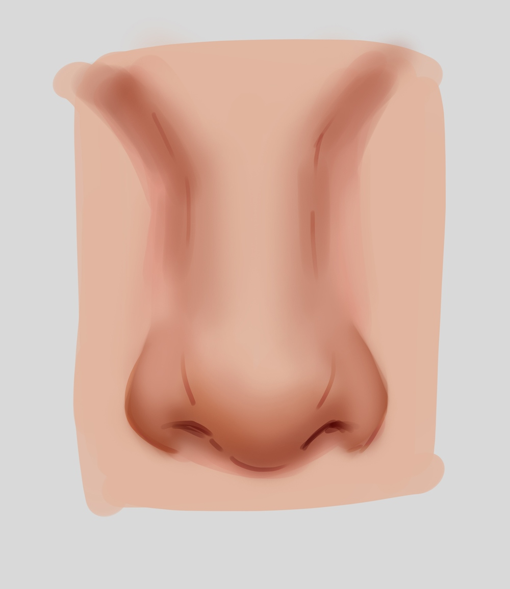
What is this and what does it do?
The nose is a primary organ for respiration and sensory perception
Breathing and Airway Pathway
Air Conditioning: Turbinates and mucous membranes heat and add moisture to incoming air so it matches body temperature before reaching the lungs.
Filtering and Defense: Nasal hairs, mucus, and microscopic cilia trap dust, pollen, germs, and particulate matter, sweeping them toward the throat to be neutralized.
Sense of Smell
Sinus and Tear Drainage

What is this and what does it do?
Pharynx
Nasopharynx: The upper section behind the nasal cavity that handles airflow and contains the adenoids and the opening of the Eustachian tube.
Oropharynx: The middle section behind the mouth that passes both air and food, containing the palatine tonsils.
Laryngopharynx (Hypopharynx): The lower section near the larynx that guides food into the esophagus and air into the voice box.

What is this and what does it do?
The trachea (commonly known as the windpipe) is a flexible, hollow tube that connects your larynx (voice box) to your bronchi (airways that lead to your lungs), serving as a primary pathway for air during respiration.

What is this?
The bronchial tree is the network of branching air passages in the lower respiratory system that transports air from the trachea to the gas-exchange units of the lungs.

What is this?
Your lungs are a pair of spongy, cone-shaped organs in your chest that handle gas exchange. They bring oxygen into your body and remove carbon dioxide waste
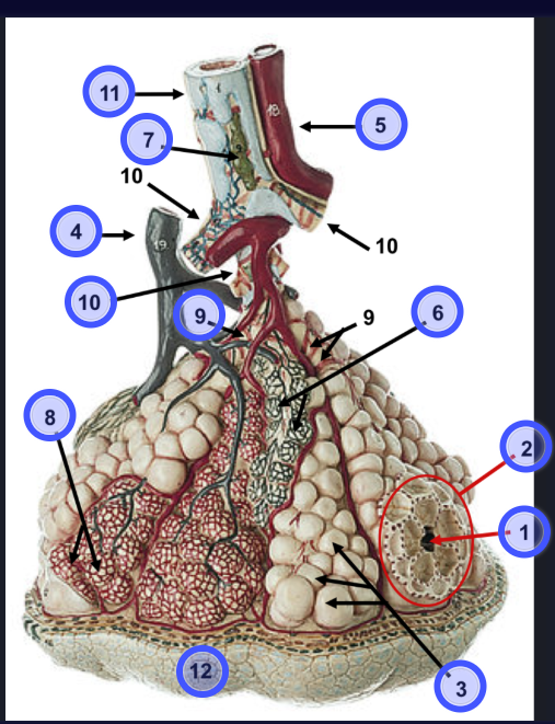
What is 11?
Bronchus

What is 5
pulmonary venule
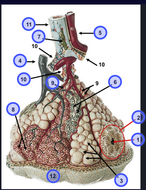
What is 7?
Mucous Cell

What is 4?
Branch of pulmonary artery

What is 10?
Terminal of bronchiole

What is 9?
smooth muscle around bronchiole?

What is 6?
elastic fibers

What is 8?
pulmonary capillaries

What is 2?
alveolar sac

What is 1?
Alveolar duct

What is 3?
alveoli

What is 12?
visceral pleura
What is boyles law?
the pressure of a given mass of an ideal gas is inversely proportional to its volume, provided the temperature and the amount of gas remain constant
how does boyles law relate to inhalation?
Volume Increases: Your diaphragm contracts and moves downward, while your rib muscles lift your ribs upward and outward. This expands the size of your chest (thoracic) cavity and your lungs.
Pressure Drops: According to Boyle's Law, increasing the volume of the lungs causes the air pressure inside them to drop below the outside (atmospheric) pressure.
Air Rushes In: Gas naturally flows from an area of higher pressure (the outside air) to an area of lower pressure (your lungs), filling them with air.
how does boyles law relate to exhalation?
Volume Decreases: Your diaphragm and rib muscles relax, allowing the chest cavity and lungs to shrink back to their smaller resting size.
Pressure Rises: According to Boyle's Law, decreasing the volume of the lungs causes the air pressure inside them to rise above the outside atmospheric pressure.
Air Rushes Out: The higher pressure inside the lungs forces air outward into the lower-pressure atmosphere.
how does the exchange of oxygen and carbon dioxide occur during external and internal respiration?
Gas exchange in the body happens via simple diffusion, where oxygen (\(O_{2}\)) and carbon dioxide (\(CO_{2}\)) move naturally from an area of higher concentration (or partial pressure) to an area of lower concentration, requiring no cellular energy.
Where does external respiration occur?
Across the respiratory membrane between the alveoli (tiny air sacs in the lungs) and the surrounding blood capillaries.
What is the movement of oxygen during external respiration?
Inhaled air in the alveoli has a high concentration of \(O_{2}\).
Deoxygenated blood arriving from the heart has a low concentration of \(O_{2}\).
\(O_{2}\) diffuses across the thin alveolar-capillary membrane from the alveoli into the blood, binding to hemoglobin in red blood cells.
What is the movement of carbon dioxide during external respiration?
The blood arriving at the lungs has a high concentration of \(CO_{2}\) (a cellular waste product).
The air in the alveoli has a lower concentration of \(CO_{2}\).
\(CO_{2}\) diffuses from the blood into the alveoli and is then removed from the body when you exhale. [1, 2, 3]
What is Daltons law?
states that the total pressure exerted by a mixture of non-reacting gases is equal to the sum of the partial pressures of the individual component gases.
How does Daltons law relate to external and internal respiration?
In respiration, this law defines how individual gases contribute to the overall pressure in the air and establish the partial pressure gradients that drive gas exchange during both external and internal respiration.
Watch a vid on this
How is O2 transported by blood?
Oxygen is not very soluble in blood plasma, so the body relies heavily on red blood cells (erythrocytes) to carry it from the lungs to the tissues. 98.5% of oxygen is bonded to hemoglobin oxygen molecules diffuse across the alveolar-capillary membrane and bind reversibly to the iron-containing heme groups of hemoglobin inside red blood cells, forming oxyhemoglobin. Each hemoglobin molecule can carry up to four O2 molecules.
1.5% of oxygen is dissolved in plasma
How is CO2 transported by blood?
Carbon dioxide is more soluble in blood than oxygen and is transported from peripheral tissues back to the lungs through three main mechanisms
As Bicarbonate Ions 70%- 85%:
CO2 enters the red blood cells from the tissues, the enzyme carbonic anhydrase rapidly converts it into carbonic acid, which immediately dissociates into bicarbonate and hydrogen ions.
Bicarbonate moves out of the red blood cells into the plasma in exchange for chloride ions (a process called the chloride shift)
Bound to hemoglobin ~10%-20%:
CO2 binds directly to the amino acid (globin) portions of the hemoglobin molecule.
Dissolved in Plasma ~5% to 10%:
A small percentage of CO2 simply dissolves directly into the blood plasma.
What are the various factors that control the rate of respiration?
1. The Respiratory Center in the Brain Stem
2. Chemical Factors (Chemoreceptors)
3. Pulmonary and Mechanical Factors (Mechanoreceptors)
4. Higher Brain Centers and Voluntary Control
5. Proprioceptors and Exercise Input
How does the Respiratory Center in the Brain Stem control the rate of respiration?
The primary neural control of breathing resides in the brain stem, which is split into two main regions: the medulla oblongata and the pons
What does the Medullary Respiratory Center do in the Respiratory center in the brain?
Dorsal Respiratory Group (DRG): Located in the nucleus tractus solitarius, the DRG acts as the primary driver of quiet inspiration. It sends rhythmic nerve impulses via the phrenic and intercostal nerves to the diaphragm and external intercostal muscles, causing them to contract.
Ventral Respiratory Group (VRG): Located in the ventrolateral medulla, the VRG contains both inspiratory and expiratory neurons. It remains mostly inactive during quiet breathing but is heavily recruited during forceful, active exhalation or deep inhalation (such as during exercise).
Pre-Bötzinger Complex: Found within the VRG, this cluster of pacemaker neurons acts as the intrinsic rhythm generator for the basic respiratory pace (establishing normal resting breathing of about 12–16 breaths per minute)
What does the Pontine Respiratory Centers (in the Pons) do in the Respiratory center in the brain?
Pneumotaxic (Pontine Respiratory Group): Located in the upper pons, it transmits inhibitory impulses to the medullary inspiratory neurons. This "switches off" inspiration, controlling the transition from inhalation to exhalation and fine-tuning respiratory rate and depth.
Apneustic Center: Located in the lower pons, it sends stimulatory signals to the medullary centers to promote prolonged inspiration (preventing premature switch-off), working in balance with the pneumotaxic center.
How does Chemical Factors (Chemoreceptors) control the rate of respiration?
Chemical composition in the blood and cerebrospinal fluid (CSF) is the most powerful influence on normal ventilation drive.
Carbon Dioxide and Hydrogen Ions:
Central Chemoreceptors: Located on the ventrolateral surface of the medulla, these sensors monitor changes in \(pH\) of the cerebrospinal fluid. Because easily crosses the blood-brain barrier, a rise in blood increases ions in the CSF (making it more acidic). This strongly stimulates the respiratory center to increase ventilation rate and depth to "blow off" is the single strongest day-to-day stimulus for breathing.
Peripheral Chemoreceptors: Located in the carotid bodies (at the bifurcation of the common carotid arteries) and aortic bodies (in the aortic arch). They detect increases in arterial and ions, as well as severe drops in blood oxygen.
Oxygen:
Peripheral chemoreceptors also monitor arterial oxygen. However, minor or moderate drops in have little effect on breathing. Oxygen levels must drop drastically (hypoxemia, typically \(P_{a}O_{2}\) falling below 60 mmHg) before peripheral chemoreceptors trigger a significant surge in the respiratory drive.
How does Pulmonary and Mechanical Factors (Mechanoreceptors) control the rate of respiration?
Inflation Reflex (Hering-Breuer Reflex):
Stretch receptors located in the visceral pleura and smooth muscle of the airways fire when the lungs become overly stretched during deep inhalation. They send inhibitory signals via the vagus nerve to the medullary centers to stop further inspiration and prevent lung over-inflation and damage.
Irritant Receptors:
Nerve endings in the airway epithelium respond to smoke, dust, pollen, or cold air, triggering reflexes like coughing, sneezing, or transient breath-holding/bronchoconstriction.
(J)-receptors (Juxtacapillary Receptors):
Located in the alveolar walls near capillaries, they respond to pulmonary congestion, edema, or increased interstitial fluid pressure, resulting in rapid, shallow breathing.
How does Higher Brain Centers and Voluntary Control, control the rate of respiration?
Cerebral Cortex: Allows for conscious, voluntary overrides of the brain stem's automatic rhythm (e.g., holding your breath, singing, or speaking). However, this voluntary control has strict limits; eventually, rising CO2 and H+ levels in the blood override conscious control, forcing the brain stem to resume automatic respiration.
Limbic System and Hypothalamus: Emotional states (fear, excitement, anxiety, pain) feed into the respiratory centers, which is why fear or sudden pain causes sharp changes in breathing rate.
How does Proprioceptors and Exercise Input control the rate of respiration?
Muscle and Joint Proprioceptors:
During physical activity, movement in the limbs and joints sends immediate signals to the respiratory center. This neural feedforward mechanism quickly increases the breathing rate before blood chemistry even changes, matching ventilation to metabolic demand during exercise.

What is this?
external nares (nostrils)
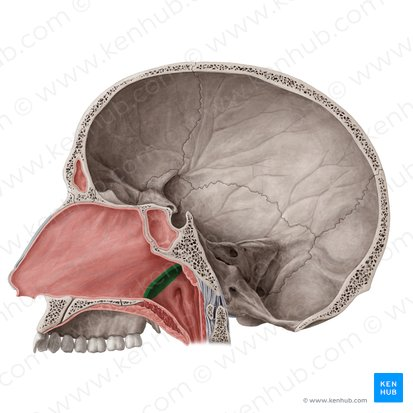
What is this?
internal nares (choanae)

What is this?
nasal septum

What is this?
Vestibule

What is this?
superior nasal concha (turbinate)
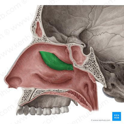
What is this?
middle nasal concha (turbinate)

What is this?
inferior nasal concha (turbinate)
What is the passage under the superior nasal concha called?
superior nasal meatus
What is the passage under the middle nasal concha called?
middle nasal meatus
What is the passage under the inferior nasal concha called?
inferior nasal meatus

What is this?
capillaries

What is this?


What is this?
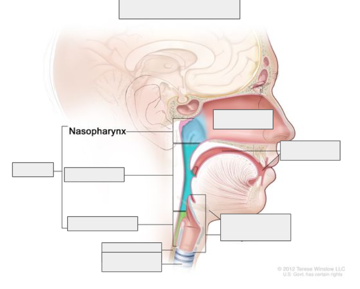

What is this?


What is this?
laryngopharynx or hypopharynx

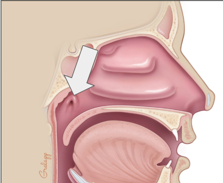
What is this?
eustachian tube in nasal cavity

What is this?
pharyngeal tonsil