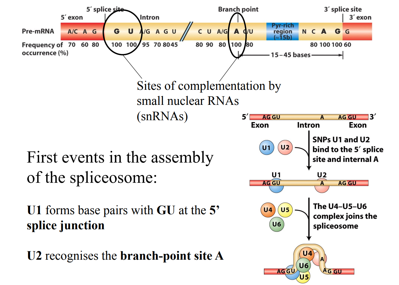
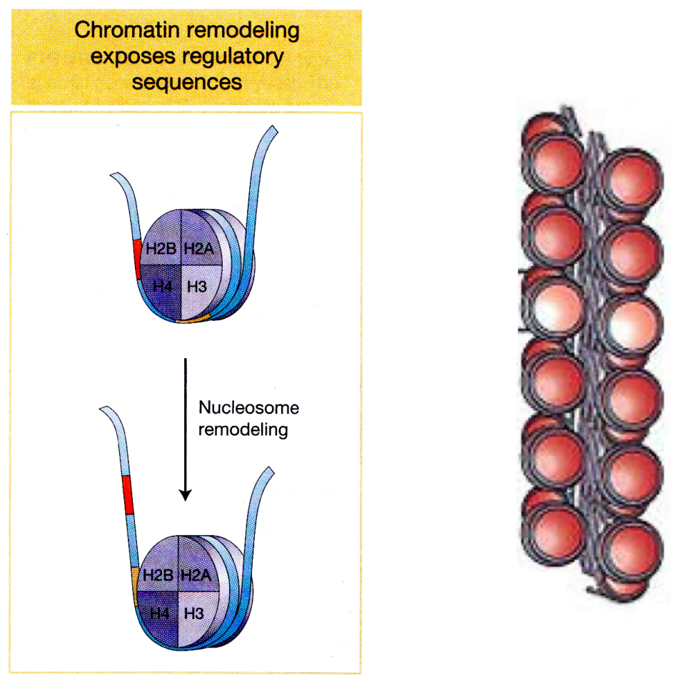
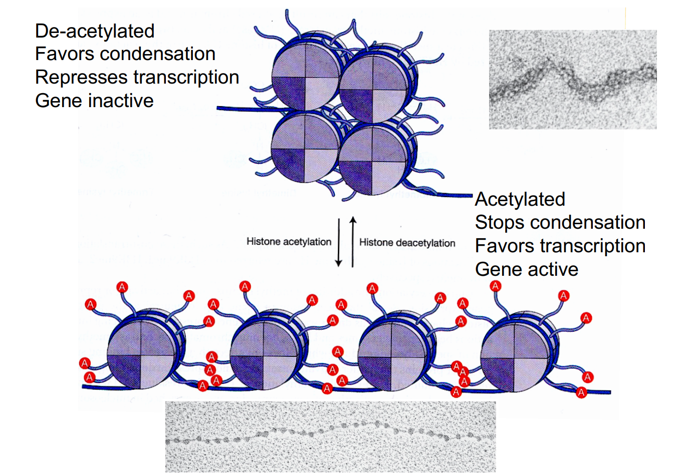
Theme IV-V

0.0(0)
Studied by 0 people
call kaiCall Kai
learnLearn
examPractice Test
spaced repetitionSpaced Repetition
heart puzzleMatch
flashcardsFlashcards
GameKnowt Play
Card Sorting

1/258

encourage image

There's no tags or description

Looks like no tags are added yet.

259 Terms

1
New cards

E. Coli Info

  • E. coli can use glucose or lactose as a carbon source

    • Glucose is utilized exclusively during the initial growth phase

    • Lactose (glucose + galactose) is utilised during the second growth phase once the glucose has been depleted

  • beta-galactosidase enzyme is required for growth because it hydrolyses lactose and converts it into the active inducer of the lactose (lac) operon. Without this enzymatic activity, the cells cannot properly induce the operon or metabolise lactose for growth

  • This means E. coli grows faster on glucose than on lactose. As Glucose enters glycolysis directly. Lactose requires lac operon induction plus hydrolysis into glucose + galactose, so growth on lactose is slower.


  • Diauxic (or diphasic growth) is defined as the growth of bacteria in media containing two carbon sources (at concentrations insufficient to support full growth), which results in a double-phased (diphasic) growth curve separated by a lag period

    • First Growth Phase: The bacteria grow exponentially by utilising glucose exclusively, while the synthesis of beta-galactosidase is completely prevented

    • Lag Phase: When glucose is depleted, growth temporarily stops (lags), during which time the cells begin synthesising beta-galactosidase

    • Second Growth Phase: Growth resumes at a rapid rate as the bacteria begin utilising lactose


  • Catabolite repression: Glucose acts through a mechanism dependent on a functional CR gene (catabolite repression gene), which reduces the basal level of beta-galactosidase permease in glucose-grown cells, thereby preventing them from taking up enough lactose to overcome the glucose inhibition


<ul><li><p>E. coli can use glucose or lactose as a carbon source </p><ul><li><p>Glucose is utilized exclusively during the initial growth phase </p></li><li><p><span>L</span>actose (glucose + galactose) is utilised during the second growth phase<span> once the glucose has been depleted</span></p></li></ul></li><li><p>beta<strong>-galactosidase enzyme is required for growth </strong>because <strong>it hydrolyses lactose and converts it into the active inducer</strong> of the lactose (<em>lac</em>) operon. Without this enzymatic activity, the cells cannot properly induce the operon or metabolise lactose for growth</p></li></ul><ul><li><p>This means E. coli grows faster on glucose than on lactose. As Glucose enters glycolysis directly. Lactose requires lac operon induction plus hydrolysis into glucose + galactose, so growth on lactose is slower.</p></li></ul><p></p><ul><li><p>Diauxic (or diphasic growth) is defined as the growth of bacteria in media containing<strong> two carbon sources</strong><span><strong> </strong>(at concentrations insufficient to support full growth), which results in a </span>double-phased (diphasic) growth curve<span> separated by a lag period</span></p><ul><li><p><strong>First Growth Phase:</strong> The bacteria grow exponentially by <strong>utilising glucose exclusively</strong>, while the synthesis of beta-galactosidase is completely prevented</p></li><li><p><strong>Lag Phase:</strong> When glucose is depleted, <strong>growth temporarily stops (lags)</strong>, during which time the cells begin synthesising beta-galactosidase</p></li><li><p><strong>Second Growth Phase:</strong> Growth <strong>resumes at a rapid rate</strong> as the bacteria begin utilising lactose</p></li></ul></li></ul><p></p><ul><li><p><strong>Catabolite repression:</strong> Glucose acts through a mechanism dependent on a functional CR gene (catabolite repression gene), which reduces the basal level of beta-galactosidase permease in glucose-grown cells, thereby preventing them from taking up enough lactose to overcome the glucose inhibition</p></li></ul><p></p>
2
New cards
<p>Stripe 2</p>

Stripe 2

  • Image 1 (top, only faint pale stripes): the stripe-2 signal is essentially gone. This is the Bicoid-minus mutant, since without Bicoid there is no activator driving the stripe.

  • Image 2 (dark, thick expression at the very anterior end): the stripe has spread forward. This is the Giant-minus mutant, since Giant normally represses the anterior border of stripe 2.

  • Image 3 (single sharp dark stripe): this is the normal, native pattern.

  • Image 4 (broadened stripe that is smeared toward the posterior): the stripe has spread backward. This is the Krüppel-minus mutant, since Krüppel normally represses the posterior border.


*Hunchback-minus: there are four images for five conditions, so one image has to serve two answers. Hunchback is a co-activator with Bicoid, so losing it also strongly reduces stripe 2. That means it matches Image 1 as well, though in the real embryo the loss is usually less complete than in a Bicoid mutant. If your course material says the Hunchback mutant should look different, the "weak or absent stripe" image is still the closest fit

3
New cards

Is the arabinose operon under positive or negative regulation?

  • Dual positive and negative control

  • The regulation of the arabinose operon provides an example in which a single DNA-binding protein may act as either a repressor or an activator

  • Repressor:

    • In the absence of arabinose, the AraC protein adopts a different conformation and binds to both the araI and the araO regions, forming a DNA loop that prevents transcription

    • This binding prevents transcription of the ara operon. I.e. in this situation, AraC represses the operon

  • Activator:

    • Transcription is activated at araI, the initiator region, which contains both an operator site and a promoter

    • The araC gene, which maps immediately upstream of the operon encodes an activator protein

    • When bound to arabinose, this protein activates transcription of the ara operon, perhaps by helping RNA polymerase bind to the promoter

    • In addition, the CAP–cAMP catabolite repression system regulates expression of the ara operon

    • In the presence of arabinose, the AraC protein binds to the araI region. In the absence of glucose, the CAP – cAMP complex binds to an ajacent site of araI

    • This binding stimulates the transcription of the araB, araA, and araD genes. Both the CAP–cAMP complex and the AraC–arabinose complex must bind to araI in order for RNA polymerase to bind to the promoter and transcribe the ara operon


  • araC protein=Repressor (arabinose absent)

  • araC protein=Activator (arabinose present)

  • Arabinose=inducer

  • CAP = Acts as a transcriptional activator that is controlled by the levels of glucose

    • Need both CAP+cAMP AND araC binding for transcription to occur

    • When glucose is absent, there is plenty of cAMP, CAP protein/cAMP complex binds, opens up ds-DNA, increasing the binding affinity of AraC and stimulating RNA polymerase to bind to the promoter

    • Proteins B, A, D are made, allowing the cells to use arabinose as the energy source. When glucose is present, CAP does not bind, and the arabinose operon is not activated. Glucose is used as the energy source, arabinose is not needed.


<ul><li><p>Dual positive and negative control</p></li><li><p>The regulation of the arabinose operon provides an example in which a single DNA-binding protein may act as either a repressor or an activator</p></li><li><p><strong><u>Repressor</u></strong>:</p><ul><li><p>In the absence of arabinose, the AraC protein adopts a different conformation and binds to both the araI and the araO regions, forming a DNA loop that prevents transcription</p></li><li><p>This binding prevents transcription of the ara operon. I.e. in this situation, AraC represses the operon</p></li></ul></li><li><p><strong><u>Activator</u></strong>:</p><ul><li><p>Transcription is activated at araI, the initiator region, which contains both an operator site and a promoter</p></li><li><p>The araC gene, which maps immediately upstream of the operon encodes an activator protein</p></li><li><p>When bound to arabinose, this protein activates transcription of the ara operon, perhaps by helping RNA polymerase bind to the promoter</p></li><li><p>In addition, the CAP–cAMP catabolite repression system regulates expression of the ara operon</p></li><li><p>In the presence of arabinose, the AraC protein binds to the araI region. In the absence of glucose, the CAP – cAMP complex binds to an ajacent site of araI</p></li><li><p>This binding stimulates the transcription of the araB, araA, and araD genes. Both the CAP–cAMP complex and the AraC–arabinose complex must bind to araI in order for RNA polymerase to bind to the promoter and transcribe the ara operon</p></li></ul></li></ul><p></p><div data-type="horizontalRule"><hr></div><ul><li><p>araC protein=Repressor (arabinose absent)</p></li><li><p>araC protein=Activator (arabinose present)</p></li><li><p>Arabinose=inducer</p></li><li><p>CAP = Acts as a transcriptional <strong>activator </strong>that is controlled by the levels of glucose</p><ul><li><p>Need both CAP+cAMP AND araC binding for transcription to occur </p></li><li><p>When glucose is absent, there is plenty of cAMP, CAP protein/cAMP complex binds, opens up ds-DNA, increasing the binding affinity of AraC and stimulating RNA polymerase to bind to the promoter</p></li><li><p>Proteins B, A, D are made, allowing the cells to use arabinose as the energy source. When glucose is present, CAP does not bind, and the arabinose operon is not activated. Glucose is used as the energy source, arabinose is not needed.</p></li></ul></li></ul><p></p>
4
New cards

Comparison between transcription in Prokaryotes and Eukaryotes

knowt flashcard image
5
New cards

Test note

  • Always assume strands are CODING STRAND going 5’ to 3’ direction

    • Therefore the mRNA produced is literally the same just the T is swapped with U

  • There is no sigma factors or Rho-dependent termination in eukaryotes → only in prokaryotes


<ul><li><p>Always assume strands are CODING STRAND going 5’ to 3’ direction</p><ul><li><p>Therefore the mRNA produced is literally the same just the T is swapped with U</p></li></ul></li><li><p>There is no sigma factors or Rho-dependent termination in eukaryotes → only in prokaryotes</p></li></ul><p></p>
6
New cards

Class stuff

  • Complementation → a technique used to test whether a gene actually works? By placing that gene in another spot

  • Insertional inactivation → add a selective marker or something into the gene, this makes the protein unable to fold or something?

  • VERY COMMON QS IS TO MOVE GENE AROUND, DELETE, ADD, SWAP ETC.

    • Very common for Taq polymerisation attenuation, what happens if you delete or elongate one of the leader sequences, what happens to attenuation

  • A gene can be mutated but a protein is a variant (not a mutant) - terminology


7
New cards
<p>How do other sigma factors not relevant not interfere? </p>

How do other sigma factors not relevant not interfere?

  • Sigma factor 32 is present at constant amounts at normal temperature

  • It is however in a complex with the molecular chaperone DnaK thus is not able to recruit RNA polymerase - and it does not need to as the cell is not under heat shot

  • If the cell were to be heat shocked, folded proteins would start to unfold, DnaK will then bind to those unfolded proteins, releasing sigma factor 32 to bind to RNA polymerase and start transcription to help deal with cytoplasmic stress

  • The sigma factor 32 recognises promoters of the genes encoding many different stress response proteins, INCLUDING DnaK


<ul><li><p>Sigma factor 32 is present at constant amounts at normal temperature</p></li><li><p>It is however in a complex with the molecular <span style="color: yellow;">chaperone </span>DnaK thus is not able to recruit RNA polymerase - and it does not need to as the cell is not under heat shot</p></li><li><p>If the cell were to be heat shocked, folded proteins would start to unfold, DnaK will then bind to those unfolded proteins, releasing sigma factor 32 to bind to RNA polymerase and start transcription to help deal with cytoplasmic stress</p></li><li><p>The sigma factor 32 recognises promoters of the genes encoding many different stress response proteins, INCLUDING DnaK</p></li></ul><p></p>
8
New cards
<p>How to get bacteria to make any protein?</p>

How to get bacteria to make any protein?

  • Cannot use linear DNA because as cells divide there is no way to retain the linear DNA copy

  • Plasmids can be retained and passed down as the cell divides

  • Allo-lactose is a bit impractical since it requires metabolite (lactose) the entire time

  • Image above: In the normal state the Lac I(?) molecule binds and blocks both the lac promoter and T7 promoter, when you add IPTG it will bind and cause the Lac I to fall off, allowing first the transcription of the T7 gene and then the target gene → this leads to the overexpression of the target gene

    • pLysS binds to the RNA polymerase to stop leakiness , mock up unwanted T7 RNA polymerase (?)


<ul><li><p>Cannot use linear DNA because as cells divide there is no way to retain the linear DNA copy </p></li><li><p>Plasmids can be retained and passed down as the cell divides </p></li><li><p>Allo-lactose is a bit impractical since it requires metabolite (lactose) the entire time </p></li><li><p>Image above: In the normal state the Lac I(?) molecule binds and blocks both the lac promoter and T7 promoter, when you add IPTG it will bind and cause the Lac I to fall off, allowing first the transcription of the T7 gene and then the target gene → this leads to the overexpression of the target gene </p><ul><li><p>pLysS binds to the RNA polymerase to stop leakiness , mock up unwanted T7 RNA polymerase (?)</p></li></ul></li></ul><p></p>
9
New cards

Are all genes expressed at the same time?

Prokaryotes

  • Different genes expressed in response to different environments

    • Presence of a food substrate (lactose)

  • Don’t waste energy expressing genes which are of no use in a particular environment

  • These genes are always on the ‘on’ state, thus we only mostly need a RNA poly to bind to begin transcription


Eukaryotes

  • All cells retain entire hereditary potential of the organism

  • BUT differentiate into different types (brain vs muscle) so need to express different factors

  • These genes are default turned ‘off’, you first need a large number of transcription factors and enhancers to help the RNA poly bind to the promoter and begin transcription

    • This is because the DNA is very compacted, tightly coiled and condensed and thus not available for RNA poly


<p><span style="color: rgb(162, 144, 183);"><strong>Prokaryotes</strong></span></p><ul><li><p>Different genes expressed in response to <em>different environments</em></p><ul><li><p>Presence of a food substrate (lactose)</p></li></ul></li><li><p>Don’t waste energy expressing genes which are of no use in a particular environment</p></li><li><p>These genes are always on the ‘<u>on</u>’ state, thus we only mostly need a RNA poly to bind to begin transcription </p></li></ul><p></p><p><span style="color: rgb(162, 144, 183);"><strong>Eukaryotes</strong></span></p><ul><li><p>All cells retain entire hereditary potential of the organism</p></li><li><p>BUT differentiate into different types (brain vs muscle) so need to express <em>different factors</em></p></li><li><p>These genes are default turned ‘<u>off</u>’, you first need a large number of transcription factors and enhancers to help the RNA poly bind to the promoter and begin transcription </p><ul><li><p>This is because the DNA is very compacted, tightly coiled and condensed and thus not available for RNA poly  </p></li></ul></li></ul><p></p>
10
New cards

What is the process of going from DNA to active proteins?

  • All of these steps could be subject to regulation


<ul><li><p>All of these steps could be subject to regulation </p></li></ul><p></p>
11
New cards

Prokaryotic gene expression

  • Bacteria sense and respond to particular stimuli often very rapidly (within minutes)

  • Example

    • Bacteria can utilise many different sugars for growth

    • Only express the genes necessary for metabolism of the sugar when it is present


  • Must be able to

    • Sense when/what the sugar is present

    • Up-regulate necessary genes in response

    • Down-regulate necessary genes when sugar is absent


12
New cards

How can prokaryotes rapidly respond to changes in the environment, how is this rapid response regulated?

  • Different genes can be regulated in different ways

    • But use a few common mechanisms


  • Regulation at the level of transcription

    • Most common form of regulation

    • Transcription of DNA into mRNA

    • Can regulate how much mRNA is made from a gene

    • If no mRNA then final protein can’t be expressed


  • Regulation at the level of translation

    • Translation is the synthesis of protein from an mRNA message

    • Regulation of how much active protein is made from an mRNA transcript

13
New cards
<p>Regulation of <span style="color: rgb(43, 187, 215);"><strong>transcription</strong></span></p>

Regulation of transcription

  • Promoter sequences determine where RNA polymerase starts transcription

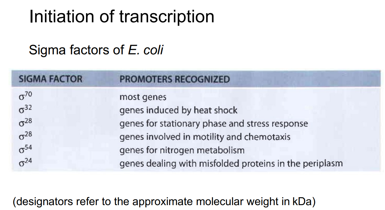
    • E.g. TTGACA-17bp-TATAAT this is a promoter sequence in E. Coli (these are usually preserved) (upstream of the gene)

    • All genes have slightly different promoters, these determine the efficiency (the one above is very efficient)

    • The sigma factor subunit that defines the specificity to the promoter comes in many different ‘flavours’

      • Sigma factor binds to the RNA polymerase to create the holoenzyme, then guides it towards the specific promoter

      • There are many different sigma factors as promoter sequences differ a bit here and there, sigma factors can regulate a whole group of genes (so not just one gene)

    • The bacterial cell has many alternative sigma factors, all competing for binding to the core RNA polymerase

    • What genes are transcribed is determined by how abundant this or that sigma factor is at this particular moment

    • How can bacteria turn promoters ON and OFF


  • Negative regulation (repressor)

    • A regulatory protein is used to stop transcription of a gene (turns promoter OFF)

    • When genes need to be turned on in response to a stimulus the negative regulator must be inactivated


  • Positive regulation (activator)

    • A regulatory protein is required before the gene is efficiently transcribed (turn promoter ON)

    • When genes need to be turned on in response to a stimulus the positive activator must be activated


<ul><li><p>Promoter sequences determine where RNA polymerase starts transcription</p><ul><li><p>E.g. <em>TTGACA-17bp-TATAAT</em> this is a promoter sequence in E. Coli (these are usually preserved) (upstream of the gene)</p></li><li><p>All genes have slightly different promoters, these determine the efficiency (the one above is very efficient)</p></li><li><p>The sigma factor subunit that defines the specificity to the promoter comes in many different ‘flavours’</p><ul><li><p>Sigma factor binds to the RNA polymerase to create the holoenzyme, then guides it towards the specific promoter </p></li><li><p>There are many different sigma factors as promoter sequences differ a bit here and there, sigma factors can regulate a whole group of genes (so not just one gene) </p></li></ul></li><li><p>The bacterial cell has many alternative sigma factors, all competing for binding to the core RNA polymerase</p></li><li><p>What genes are transcribed is determined by how abundant this or that sigma factor is at this particular moment</p></li><li><p>How can bacteria turn promoters ON and OFF</p></li></ul></li></ul><p></p><ul><li><p><span style="color: rgb(217, 239, 189);">Negative regulation (<strong>repressor</strong>)</span></p><ul><li><p>A regulatory protein is used to stop transcription of a gene (turns promoter OFF)</p></li><li><p>When genes need to be turned on in response to a stimulus the negative regulator must be inactivated</p></li></ul></li></ul><p></p><ul><li><p><span style="color: rgb(217, 239, 189);">Positive regulation (<strong>activator</strong>)</span></p><ul><li><p>A regulatory protein is required before the gene is efficiently transcribed (turn promoter ON)</p></li><li><p>When genes need to be turned on in response to a stimulus the positive activator must be activated</p></li></ul></li></ul><p></p>
14
New cards

How can bacteria turn promoters OFF (repressor protein)

  • Negative regulation of transcription

  • A repressor protein binds to the DNA turning a promoter off

    • A promoter is “off” when RNA polymerase can’t bind and begin transcription

    • The repressor usually binds over the promoter sequence so RNA pol can’t bind to the DNA

  • The place where the repressor binds is called the “operator site”

    • The operator site usually partially overlaps the promoter sequence

  • The repressor may bind an “inducer” molecule

    • Repressor plus inducer becomes inactive for binding (repressor falls off)

  • In some instances a co-repressor molecule may be required to allow the repressor to bind

    • Repressor plus co-repressor becomes active for binding


<ul><li><p>Negative regulation of transcription</p></li><li><p>A repressor protein binds to the DNA turning a promoter off</p><ul><li><p>A promoter is “off” when RNA polymerase can’t bind and begin transcription</p></li><li><p>The repressor usually binds over the promoter sequence so RNA pol can’t bind to the DNA</p></li></ul></li><li><p><span style="color: rgb(252, 148, 234);">The place where the repressor binds is called the “<strong>operator site</strong>”</span></p><ul><li><p>The operator site usually partially overlaps the promoter sequence </p></li></ul></li><li><p>The repressor may bind an “inducer” molecule</p><ul><li><p>Repressor plus inducer becomes inactive for binding (repressor falls off) </p></li></ul></li><li><p>In some instances a <strong>co-repressor </strong>molecule may be required to allow the repressor to bind</p><ul><li><p>Repressor plus co-repressor becomes active for binding</p></li></ul></li></ul><p></p>
15
New cards
<p>Repression is relieved by an inducer</p>

Repression is relieved by an inducer

  • 1) The repressor binds to the operator site and stops transcription

    • Operator site is -5 to +21 (transcription should start at +1)

    • When the repressor binds it covers this +1 and therefore the RNA poly cannot bind as efficiently and even if it could the gene cannot unwind

  • 2) When the inducer is present it binds to the repressor and the repressor falls off the DNA

    • This inducer is called allo-lactose and it is a derivative of lactose

  • 3) RNA polymerase can now access the promoter and perform transcription


<ul><li><p>1) The repressor binds to the operator site and stops transcription</p><ul><li><p>Operator site is -5 to +21 (transcription should start at +1)</p></li><li><p>When the repressor binds it covers this +1 and therefore the RNA poly cannot bind as efficiently and even if it could the gene cannot unwind </p></li></ul></li><li><p>2) When the inducer is present it binds to the repressor and the repressor falls off the DNA</p><ul><li><p>This inducer is called allo-lactose and it is a derivative of lactose </p></li></ul></li><li><p>3) RNA polymerase can now access the promoter and perform transcription</p></li></ul><p></p>
16
New cards

How can bacteria turn promoters ON (activator protein)

  • Positive regulation of transcription

  • An “activator” protein is required to bind to the DNA to turn a promoter on

    • The promoter is only active when the activator binds

  • The activator can only bind DNA in the presence of an inducer molecule

    • The inducer interacts with the activator allowing it to bind to the DNA

    • Most likely to bind at an initiator site (?)

  • In the absence of an inducer molecule

    • The activator does not bind to DNA so the gene is off


17
New cards
<p>Activation is induced by inducer</p>

Activation is induced by inducer

  • 1) The activator must bind to the promoter to allow transcription to begin

    • Activators mostly bind upstream of the promoter sequence

    • Therefore they can interact with the RNA poly and help it to bind to the DNA sequence

    • Thus increasing the amount of bound RNA poly → increases gene expression

    • The TATA box is intact, so the general transcription factors and RNA polymerase II can still assemble, but without an activator they give only low-level transcription


  • 2) The activator can only bind to the DNA in the presence of the inducer

    • Inducer => able to induce gene expression


<ul><li><p>1) The activator must bind to the promoter to allow transcription to begin</p><ul><li><p>Activators mostly bind upstream of the promoter sequence</p></li><li><p>Therefore they can interact with the RNA poly and help it to bind to the DNA sequence</p></li><li><p>Thus increasing the amount of bound RNA poly → increases gene expression</p></li><li><p>The TATA box is intact, so the general transcription factors and RNA polymerase II can still assemble, but without an activator they give only low-level transcription</p></li></ul></li></ul><p></p><ul><li><p>2) The activator can only bind to the DNA in the presence of the inducer</p><ul><li><p>Inducer =&gt; able to induce gene expression</p></li></ul></li></ul><p></p>
18
New cards

What are Allosteric Proteins?

  • Allosteric proteins are proteins that exist in two conformational forms (different shapes)

    • Transcriptional repressors and activators are allosteric proteins

  • These proteins change conformation when they bind the inducer

    • Change in conformation alters ability to bind DNA

  • These allosteric proteins have at least two different binding sites

    • One for binding the inducer (effector)

    • One for binding DNA


<ul><li><p>Allosteric proteins are proteins that <span style="color: rgb(167, 247, 232);">exist in two conformational forms</span> (different shapes)</p><ul><li><p>Transcriptional <strong>repressors </strong>and <strong>activators </strong>are allosteric proteins</p></li></ul></li><li><p>These proteins change conformation when they bind the inducer</p><ul><li><p>Change in conformation alters ability to bind DNA</p></li></ul></li><li><p>These allosteric proteins have at least two <em>different binding sites</em></p><ul><li><p>One for binding the inducer (effector)</p></li><li><p>One for binding DNA</p></li></ul></li></ul><p></p>
19
New cards

What is an operon?

  • A region of DNA that consists of three key elements: a promoter, an operator and a set of related genes referred to as ‘structural genes’

  • Usually an operon includes 2-6 structural genes but can range from 1 to over 20

  • The genes within an operon are under the control of the same promoter and as such, are transcribed together when the promoter is turned ‘on’

  • Regulatory proteins bind to sequences within the operon allow for control of transcription

  • E. coli: the lac operon and the trp operon

    • The lac operon is a classic example of an inducible operon where the binding of a regulatory molecule increases gene transcription

    • The trp operon is a repressible operon which means that gene transcription can be turned off or repressed by the binding of a regulatory molecule

    • It’s very important to note that different operons are regulated in different ways. That being the case, there are still some common elements


<ul><li><p>A region of DNA that consists of three key elements: a promoter, an operator and a set of related genes referred to as ‘structural genes’</p></li><li><p>Usually an operon includes 2-6 structural genes but can range from 1 to over 20</p></li><li><p>The genes within an operon are under the control of the same promoter and as such, are transcribed together when the promoter is turned ‘on’</p></li><li><p>Regulatory proteins bind to sequences within the operon allow for control of transcription</p></li><li><p><em>E. coli</em>: the <em>lac</em> operon and the <em>trp</em> operon</p><ul><li><p>The <em>lac</em> operon is a classic example of an inducible operon where the binding of a regulatory molecule increases gene transcription</p></li><li><p>The <em>trp</em> operon is a repressible operon which means that gene transcription can be turned off or repressed by the binding of a regulatory molecule</p></li><li><p>It’s very important to note that different operons are regulated in different ways. That being the case, there are still some common elements</p></li></ul></li></ul><p></p>
20
New cards

What is Negative Regulation?

  • Negative regulation is modulated by a repressor protein. When bound to an operator site, a repressor protein inhibits the binding of RNA polymerase, effectively turning off the promoter. This results in a decrease in transcription (2a)

  • In inducible operons, this is the default state. The repressor is constitutively expressed, bound to the operator and the expression is turned off. Repression stops when an inducer binds to the repressor and allows transcription to begin

  • In a repressible operon, transcription remains active until a repressor protein binds to the operator (2b)

    • Repression occurs when a co-repressor binds to a repressor protein, inducing a conformational change, allowing it to bind to the operator and prevent transcription.  Until the co-repressor binds, a repressor protein is in an inactive state, unable to bind to the operator


<ul><li><p><strong>Negative regulation</strong> is modulated by a <strong>repressor protein</strong>. When bound to an operator site, a repressor protein inhibits the binding of RNA polymerase, effectively turning off the promoter. This results in a decrease in transcription (2a)</p></li><li><p>In<strong> inducible operons</strong>, this is the default state. The repressor is constitutively expressed, bound to the operator and the expression is turned off. Repression stops when an <strong>inducer</strong> binds to the repressor and allows transcription to begin </p></li><li><p>In a <strong>repressible operon</strong>, transcription remains active until a repressor protein binds to the operator (2b)</p><ul><li><p>Repression occurs when a <strong>co-repressor</strong> binds to a repressor protein, inducing a conformational change, allowing it to bind to the operator and prevent transcription.&nbsp; Until the co-repressor binds, a repressor protein is in an inactive state, unable to bind to the operator</p></li></ul></li></ul><p></p>
21
New cards

What is Positive Regulation?

  • Positive regulation is modulated by activator proteins that bind to an activator binding site located within or within close proximity of a promoter region. When bound, activator proteins increase the efficiency of transcription and thus the amount of mRNA

  • For an inducible operon, activator proteins remain unbound until an inducer molecule binds to the activator and increases its affinity for it’s specific binding site in the DNA (Figure 2d)

  • In a repressible operon, an activator protein is normally bound to DNA allowing gene transcription. Positive regulation of this system involves a signalling molecule that  binds to the activator leading to its dissociation from DNA (Figure 2c). This results in reduced transcription.


<ul><li><p><strong>Positive regulation</strong> is modulated by <strong>activator</strong> proteins that bind to an activator binding site located within or within close proximity of a promoter region. When bound, activator proteins increase the efficiency of transcription and thus the amount of mRNA</p></li><li><p>For an <strong>inducible operon</strong>, activator proteins remain unbound until an <strong>inducer</strong> molecule binds to the activator and increases its affinity for it’s specific binding site in the DNA (Figure 2d)</p></li><li><p>In a<strong> repressible operon</strong>, an activator protein is normally bound to DNA allowing gene transcription. Positive regulation of this system involves a signalling molecule that &nbsp;binds to the activator leading to its dissociation from DNA (Figure 2c). This results in reduced transcription.</p><p>&nbsp;</p></li></ul><p></p>
22
New cards

What is Catabolite Repression?

  • Many prokaryotes can use a number of different energy sources (catabolites) to generate ATP but need to express the relevant enzymes to do so

  • There is no point expressing all the enzymes for the catabolism of a particular energy source if it is not present

    • For example, if glucose is present but not lactose, it would be inefficient to express the enzymes required for lactose metabolism

    • In addition, glucose is the preferred energy source

    • So, when both glucose and lactose are present, glucose would be preferentially utilised and again, there would be no requirement of the lactose metabolism enzymes


  • So how is this controlled? How does a cell only transcribe the set of enzymes for the present or even preferred catabolite?

    • The answer is catabolite repression

    • Some operons have a regulatory site known as a CAP-binding site that binds a protein called catabolite activator protein (CAP)

    • When glucose is high, the levels of ATP in the cell are high

    • When glucose is low, ATP is converted to cAMP via an enzyme called adenylate cyclase and the cAMP levels in the cell rise

    • In the absence of glucose, cAMP binds to CAP

    • This complex then binds to a specific regulatory sequence in the operon known as the CAP-binding site, and increases the efficiency of transcription


23
New cards
<p>Regulation of the lactose operon</p>

Regulation of the lactose operon

  • Many genes are controlled by both negative and positive regulation

  • Lactose (lac) operon of E. coli

    • Controls metabolism of lactose (galactose + glucose) → we need glucose


  • Negative regulation

    • By the lactose repressor (product of lacI gene)

      • Can sense how much lactose is in the environment

    • Inducers are low MW b-galactosidase (allo-lactose)


  • Positive regulation

    • By CAP activator protein

    • Inducer is cAMP (which responds to glucose)

      • Aka can sense how much glucose is in the environment


<ul><li><p>Many genes are controlled by both negative and positive regulation</p></li><li><p>Lactose (lac) operon of E. coli</p><ul><li><p>Controls metabolism of lactose (galactose + glucose) → we need glucose </p></li></ul></li></ul><p></p><ul><li><p>Negative regulation</p><ul><li><p>By the <span style="color: rgb(225, 139, 139);">lactose repressor </span>(product of lacI gene)</p><ul><li><p>Can sense how much <u>lactose</u> is in the environment</p></li></ul></li><li><p>Inducers are low MW b-galactosidase (allo-lactose)</p></li></ul></li></ul><p></p><ul><li><p>Positive regulation</p><ul><li><p>By <span style="color: rgb(132, 241, 186);">CAP activator</span> protein</p></li><li><p>Inducer is cAMP (which responds to glucose)</p><ul><li><p>Aka can sense how much <u>glucose</u> is in the environment </p></li></ul></li></ul></li></ul><p></p>
24
New cards

Negative regulation of the lac operon

  • I gene → the Lacl gene (code for the Lactose repressor protein), found elsewhere in the chromosome and is made all the time

  • The Lac operon means these gene share the same promoter and are either ALL turned on/off at the same time, this is because all these gene have a very similar function in metabolising lactose and we need the translated proteins at roughly the same level

  • In the absence of lactose, the repressor is active and will bind to the operate and block unwinding of the DNA and progression the RNA poly

    • No lactose present → no need for Lac operon genes as no need for lactose metabolism → turn gene OFF

  • In the presence of lactose (converted to allo-lactose), it can bind to the allosteric site on the Lac repressor protein which causes a confirmational change such that it can no longer bind to the Lac operator site, thus RNA poly can no transcribe the operon

    • Lactose present → NEED Lac operon associated protein to metabolise lactose in environment → turn gene ON


<ul><li><p>I gene → the Lacl gene (code for the Lactose repressor protein), found elsewhere in the chromosome and is made all the time </p></li><li><p>The Lac operon means these gene share the same promoter and are either ALL turned on/off at the same time, this is because all these gene have a very similar function in metabolising lactose and we need the translated proteins at roughly the same level </p></li><li><p>In the absence of lactose, the repressor is active and will bind to the operate and block unwinding of the DNA and progression the RNA poly </p><ul><li><p>No lactose present → no need for Lac operon genes as no need for lactose metabolism → turn gene OFF </p></li></ul></li><li><p>In the presence of lactose (converted to allo-lactose), it can bind to the allosteric site on the Lac repressor protein which causes a confirmational change such that it can no longer bind to the Lac operator site, thus RNA poly can no transcribe the operon </p><ul><li><p>Lactose present → NEED Lac operon associated protein to metabolise lactose in environment → turn gene ON </p></li></ul></li></ul><p></p>
25
New cards

Positive regulation of the lac operon

  • As well as negative regulation of lac operon there is also positive regulation

  • Glucose is the most efficient carbon source for bacteria

    • So if glucose is present it is metabolised preferentially to other sugars including lactose

    • Lac operon will not be expressed when glucose is present (see table for the experimental evidence for this)

      • This is called Diauxic growth (includes the lag period)


  • Mediated by levels of cAMP and the positive regulator CAP (catabolite activator protein)

    • Called “catabolite repression”


<ul><li><p>As well as negative regulation of lac operon there is also positive regulation</p></li><li><p>Glucose is the most efficient carbon source for bacteria</p><ul><li><p>So if glucose is present it is metabolised <em>preferentially </em>to other sugars including lactose</p></li><li><p>Lac operon will not be expressed when glucose is present (see table for the experimental evidence for this)</p><ul><li><p>This is called Diauxic growth (includes the lag period)</p></li></ul></li></ul></li></ul><p></p><ul><li><p>Mediated by levels of cAMP and the positive regulator CAP (catabolite activator protein)</p><ul><li><p>Called “catabolite repression”</p></li></ul></li></ul><p></p>
26
New cards

Catabolite repression of the Lac operon

  • High lvls of glucose → no cAMP

  • Low lvls of glucose → lots of cAMP

  • cAMP is the inducer of the CAP activator protein

    • Only when the inducer binds to the CAP activator protein will it bind to the DNA UPSTREAM of the lac operon

    • Low glucose → we need lactose metabolism now → low cAMP → binds to CAP → transcribes Lac operon


<ul><li><p>High lvls of glucose → no cAMP </p></li><li><p>Low lvls of glucose → lots of cAMP</p></li><li><p>cAMP is the inducer of the CAP activator protein </p><ul><li><p>Only when the inducer binds to the CAP activator protein will it bind to the DNA UPSTREAM of the lac operon </p></li><li><p>Low glucose → we need lactose metabolism now → low cAMP → binds to CAP → transcribes Lac operon </p></li></ul></li></ul><p></p>
27
New cards
<p>What is the mode of action of CAP?</p>

What is the mode of action of CAP?

  • CAP activates transcription after binding cAMP

    • CAP is an allosteric protein

    • Induces DNA bending

    • Bent DNA allows RNA polymerase to access the promoter more efficiently

      • This is because DNA is a double helix, thus when bent DNA you create torsional pressure that causes partial unwinding

  • CAP only activates transcription efficiently in the absence of Lac repressor

    • No glucose, no lactose: cAMP is high, so CAP is active and bound to the DNA. But with no lactose, the Lac repressor stays bound to the operator and blocks RNA polymerase. Only a small amount of Lac mRNA is made

  • So high levels of lac operon expression when

    • Glucose is absent and Lactose is present


<ul><li><p>CAP activates transcription after binding cAMP</p><ul><li><p>CAP is an allosteric protein</p></li><li><p><u>Induces DNA bending</u></p></li><li><p>Bent DNA allows RNA polymerase to access the promoter more efficiently</p><ul><li><p>This is because DNA is a double helix, thus when bent DNA you create torsional pressure that causes partial unwinding</p></li></ul></li></ul></li><li><p>CAP only activates transcription efficiently in the <em>absence </em>of Lac repressor</p><ul><li><p><strong>No glucose, no lactose:</strong> cAMP is high, so CAP is active and bound to the DNA. But with no lactose, the Lac repressor stays bound to the operator and blocks RNA polymerase. Only a small amount of Lac mRNA is made</p></li></ul></li><li><p>So high levels of lac operon expression when</p><ul><li><p>Glucose is absent and Lactose is present</p></li></ul></li></ul><p></p>
28
New cards

Difference between Inducible and repressible genes?

  • Catabolism → the proteins encoded by the lac operon were used to break down lactose

    • Lactose acts as an inducer

  • Anabolism → the protein encoded by the Trp operon are used to help synthesis Tryptophan

    • Trp acts as a co-repressor


<ul><li><p>Catabolism → the proteins encoded by the lac operon were used to break down lactose</p><ul><li><p>Lactose acts as an inducer </p></li></ul></li><li><p>Anabolism → the protein encoded by the Trp operon are used to help synthesis Tryptophan </p><ul><li><p>Trp acts as a co-repressor </p></li></ul></li></ul><p></p>
29
New cards

Why are some genes inducible and some repressible?

  • If the function of the gene’s product is to break a molecule down, and this molecule is present in abundance, that molecule will serve as an inducer of this inducible gene, it will switch the gene on

    • As is the case with lactose and the lac operon


  • If the function of the gene’s product is to synthesize a molecule, and this molecule is present in abundance, that molecule will serve as a co-repressor of that repressible gene, it will switch the gene off

    • As is the case with the trp operon


30
New cards

What is the Tryptophan Biosynthetic Pathway in E. coli

  • Multi-step pathway, all enzymes are encoded by genes that belong to a single operon

  • The final product is amino acid tryptophan

  • There are 2 ways to repress Trp:

    • 1) Through a repressor protein

    • 2) Through transcriptional attenuation


<ul><li><p>Multi-step pathway, all enzymes are encoded by genes that belong to a single operon</p></li><li><p>The final product is amino acid <span style="color: rgb(128, 164, 246);"><strong>tryptophan</strong></span></p></li><li><p><span style="color: rgb(255, 255, 255);">There are 2 ways to repress Trp:</span></p><ul><li><p>1) Through a repressor protein </p></li><li><p>2) Through transcriptional attenuation </p></li></ul></li></ul><p></p>
31
New cards

What is Feedback Inhibition (regulating protein activity)?

  • Where the product of a pathway inhibits the activity of an enzyme earlier in the pathway

  • Often the end product of the pathway will bind to an enzyme for the first step reducing its activity

  • The end product (tryptophan) binds to the allosteric site of the first enzyme, TrpE, and inhibits activity

  • All the genes are all still being expressed to make each of these enzymes, but without the first intermediate — the enzymes sit idle

    • This may seem like a waste of energy as you make all the enzymes anyways, but the advantage of this is that when this pathway is needed, it is ready to go straight away

      • Also no energy is being used in the subsequent steps because by blocking one step no cofactors/energy is being consumed as nothing is being made

    • Trp decreases → TrpE enzyme no longer inhibited → production resumes and goes all the way through the pathway straight away (responds more rapidly that gene expression regulation)


<ul><li><p>Where the product of a pathway inhibits the <span style="color: rgb(255, 204, 77);"><strong>activity </strong></span>of an <span style="color: rgb(255, 204, 77);"><strong>enzyme</strong></span><strong> </strong>earlier in the pathway</p></li><li><p>Often the end product of the pathway will bind to an enzyme for the first step reducing its activity</p></li><li><p>The end product (tryptophan) binds to the allosteric site of the first enzyme, TrpE, and inhibits activity</p></li><li><p>All the genes are all still being expressed to make each of these enzymes, but without the first intermediate — the enzymes sit idle</p><ul><li><p>This may seem like a waste of energy as you make all the enzymes anyways, but the advantage of this is that when this pathway is needed, it is ready to go straight away</p><ul><li><p>Also no energy is being used in the subsequent steps because by blocking one step no cofactors/energy is being consumed as nothing is being made </p></li></ul></li><li><p>Trp decreases → TrpE enzyme no longer inhibited → production resumes and goes all the way through the pathway straight away (responds more rapidly that gene expression regulation) </p></li></ul></li></ul><p></p>
32
New cards

What is Feedback Repression (gene expression)?

  • Where the product of a pathway interacts with a regulatory protein (e.g. a repressor)

  • To stop transcription of genes encoding enzymes in the pathway

  • Involves the action of an allosteric repressor protein

  • Repressor normally cannot bind to the operator site on the DNA (Regulating transcription initiation)

    • No repression of transcription

  • Repressor can bind the molecule that is the end product of the pathway (Regulating transcription termination)

    • Causes a change in conformation

    • Can bind to the operator site on DNA

    • Represses transcription


<ul><li><p>Where the product of a pathway interacts with a regulatory protein (e.g. a repressor)</p></li><li><p>To stop <span style="color: rgb(245, 153, 198);"><strong>transcription </strong></span>of <span style="color: rgb(245, 153, 198);"><strong>genes </strong></span>encoding enzymes in the pathway</p></li><li><p>Involves the action of an allosteric repressor protein</p></li><li><p>Repressor normally cannot bind to the operator site on the DNA (Regulating transcription initiation) </p><ul><li><p>No repression of transcription</p></li></ul></li><li><p>Repressor can bind the molecule that is the end product of the pathway (Regulating transcription termination) </p><ul><li><p>Causes a change in conformation</p></li><li><p>Can bind to the operator site on DNA</p></li><li><p>Represses transcription</p></li></ul></li></ul><p></p>
33
New cards

Regulation after transcription initiation

  • Most of the mechanisms we have looked at so far involve altering promoter activity

  • These mechanisms alter transcription initiation

    • Repressors turn promoters off (or down)

    • Activators turn promoters on (or up)

  • Some regulatory mechanisms function after initiation of transcription

    • Transcriptional attenuation of the tryptophan biosynthesis operon


34
New cards

Where is the transcription attenuation sequence in the Trp Operon?

  • After TrpR, there is also a second level of control

  • Involves a process known as transcriptional attenuation

  • Involves an upstream leader sequence encoding a peptide (L, TrpL)

    • Located between O (operator) and the first structural gene TrpE

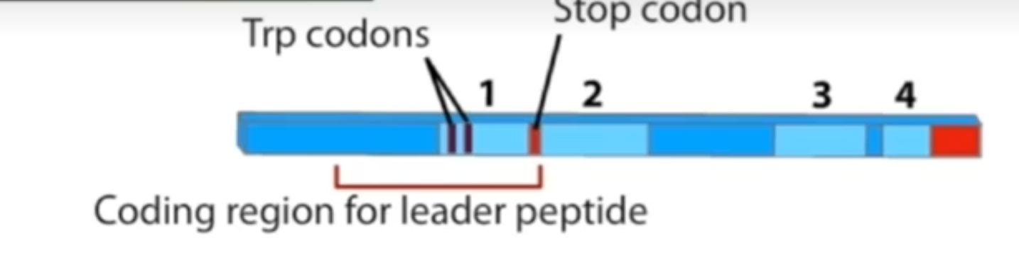
    • Has 2 tryptophan codons close to the start and an attenuator stem loop (A) close to the end

  • The trp operon is transcribed as a single polycistronic mRNA

  • In a normal situation, RNA polymerase would bind to promoter P, transcribe all the way through the operon and finish at the hairpin terminator sequence at the end of the operon

  • However, it would be beneficial for the cell if these genes were not expressed when tryptophan is present

  • And it is the presence of this leader sequence that allows this to happen
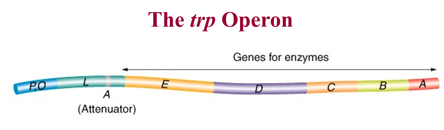

<ul><li><p>After TrpR, there is also a second level of control</p></li><li><p>Involves a process known as <span style="color: rgb(247, 147, 147);"><strong>transcriptional attenuation</strong></span></p></li><li><p>Involves an upstream leader sequence encoding a peptide (L, TrpL)</p><ul><li><p>Located between O (operator) and the first structural gene TrpE</p></li><li><p>Has 2 tryptophan codons close to the start and an attenuator stem loop (A) close to the end</p></li></ul></li><li><p>The trp operon is transcribed as a single polycistronic mRNA</p></li><li><p>In a normal situation, RNA polymerase would bind to promoter P, transcribe all the way through the operon and finish at the hairpin terminator sequence at the end of the operon</p></li><li><p>However, it would be beneficial for the cell if these genes were not expressed when tryptophan is present</p></li><li><p>And it is the presence of this leader sequence that allows this to happen</p></li></ul><p></p>
35
New cards
<p>Transcriptional attenuation</p>

Transcriptional attenuation

  • Leader sequence

    • Contains 2 tryptophan codons close to the start and an attenuator stem loop (A) close to the end

    • When region 3 and 4 pair → attenuation

    • When regions 2 and 3 pair → no attenuation (hair pin stem loop)


  • When tryptophan is absent from the medium the whole mRNA is transcribed

    • The ribosome will stall at the trp codon in region 1 (hence region 2 and 3 pair together), the RNA poly continues on

    • And only then transcription stops


  • When tryptophan is present only the leader sequence is transcribed

    • The ribosome rushes past the 2 trp codons in region 1 and comes to a stop at the stop codon between region 1 and 2, therefore region 3 and 4 can bind together and from an attenuator loop which pull the RNA from out of the RNA poly and then transcription stops


<ul><li><p>Leader sequence</p><ul><li><p>Contains 2 tryptophan codons close to the start and an attenuator stem loop (A) close to the end</p></li><li><p>When region 3 and 4 pair → attenuation</p></li><li><p>When regions 2 and 3 pair → no attenuation (hair pin stem loop)</p></li></ul></li></ul><p></p><ul><li><p>When tryptophan is <strong>absent </strong>from the medium the whole mRNA is transcribed</p><ul><li><p>The ribosome will stall at the trp codon in region 1 (hence region 2 and 3 pair together), the RNA poly continues on</p></li><li><p>And only then transcription stops</p></li></ul></li></ul><p></p><ul><li><p>When tryptophan is <strong>present </strong>only the leader sequence is transcribed</p><ul><li><p>The ribosome rushes past the 2 trp codons in region 1 and comes to a stop at the stop codon between region 1 and 2, therefore region 3 and 4 can bind together and from an attenuator loop which pull the RNA from out of the RNA poly and then transcription stops</p></li></ul></li></ul><p></p>
36
New cards

Secondary Structure of the Trp Leader mRNA

  • Four regions that can form stem and loop structures by hydrogen bonding

  • Segments 1, 2, 3 & 4

  • Can form 1:2, 2:3 and 3:4 stem and loop structures

  • UGG/UGG that encode two Trp are in segment 1 back to back

  • Look at the string of U’s at the end of region 4 → weak bonding


<ul><li><p>Four regions that can form stem and loop structures by hydrogen bonding</p></li><li><p>Segments 1, 2, 3 &amp; 4</p></li><li><p>Can form 1:2, 2:3 and 3:4 stem and loop structures</p></li><li><p>UGG/UGG that encode two Trp are in segment 1 back to back</p></li><li><p>Look at the string of U’s at the end of region 4 → weak bonding </p></li></ul><p></p>
37
New cards
<p>How does this leader sequence cause attenuation of transcription when tryptophan is present?</p>

How does this leader sequence cause attenuation of transcription when tryptophan is present?
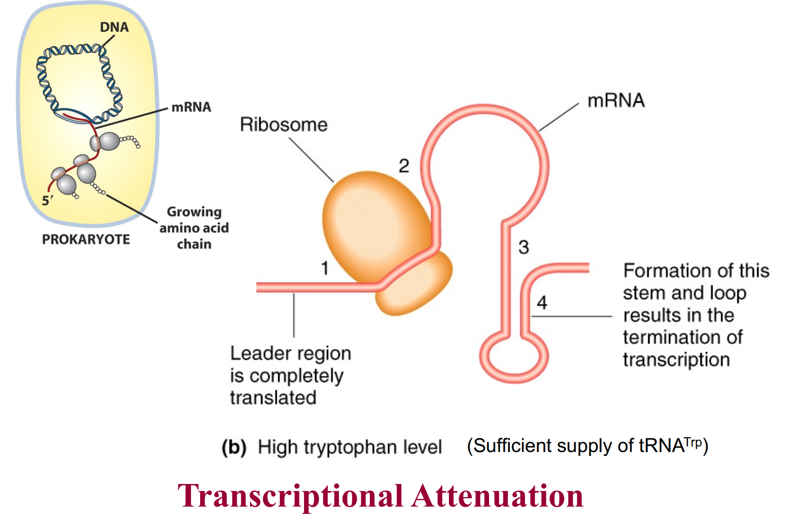
knowt flashcard image
38
New cards

Overview of what happens during the post-transcription of mRNA in Eukaryotes

  • The mRNA that is formed directly after transcription is known as pre-mRNA

  • 1) Addition of 7-methylguanosine cap at 5’ end (via a 5’ to 5’ linkage of the triphosphates)

  • 2) Splicing to remove introns and join exons together + Alternative splicing

  • 3) Addition of 3’ polyA tail


39
New cards
<p>mRNA Synthesis and Processing in Prokaryotes and Eukaryotes</p>

mRNA Synthesis and Processing in Prokaryotes and Eukaryotes

Prokaryotes

  • Transcription & translation occur concurrently in cytoplasm

  • An average protein takes about 1 min to synthesis, this is quite slow for a prokaryote life, therefore the prokaryote must have multiple ribosomes transcribing at ones to make 100s of copies of the same protein. Prokaryotic mRNA is also unstable and breaks down quick, that is why it so important the transcription and translation happens concurrently


Eukaryotes

  • Transcription occurs in nucleus

  • Translation occurs in cytoplasm

  • Processes are not concurrent

  • Undergoes mRNA processing unlike prokaryotes


40
New cards

What is the 7-methylguanosine cap at 5’ end

  • When mRNA is synthesised the first nucleotide actually retains its 5’ triphosphate group, but because of this phosphate at the end the mRNA is susceptible to attack by exonucleases

  • Thus to protect from hydrolysis by exonucleases (ensures the quality of the mRNA)

  • A 7-methylguanosine cap is added at the 5’ end (in the black circle) - this causes a 5’ to 5’ linkage

  • This tells the cell that the 5’ end is intact

  • It also functions as an ‘attach here’ signal for ribosomes
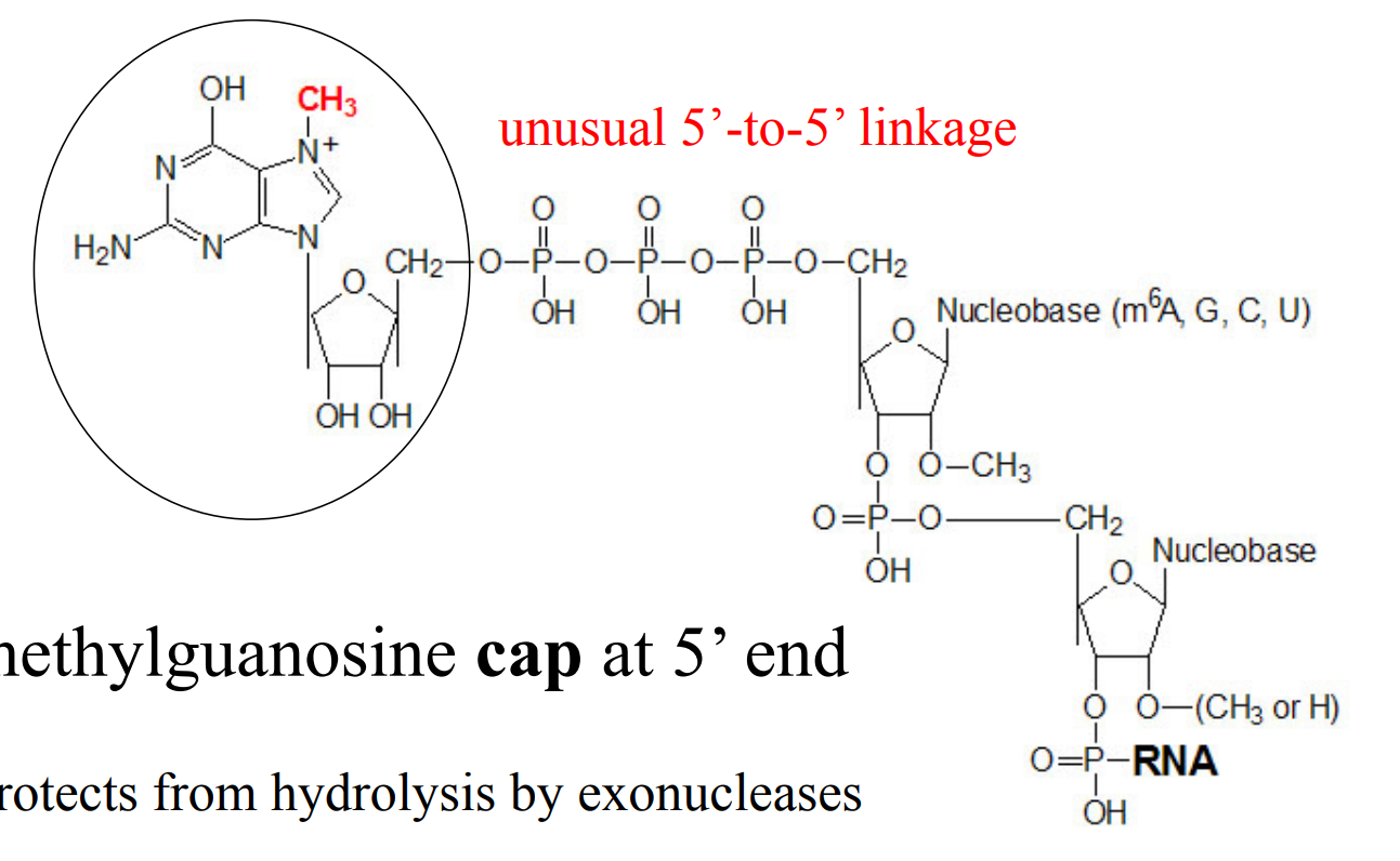

<ul><li><p>When mRNA is synthesised the first nucleotide actually retains its 5’ triphosphate group, but because of this phosphate at the end the mRNA is susceptible to attack by exonucleases </p></li><li><p>Thus to <span style="color: rgb(255, 127, 80);"><strong>protect </strong></span>from hydrolysis by exonucleases (ensures the quality of the mRNA) </p></li><li><p>A 7-methylguanosine cap is added at the 5’ end (in the black circle) - this causes a 5’ to 5’ linkage </p></li><li><p>This tells the cell that the 5’ end is intact</p></li><li><p>It also functions as an ‘attach here’ <span style="color: rgb(255, 127, 80);"><strong>signal for ribosomes</strong></span></p></li></ul><p></p>
41
New cards

Polyadenylation of the 3’terminus of eukaryotic mRNA

  • Approx. 200 A residues are added enzymatically to 3’ end of mRNA

  • Function

    • Increased stability of mRNA (prevents its degradation)

    • Has a role in export out of nucleus (quality control - that the 3’ end is intact)

      • Export protein in the nucleus that bind to the poly A tail to help export the mRNA out of the nucleus

    • Essential for initiating translation of some mRNA from the 5’ end

      • Some protein bound the the poly A tail from the nucleus are able to recruit initiation factors to the 5’ end

      • This forms the closed‑loop mRNA structure, which enhances initiation

    • Can be about 100-200 bp long


<ul><li><p>Approx. 200 A residues are added enzymatically to 3’ end of mRNA</p></li><li><p>Function</p><ul><li><p><span style="color: yellow;">Increased stability</span> of mRNA (prevents its degradation)</p></li><li><p>Has a role in <span style="color: yellow;">export out of nucleus</span> (quality control - that the 3’ end is intact)</p><ul><li><p>Export protein in the nucleus that bind to the poly A tail to help export the mRNA out of the nucleus</p></li></ul></li><li><p>Essential for <span style="color: yellow;">initiating translation</span> of some mRNA from the 5’ end</p><ul><li><p>Some protein bound the the poly A tail from the nucleus are able to recruit initiation factors to the 5’ end</p></li><li><p>This forms the closed‑loop mRNA structure, which enhances initiation</p></li></ul></li><li><p>Can be about 100-200 bp long</p></li></ul></li></ul><p></p>
42
New cards

A comparison of the structures of prokaryotic and eukaryotic mRNA

  • In prokaryotes we can see the exposed 5’ phosphate ends with no protection (no cap), this means mRNA gets degraded very quickly here

  • Prokaryote is also polycistronic (1 mRNA => many genes)


<ul><li><p>In prokaryotes we can see the exposed 5’ phosphate ends with no protection (no cap), this means mRNA gets degraded very quickly here </p></li><li><p>Prokaryote is also polycistronic (1 mRNA =&gt; many genes)  </p></li></ul><p></p>
43
New cards

What are the functions of the 3 different RNA polymerases in Eukaryotes?

RNA Polymerase I

  • Transcribes rRNA genes (except for 5S rRNA)


*RNA Polymerase II

  • Produces mRNA

  • Also transcribes some snRNA genes and miRNAs

  • Eukaryotic RNA polymerase II is much larger and more complex than its bacterial counterpart, as it not only synthesizes mRNA, but also coordinates its processing

  • The Carboxyl Tail Domain (CTD) of RNAP II plays a central role in coordinating all three processing events (5’-capping, 3’-polyadenylation and splicing)


RNA Polymerase III

  • Transcribes most small RNA genes (tRNA, some snRNAs, 5S rRNA)


*Kinda makes sense = more genes in eukaryotes than prokaryotes = more RNAP

44
New cards

7-methylguanosine cap at 5’ end - role of CTD domain of RNAP II

  • Co-transcriptional - takes place while transcription is still occurring

  • The CTD (Carboxyl Tail Domain) of RNAP II is located near the site where newly synthesized RNA emerges from the polymerase

  • It is an ideal place to orchestrate the binding and release of proteins needed to process RNA as it is being synthesized

  • Thus the CTD plays a central role in coordinating the processing events

  • As the 5’ end of RNA first emerges from RNA polymerase, the CTD recruits capping enzymes that add a 7- methyl guanosine residue to its 5’ triphosphate group of the mRNA

  • Transcription is not complete, but the 5’ cap has been added now


<ul><li><p><em>Co-transcriptional </em>- takes place while transcription is still occurring</p></li><li><p>The CTD (Carboxyl Tail Domain) of RNAP II is located near the site where newly synthesized RNA emerges from the polymerase</p></li><li><p>It is an ideal place to orchestrate the binding and release of proteins needed to process RNA as it is being synthesized</p></li><li><p>Thus the CTD plays a central role in coordinating the processing events</p></li><li><p>As the 5’ end of RNA first emerges from RNA polymerase, the CTD recruits capping enzymes that add a 7- methyl guanosine residue to its 5’ triphosphate group of the mRNA </p></li><li><p>Transcription is not complete, but the 5’ cap has been added now </p></li></ul><p></p>
45
New cards
<p>Polyadenylation at 3’ terminus - role of CTD domain of RNAP II</p>

Polyadenylation at 3’ terminus - role of CTD domain of RNAP II

  • Recent data suggests that poly-adenylation of the 3’-end occurs concurrently and in coordination with splicing of mRNA (image above)

  • Catalysed by enzyme poly(A) polymerase (PAP) and involves CPSF protein (cleavage and polyadenylation specificity factor)

  • Transcription termination signal AAUAAA (11-30 nt upstream of the polyadenylation site)


  • 1) Once transcription started, CPSF is recruited to the CTD and ‘rides’ on the RNA polymerase until it reads in AATAAA termination signal and transcribes AAUAAA on the mRNA

  • 2) At which point CPSF dissociates from the CTD and binds to the AAUAAA sequence instead (high affinity of the AAUAAA)

  • 3) Additional cleavage factors are recruited that to cleave the mRNA cleaved ~11-13 nt downstream of the of termination sequence

  • 4) Polymerase PAP then adds ~200 adenylate residues to the 3' end of the transcript


<ul><li><p>Recent data suggests that poly-adenylation of the 3’-end occurs concurrently and in coordination with <em>splicing </em>of mRNA (image above)</p></li><li><p>Catalysed by enzyme <span style="color: rgb(216, 174, 225);"><strong>poly(A) polymerase (PAP) </strong></span>and involves <strong><em>CPSF </em></strong>protein (cleavage and polyadenylation specificity factor)</p></li><li><p>Transcription termination signal AAUAAA (11-30 nt upstream of the polyadenylation site)</p></li></ul><p></p><ul><li><p>1) Once transcription started, CPSF is recruited to the CTD and ‘rides’ on the RNA polymerase until it reads in AATAAA termination signal and transcribes AAUAAA on the mRNA </p></li><li><p>2) At which point CPSF dissociates from the CTD and binds to the AAUAAA sequence instead (high affinity of the AAUAAA)</p></li><li><p>3) Additional cleavage factors are recruited that to cleave the mRNA cleaved ~11-13 nt downstream of the of termination sequence </p></li><li><p>4) Polymerase PAP then adds ~200 adenylate residues to the 3' end of the transcript</p></li></ul><p></p>
46
New cards
<p>Comparison between transcription in Prokaryotes and Eukaryotes</p>

Comparison between transcription in Prokaryotes and Eukaryotes

  • Prokaryotic genes are always ready to be expressed - RNAP just needs to bind

    • Activator and repressors adjust levels but are not 100% efficient

  • Eukaryotes genes are usually off

    • Cuz DNA is tightly compacted in the chromatin/chromone thus not accessible to the RNAP

    • Gene expression is also stringently controlled because eukaryotic genes need much MORE than just RNA polymerase (e.g. transcription regulators etc.)


<ul><li><p>Prokaryotic genes are always ready to be expressed - RNAP just needs to bind </p><ul><li><p>Activator and repressors adjust levels but are not 100% efficient </p></li></ul></li><li><p>Eukaryotes genes are usually off</p><ul><li><p>Cuz DNA is tightly compacted in the chromatin/chromone thus not accessible to the RNAP</p></li><li><p>Gene expression is also stringently controlled because eukaryotic genes need much MORE than just RNA polymerase (e.g. transcription regulators etc.) </p></li></ul></li></ul><p></p>
47
New cards

RNAP II activity

  • Heavy reliance on transcription regulators

  • RNAP II alone only gives low level of gene transcription in the absence of regulatory proteins

  • Usually multiple activators are required for maximal promoter activity and good expression

  • Regulatory proteins bind to cis-acting regulatory elements (cis-regulatory sequences)

    • Regions of non-coding DNA on the same chromosome (that is, in cis) as the gene

    • They are typically located near the gene, often in the intergenic region directly upstream from the transcription start point. Sometimes downstream

    • Most genes have multiple cis-regulatory elements, each recognized by a different transcription regulator

    • Can be 10,000 – 100,000 bp from promoter (due to DNA looping)

    • These serve as binding site for the regulatory proteins, most cells have many different types of cis-acting regulatory sequences each recognising a different translation factor/regulator


48
New cards

Transcription Initiation in Eukaryotes

  • In prokaryotes, RNA polymerase recognizes and binds directly to the promoter region via its sigma-factor subunit (-35 and -10 regions in E. coli promoters)

  • In eukaryotes, RNA polymerase II requires a set of proteins called general transcription factors that bind to the promoter and promoter-proximal cis-regulatory elements. Called TFIID, TFIIF etc.

  • In addition, there are gene-specific transcription regulators (activators and repressors) that bind to different cis-regulatory elements


49
New cards
<p>Steps in the Initiation of mRNA synthesis in Eukaryotes? </p>

Steps in the Initiation of mRNA synthesis in Eukaryotes?

  • Transcription activator binding to enhancers and assembly of the enhanceosome occurs first, before TATA box can get exposed

  • Promoter recognition involves TATA box located ~30 bp upstream from the transcription start site (TATA is part of the promoter)

  • 1st event - binding of the TATA box binding protein (TBP) which is in fact part of the TFIID (TF2D)

    • Since TATA sequence is conserved → TBP and general transcription factors are the same for all genes

  • Binding of TBP to the TATA box causes bending of DNA which is though to serve as a landmark that helps to attract the other general transcription factors

  • This kink that forms from the bending signals that this is an active promoter, it also brings the DNA strands closer together which allows the assembly of the multi-protein complex of the DNA called:

  • Formation of pre initiation complex:

    • When bound to the TATA box, TBP attracts other general transcription factors (GTFs) and the RNA polymerase II, thus forming the pre-initiation complex, which kinda acts as a lock not allowing the RNAPII to start transcription

  • Transcription can start after Carboxyl Tail Domain (CTD) of RNAP II is phosphorylated by one of the GTFs

    • The CTD is composed of many repeats of a sequence of 7 aa that be be phosphorylated or dephosphorylated in different combinations, the phosphorylation state of the CTD determines which proteins can bind

    • And it is thus hypothesised that this then weakens the binding between the RNAPII to the transcription factors and allows RNAPII to slip out and start transcription


<ul><li><p>Transcription activator binding to enhancers and assembly of the enhanceosome occurs first, before TATA box can get exposed</p></li><li><p>Promoter recognition involves <span style="color: rgb(255, 235, 184);"><strong>TATA box</strong></span> located ~30 bp upstream from the transcription start site (TATA is part of the promoter)</p></li><li><p>1st event - binding of the<span style="color: rgb(255, 235, 184);"> <strong><u>T</u></strong>ATA box<strong> <u>b</u></strong>inding <strong><u>p</u></strong>rotein (<strong>TBP</strong>) which is in fact part of the TFIID</span> (TF2D)</p><ul><li><p>Since TATA sequence is conserved → TBP and general transcription factors are the same for all genes</p></li></ul></li><li><p>Binding of TBP to the TATA box causes <span style="color: rgb(255, 235, 184);">bending </span>of DNA which is though to serve as a landmark that helps to attract the other general transcription factors</p></li><li><p>This kink that forms from the bending signals that this is an active promoter, it also brings the <span style="color: rgb(188, 186, 186);"><em>DNA strands closer together </em></span>which allows the assembly of the multi-protein complex of the DNA called:</p></li><li><p>Formation of<span style="color: rgb(255, 235, 184);"> pre initiation complex</span>:</p><ul><li><p>When bound to the TATA box, TBP attracts other general transcription factors (GTFs) and the RNA polymerase II, thus forming the pre-initiation complex, which kinda acts as a lock not allowing the RNAPII to start transcription</p></li></ul></li><li><p>Transcription can start after Carboxyl Tail Domain (CTD) of RNAP II is phosphorylated by one of the GTFs</p><ul><li><p>The CTD is composed of many repeats of a sequence of 7 aa that be be phosphorylated or dephosphorylated in different combinations, the phosphorylation state of the CTD determines which proteins can bind</p></li><li><p>And it is thus hypothesised that this then weakens the binding between the RNAPII to the transcription factors and allows RNAPII to slip out and start transcription</p></li></ul></li></ul><p></p>
50
New cards

What are general transcription factors?

  • General transcription factors are named so because they are same for all genes (which is why the promoter-proximal cis-regulatory regions are conserved)

  • In contrast, transcriptional regulators (activators and repressors) are unique to a gene or a group of genes

  • Cis-regulatory sequences that serve as binding sites for activators are called enhancers

  • A single gene may be controlled by multiple different transcriptional activators

  • A gene may have many enhancers which add to give maximal promoter activity

  • 10% of all proteins produced by a eukaryotic cells are transcriptional regulators (activators or repressors).


<ul><li><p>General transcription factors are named so because they are same for all genes (which is why the promoter-proximal cis-regulatory regions are conserved)</p></li><li><p>In contrast, transcriptional regulators (activators and repressors) are unique to a gene or a group of genes</p></li><li><p>Cis-regulatory sequences that serve as binding sites for <span style="color: rgb(176, 205, 230);"><strong>activators </strong></span>are called <span style="color: rgb(176, 205, 230);"><strong>enhancers</strong></span></p></li><li><p>A single gene may be controlled by multiple different transcriptional activators</p></li><li><p>A gene may have many enhancers which <em>add </em>to give maximal promoter activity</p></li><li><p>10% of all proteins produced by a eukaryotic cells are transcriptional regulators (activators or repressors).</p></li></ul><p></p>
51
New cards

Polymerase II also requires Activator, Mediator and Chromatin-modifying proteins

  • Eukaryotic RNAP II requires activator, mediator and chromatin modifying proteins for its activity

  • To initiate transcription of a eukaryotic gene, first a transcription activator must bind to the enhancer to help to attract DNA-bending proteins, GTFs and RNA polymerase to the start point of transcription (some activators act from very far away, this is possible due to DNA loops that form)

    • The TATA box is intact, so the general transcription factors and RNA polymerase II can still assemble, but without an activator they give only low-level transcription

  • Second, eukaryotic transcription initiation also requires the presence of a protein complex known as Mediator

    • It mediates interactions between activators, RNA polymerase and GTFs

    • For example, mediator positions the right GTF next to the carboxyl tail domain of the RNA polymerase so that it can phosphorylate it to release the polymerase and allow it to start transcription

  • Finally, ATP-driven chromatin-remodelling complexes and histone-modifying enzymes are recruited – a feature that is not present in prokaryotes

    • They increase access to the DNA region in the chromatin that needs to be transcribed, as they unwind and relax DNA in the region that needs to be transcribed


<ul><li><p>Eukaryotic RNAP II requires activator, mediator and chromatin modifying proteins for its activity</p></li><li><p>To initiate transcription of a eukaryotic gene, first a transcription <span style="color: rgb(255, 209, 102);"><strong>activator </strong></span>must bind to the enhancer to help to attract DNA-bending proteins, GTFs and RNA polymerase to the start point of transcription (some activators act from very far away, this is possible due to DNA loops that form)</p><ul><li><p>The TATA box is intact, so the general transcription factors and RNA polymerase II can still assemble, but without an activator they give only low-level transcription</p></li></ul></li><li><p>Second, eukaryotic transcription initiation also requires the presence of a protein complex known as <span style="color: rgb(255, 209, 102);"><strong>Mediator</strong></span></p><ul><li><p>It mediates interactions between activators, RNA polymerase and GTFs</p></li><li><p>For example, mediator positions the right GTF next to the carboxyl tail domain of the RNA polymerase so that it can phosphorylate it to release the polymerase and allow it to start transcription</p></li></ul></li><li><p>Finally, ATP-driven <span style="color: rgb(255, 209, 102);"><strong>chromatin-remodelling complexes</strong></span> and <span style="color: rgb(255, 209, 102);"><strong>histone-modifying enzymes</strong></span> are recruited – a feature that is not present in prokaryotes</p><ul><li><p>They increase access to the DNA region in the chromatin that needs to be transcribed, as they unwind and relax DNA in the region that needs to be transcribed</p></li></ul></li></ul><p></p>
52
New cards
<p>Transcription regulators cause genes to express in the right place (Eukaryotes) </p>

Transcription regulators cause genes to express in the right place (Eukaryotes)

  • The embryo starts as one big cell (a syncytium) and needs to work out where to build each body segment. The gene even-skipped (Eve) is expressed in 7 stripes, and that pattern helps lay out the body plan

  • The Eve gene has a promoter (TATA box) right next to it, plus 7 regulatory DNA segments spread upstream (the orange blocks). These act like switches

  • Researchers cut out the "stripe 2 regulatory segment", and spliced it in front of a reporter gene (LacZ) with its own TATA box. LacZ makes an enzyme that produces a blue stain, so it lets you see where the gene is turned on

  • Result: the embryo showed only one blue stripe, in position 2

  • Conclusion: the stripe 2 regulatory segment is sufficient to switch a gene on in stripe 2 and nowhere else. Each of the 7 stripes has its own regulatory segment that responds to its own local combination of regulators


  • The stripe 2 segment contains binding sites (cis-regulatory sequences) for four transcription regulators:

    • Activators: Bicoid and Hunchback (turn transcription on)

    • Repressors: Giant and Krüppel (turn transcription off)

  • These proteins are not evenly distributed in the embryo

  • Each is concentrated in different regions

  • Only at the stripe 2 position are both activators present and both repressors absent. Everywhere else, at least one repressor is there or an activator is missing, so the gene stays off


<ul><li><p>The <em>e</em>mbryo starts as one big cell (a syncytium) and needs to work out where to build each body segment. The gene <strong><em>even-skipped</em> (<em>Eve</em>)</strong> is expressed in <strong>7 stripes</strong>, and that pattern helps lay out the body plan</p></li><li><p>The <em>Eve</em> gene has a promoter (TATA box) right next to it, plus 7 regulatory DNA segments spread upstream (the orange blocks). These act like switches</p></li><li><p>Researchers <strong>cut out the "stripe 2 regulatory segment"</strong>, and spliced it in front of a <strong>reporter gene (<em>LacZ</em>)</strong> with its own TATA box. <em>LacZ</em> makes an enzyme that produces a blue stain, so it lets you <em>see</em> where the gene is turned on</p></li><li><p>Result: the embryo showed <strong>only one blue stripe, in position 2</strong></p></li><li><p><strong>Conclusion:</strong> the stripe 2 regulatory segment is <em>sufficient</em> to switch a gene on in stripe 2 and nowhere else. Each of the 7 stripes has its own regulatory segment that responds to its own local combination of regulators</p></li></ul><p></p><ul><li><p>The stripe 2 segment contains binding sites (<em>cis</em>-regulatory sequences) for four transcription regulators:</p><ul><li><p><strong>Activators:</strong> Bicoid and Hunchback (turn transcription on)</p></li><li><p><strong>Repressors:</strong> Giant and Krüppel (turn transcription off)</p></li></ul></li><li><p>These proteins are not evenly distributed in the embryo</p></li><li><p>Each is concentrated in different regions</p></li><li><p><span style="color: yellow;">Only at the stripe 2 position are <strong><em>both </em></strong>activators present and both repressors absent</span>. Everywhere else, at least one repressor is there or an activator is missing, so the gene stays off</p></li></ul><p></p>
53
New cards
<p>Distribution of the transcription regulators responsible for ensuring that Eve is expressed in stripe 2</p>

Distribution of the transcription regulators responsible for ensuring that Eve is expressed in stripe 2

  • Therefore stripe 2 can only form where there are activators and no repressors (yellow region)
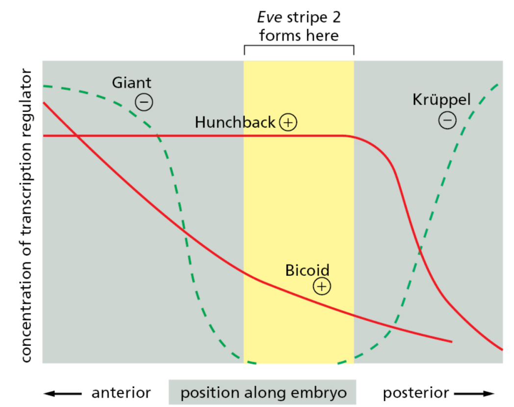

<ul><li><p>Therefore stripe 2 can only form where there are activators and no repressors (yellow region) </p></li></ul><p></p>
54
New cards

The role of transcriptional activators in regulating gene expression in response to change in physiological conditions

  • Example: the GAL system from yeast

  • Enzymes GAL1, GAL2,… convert the imported galactose into glucose that can be metabolised

    • These genes are silent if there is no galactose in the growth medium

  • Three additional genes (GAL3, GAL4 and GAL80) encode regulatory proteins

    • GAL4 is a transcriptional activator

      • Must bind its enhancer before its activation domain can recruit transcriptional machinery

    • GAL 3 senses/binds galactose and then interacts with GAL80 (negative regulator); without it the inhibitory GAL80-GAL4 state persists


<ul><li><p>Example: the GAL system from yeast</p></li><li><p>Enzymes GAL1, GAL2,… convert the imported galactose into glucose that can be metabolised</p><ul><li><p>These genes are silent if there is no galactose in the growth medium</p></li></ul></li><li><p>Three additional genes (GAL3, GAL4 and GAL80) encode regulatory proteins</p><ul><li><p>GAL4 is a transcriptional activator</p><ul><li><p>Must bind its enhancer before its activation domain can recruit transcriptional machinery </p></li></ul></li><li><p>GAL 3 senses/binds galactose and then interacts with GAL80 (negative regulator); without it the inhibitory GAL80-GAL4 state persists </p></li></ul></li></ul><p></p>
55
New cards

The Mechanism of gene repression by microRNAs

  • Some miRNA genes are transcribed by RNA polymerase II

  • Example of regulation of gene expression by noncoding RNAs (RNA interference or RNAi)

    • They are the products of transcription from independent genes or from introns of protein-coding genes

  • MiRNA produced during transcription and are capped and polyadenylated miRNA precursors are synthesized by RNAP II

  • A ~70-nucleotide precursor hairpin (pre-miRNA) is produced in the nucleus, processed into a smaller but not yet final form, and then exported to the cytoplasm

  • There, it is cleaved by an enzyme Dicer to yield ~20-bp miRNA/miRNA duplex (double stranded RNA)

  • This duplex is bound by Argonaute protein and one strand is degraded

  • Other proteins bind and a complex named RNA-induced silencing complex, or RISC, is formed

  • Example of regulation of gene expression by noncoding RNAs (RNA interference or RNAi)

    • RISC seeks out its target mRNAs by searching for complementary nucleotide sequences

    • 2 pathways:

      • If microRNA is fully complementary to the target sequence, the Argonaute protein cleaves the target sequence, then uses the energy from ATP hydrolysis to unwind this complex separating the miRNA from the mRNA, resulting in a rapid degradation of the mRNA as the 5’ and 3’ protection is missing from the cut site

      • If microRNA is only partially complementary to the target sequence, gene expression is inhibited at the level of translation, and mRNA is eventually degraded → thus both cases repression occurs


<ul><li><p>Some miRNA genes are transcribed by RNA polymerase II </p></li><li><p>Example of regulation of gene expression by <em>noncoding </em>RNAs (RNA interference or RNAi)</p><ul><li><p>They are the products of transcription from independent genes or from introns of protein-coding genes</p></li></ul></li><li><p>MiRNA produced during transcription and are capped and polyadenylated miRNA <em>precursors </em>are synthesized by RNAP II</p></li><li><p>A ~70-nucleotide precursor hairpin (pre-miRNA) is produced in the nucleus, processed into a smaller but not yet final form, and then exported to the cytoplasm</p></li><li><p>There, it is cleaved by an enzyme Dicer to yield ~20-bp miRNA/miRNA duplex (double stranded RNA)</p></li><li><p>This duplex is bound by Argonaute protein and one strand is degraded</p></li><li><p>Other proteins bind and a complex named <strong>RNA-induced silencing complex, or RISC</strong>, is formed</p></li><li><p>Example of regulation of gene expression by noncoding RNAs (RNA interference or RNAi)</p><ul><li><p>RISC seeks out its target mRNAs by searching for complementary nucleotide sequences</p></li><li><p>2 pathways:</p><ul><li><p>If microRNA is fully complementary to the target sequence, the Argonaute protein cleaves the target sequence, then uses the energy from ATP hydrolysis to unwind this complex separating the miRNA from the mRNA, resulting in a rapid <strong><u>degradation</u> </strong>of the mRNA as the 5’ and 3’ protection is missing from the cut site</p></li><li><p>If microRNA is only partially complementary to the target sequence, gene expression is inhibited at the level of <strong>translation</strong>, and mRNA is eventually <strong>degraded </strong>→ thus both cases repression occurs</p></li></ul></li></ul></li></ul><p></p>
56
New cards

Splicing in Eukaryotes vs Prokaryotes

  • Does not occur in prokaryotes - as there is no introns

  • Like 5’-capping and 3’- polyadenylation, SPLICING takes place in the nucleus, and only when all 3 processing steps are complete, mature mRNA is secreted from the nucleus into the cytosol via a nuclear pore

  • Splicing is how we get the 21,000 genes encoding proteins in the human genome to encode foe ~100,000 proteins


<ul><li><p>Does not occur in prokaryotes - as there is no introns </p></li><li><p>Like 5’-capping and 3’- polyadenylation, SPLICING takes place in the nucleus, and only when all 3 processing steps are complete, mature mRNA is secreted from the nucleus into the cytosol via a nuclear pore</p></li><li><p>Splicing is how we get the 21,000 genes encoding proteins in the human genome to encode foe ~100,000 proteins</p></li></ul><p></p>
57
New cards
<p>What is RNA splicing? </p>

What is RNA splicing?

  • Splicing to remove introns (segments of noncoding RNA) and bring coding regions (exons) together

  • Introns (in-terrupting region)

    • Segments of DNA or RNA that do not code for polypeptides

    • Sequences interrupt coding sequence of mRNA

    • Size ranges from 1 kb to several kb

    • All higher and many lower eukaryotic mRNAs have introns

    • Must be removed by splicing to form a coding mRNA molecule


  • Exons (ex-pressed region)

    • The actual coding sequences that are interrupted by introns


<ul><li><p>Splicing to remove <strong>introns </strong>(segments of noncoding RNA) and bring coding regions (exons) together</p></li><li><p><span style="color: rgb(124, 213, 199);"><strong>Introns (<em>in</em>-terrupting region)</strong></span></p><ul><li><p>Segments of DNA or RNA that do not code for polypeptides </p></li><li><p>Sequences interrupt coding sequence of mRNA</p></li><li><p>Size ranges from 1 kb to several kb </p></li><li><p>All higher and many lower eukaryotic mRNAs have introns</p></li><li><p>Must be removed by splicing to form a coding mRNA molecule</p></li></ul></li></ul><p></p><ul><li><p><span style="color: rgb(255, 146, 146);"><strong>Exons (ex-pressed region) </strong></span></p><ul><li><p>The actual coding sequences that are interrupted by introns</p><p></p></li></ul></li></ul><p></p>
58
New cards
<p>Depending on the cell type, different coding exons can be removed - Alternative Splicing </p>

Depending on the cell type, different coding exons can be removed - Alternative Splicing

  • Tropomyosin is produced in specialized forms in different types of cells

  • Research suggests that up to 95% of genes are can be alternatively spliced to some degree → indicating that splicing is a quick and common way to create diversity in cells

  • This (bottom image) is example of the regulation of RNA splicing via exon skipping which can generate different versions of a protein in different cell types, according to the needs of the cell

  • Alternative RNA splicing can also be regulated via intron retention mechanism

    • This enabling switching from production of non-functional protein to production of functional one (or other way around)

    • Difference is due to presence of intron sequence in mRNA that is removed in one cell type/tissue but retained in the other


<ul><li><p>Tropomyosin is produced in specialized forms in different types of cells</p></li><li><p>Research suggests that up to 95% of genes are can be alternatively spliced to some degree → indicating that splicing is a quick and common way to create diversity in cells </p></li><li><p>This (bottom image) is example of the <span style="color: rgb(148, 183, 239);"><strong>regulation </strong></span>of RNA splicing via<em> exon skipping </em>which can generate different versions of a protein in different cell types, according to the needs of the cell</p></li><li><p>Alternative RNA splicing can also be regulated via<em> intron retention </em>mechanism</p><ul><li><p>This enabling switching from production of non-functional protein to production of functional one (or other way around)</p></li><li><p>Difference is due to presence of intron sequence in mRNA that is removed in one cell type/tissue but retained in the other</p></li></ul></li></ul><p></p>
59
New cards

Negative and positive control of alternative splicing via intron retention

  • Negative control

    • In which a repressor protein binds to the primary RNA transcript in tissue 2

    • Thereby preventing the splicing machinery from removing an intron sequence

    • Hence you get an inactive protein, only if an repressor protein is present


  • Positive control

    • In which the splicing machinery is unable to remove a particular intron sequence without assistance from an activator protein

    • Therefore activator needs to be present to make a protein


<ul><li><p><strong>Negative control</strong></p><ul><li><p>In which a repressor protein binds to the primary RNA transcript in tissue 2</p></li><li><p>Thereby preventing the splicing machinery from removing an intron sequence</p></li><li><p>Hence you get an inactive protein, only if an repressor protein is present </p></li></ul></li></ul><p></p><ul><li><p><strong>Positive control</strong></p><ul><li><p>In which the splicing machinery is <em>unable to remove a particular intron </em>sequence without assistance from an activator protein</p></li><li><p>Therefore activator needs to be present to make a protein </p></li></ul></li></ul><p></p>
60
New cards
<p>Formation of secondary splice sites?</p>

Formation of secondary splice sites?

  • The cell will have a primary splice site that is used preferentially (the middle) when no repressor protein is present

  • However is a repressor protein binds to the primary splice junction, blocking the splicing machinery from accessing it, then the cell is forced to use this secondary splice site thereby producing an altered pattern of splicing

  • Alternative 5′ splice site → the two competing cut points sit immediately after an exon (they're choosing where the intron starts)

  • Alternative 3′ splice site → the two competing cut points sit immediately before an exon (they're choosing where the intron ends / exon resumes)

  • Each yellow block = exon, Each blue segment = intron, Each red triangle = splice junction (where cutting occurs)
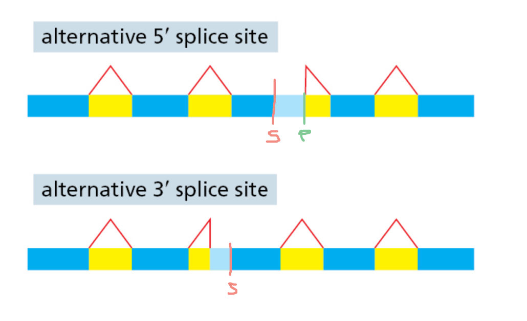

<ul><li><p>The cell will have a primary splice site that is used preferentially (the middle) when no repressor protein is present</p></li><li><p>However is a repressor protein binds to the primary splice junction, blocking the splicing machinery from accessing it, then the cell is forced to use this secondary splice site thereby producing an altered pattern of splicing</p></li><li><p>Alternative <strong>5′</strong> splice site → the two competing cut points sit <strong>immediately after an exon</strong> (they're choosing where the intron starts)</p></li><li><p>Alternative <strong>3′</strong> splice site → the two competing cut points sit <strong>immediately before an exon</strong> (they're choosing where the intron ends / exon resumes)</p></li></ul><ul><li><p>Each yellow block = <strong>exon</strong>, Each blue segment = <strong>intron</strong>, Each red triangle = <strong>splice junction</strong> (where cutting occurs)</p></li></ul><p></p>
61
New cards

Mechanisms of RNA splicing - How are introns recognised?

  • Conserved sequences related to intron splicing

  • GU(5’) AG (3’) end of the intron

  • ‘branch point’ A base 15-45 b upstream of 3’ splice site

  • Poly-pyrimidine tract upstream of the 3’ splice site (in blue)

  • The existence of conserved sequence features at splice junctions suggested that there must be a cellular machine that recognises these sequences and carries out splicing → spliceosome


<ul><li><p>Conserved sequences related to intron splicing</p></li><li><p>GU(5’) AG (3’) end of the intron </p></li><li><p>‘branch point’ A base 15-45 b upstream of 3’ splice site</p></li><li><p>Poly-pyrimidine tract upstream of the 3’ splice site (in blue) </p></li><li><p>The existence of conserved sequence features at splice junctions suggested that there must be a cellular machine that recognises these sequences and carries out splicing → spliceosome</p></li></ul><p></p>
62
New cards
<p>Spliceosome </p>

Spliceosome

  • The existence of conserved sequence features at splice junctions suggested that there must be a cellular machine that recognises these sequences and carries out splicing → spliceosome

  • he complex between small nuclear RNA and a protein is called small nuclear ribonucleoprotein (snRNP, pronounced SNURP)

  • 5 small nuclear ribonucleoproteins are components of the spliceosome (U1, U2, U4, U5, U6)

    • U3 is not involved in splicing

    • U1 binds the 5' splice site

    • U2 binds the branch point

    • The U4/U6.U5 tri-snRNP joins

    • U1 and U4 are released, and the catalytic spliceosome forms

  • Each of the 5 small nuclear RNAs within this spliceosome is found in complex with a protein


<ul><li><p>The existence of conserved sequence features at splice junctions suggested that there must be a cellular machine that recognises these sequences and carries out splicing → spliceosome</p></li><li><p>he complex between small nuclear RNA and a protein is called small nuclear ribonucleoprotein (snRNP, pronounced SNURP)</p></li><li><p>5 small nuclear ribonucleoproteins are components of the spliceosome (U1, U2, U4, U5, U6)</p><ul><li><p>U3 is not involved in splicing</p></li><li><p>U1 binds the 5' splice site</p></li><li><p>U2 binds the branch point</p></li><li><p>The U4/U6.U5 tri-snRNP joins</p></li><li><p>U1 and U4 are released, and the catalytic spliceosome forms</p></li></ul></li><li><p>Each of the 5 small nuclear RNAs within this spliceosome is found in complex with a protein</p></li></ul><p></p>
63
New cards
<p>What is the role of snRNAs in splicing </p>

What is the role of snRNAs in splicing

  • snRNA is not a protein is it RNA that acts as a catalyst

  • Small nuclear ribonucleoprotein U1 is a complex of snRNA with 4 different proteins

  • It has a site that is complementary to the 5’ end of the intron (shown in purple in the crystal structure)

  • Small nuclear RNA is drawn in black


<ul><li><p>snRNA is not a protein is it RNA that acts as a catalyst </p></li><li><p>Small nuclear ribonucleoprotein U1 is a complex of snRNA with 4 different proteins</p></li><li><p>It has a site that is complementary to the 5’ end of the intron (shown in purple in the crystal structure)</p></li><li><p>Small nuclear RNA is drawn in black</p></li></ul><p></p>
64
New cards
<p>What are the steps in splicing? </p>

What are the steps in splicing?

  • 1) Assembly of the spliceosome (image above)

    • U1 forms base pairs with GU at the 5’ splice junction

    • U2 recognises the branch-point site A

    • Then recruits other parts (U4, U5, U


  • 2) The spliceosome catalyses the removal of the intron through two consecutive splicing steps (where 2 phosphate ester bonds are broken and made)

    • In the first splicing reaction, the 5’ end of the intron is attached to the conserved A (‘branch point’)

    • This is the first transesterification step

    • Here, a reaction between the C2’ hydroxyl group of ribose in nucleotide A and a target nucleotide in the upstream exon I leads to the breaking of a 5′–3′ phosphodiester bond at the end of this exon

    • One of the cut ends is joined to the ribose C2’ in nucleotide A, closing the intron circle

    • In the second splicing reaction, the 3’-OH of the 5’-exon becomes a nucleophile, and the intron loop is cleaved from the 3’ splice site and the the two adjacent exons are joined

    • This is achieved in a second transesterification step
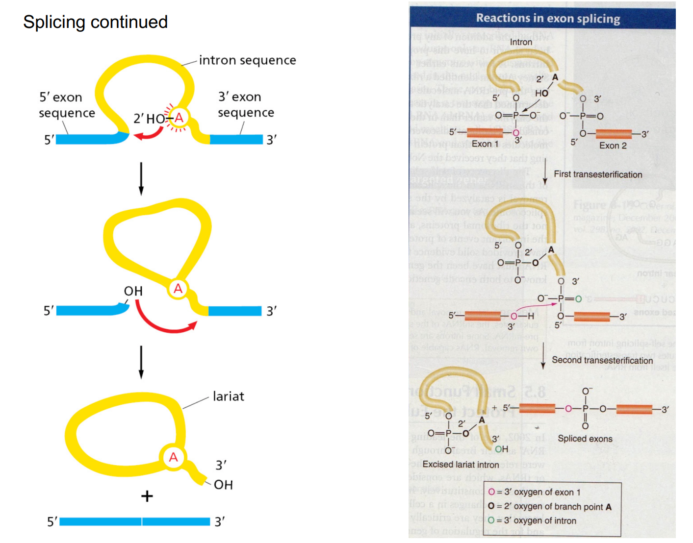

<ul><li><p>1) Assembly of the spliceosome (image above)</p><ul><li><p>U1 forms base pairs with GU at the 5’ splice junction</p></li><li><p>U2 recognises the branch-point site A</p></li><li><p>Then recruits other parts (U4, U5, U</p></li></ul></li></ul><p></p><ul><li><p>2) The spliceosome catalyses the removal of the intron through two consecutive splicing steps (where 2 phosphate ester bonds are broken and made) </p><ul><li><p>In the first splicing reaction, the 5’ end of the intron is attached to the conserved A (‘branch point’)</p></li><li><p>This is the<strong> first </strong>transesterification step</p></li><li><p>Here, a reaction between the C2’ hydroxyl group of ribose in nucleotide A and a target nucleotide in the upstream exon I leads to the breaking of a 5′–3′ phosphodiester bond at the end of this exon</p></li><li><p>One of the cut ends is joined to the ribose C2’ in nucleotide A, closing the intron circle</p></li><li><p>In the second splicing reaction, the 3’-OH of the 5’-exon becomes a nucleophile, and the intron loop is cleaved from the 3’ splice site and the the two adjacent exons are joined</p></li><li><p>This is achieved in a <strong>second </strong>transesterification step</p></li></ul></li></ul><p></p>
65
New cards

What is the difference between intercistronic regions (in bacteria) and introns (in eukaryotes)?

  • Think of key words 'inter' (between) and 'in' (inside)

  • An intercistronic region separates the RNA coding region for protein A from the coding region for protein B => It is between the genes

  • An intron would split the coding region for one protein (the gene) into two => It is inside the gene

  • It is thought that introns evolved from intercistronic regions, because the latter are found in both eukaryotes (albeit rarely) and prokaryotes whilst the former in eukaryotes only


<ul><li><p>Think of key words 'inter' (between) and 'in' (inside)</p></li><li><p>An <span style="color: rgb(43, 187, 215);"><strong>intercistronic</strong></span> region separates the RNA coding region for protein A from the coding region for protein B =&gt; It is <span style="color: rgb(43, 187, 215);"><strong>between </strong></span>the genes</p></li><li><p>An <span style="color: rgb(189, 178, 255);"><strong>intron </strong></span>would split the coding region for one protein (the gene) into two =&gt; It is <span style="color: rgb(189, 178, 255);"><strong>inside </strong></span>the gene</p></li><li><p>It is thought that introns evolved from intercistronic regions, because the latter are found in both eukaryotes (albeit rarely) and prokaryotes whilst the former in eukaryotes only</p></li></ul><p></p>
66
New cards
<p>What is the catalyst in splicing? </p>

What is the catalyst in splicing?

  • In this process, the snRNA acts as a ribozyme, an RNA molecule that functions as an enzyme

  • In a few cases, intron RNA can catalyze its own excision without proteins or extra RNA molecules (bottom image)

    • In group I self-splicing introns, the OH of a free guanine nucleoside or a nucleotide cofactor (GMP, GDP, GTP) attacks phosphate at the 5' splice site

    • 3’-OH of the 5’-exon becomes a nucleophile and the second transesterification results in the joining of the two exons

  • Not all biological catalysts are proteins - not a single catalyst here!

  • While the reaction is neutral (2 phosphodiester bounds broken and made), there needs to be some activity to bring the 5’ end of the intron to the branch A point (and RNA can form hairpin loops etc. → become rigid/hard), therefore you need lots of energy to break some parts of the branch point and do the twisting of the RNA , thus for this energy extensive process we require the help of the spliceosome


<ul><li><p>In this process, the snRNA acts as a ribozyme, an RNA molecule that functions as an enzyme</p></li><li><p>In a few cases, intron RNA can catalyze its own excision without proteins or extra RNA molecules (bottom image)</p><ul><li><p>In group I self-splicing introns, the OH of a free guanine nucleoside or a nucleotide cofactor (GMP, GDP, GTP) attacks phosphate at the 5' splice site</p></li><li><p>3’-OH of the 5’-exon becomes a nucleophile and the second transesterification results in the joining of the two exons</p></li></ul></li><li><p>Not all biological catalysts are proteins - not a single catalyst here!</p></li><li><p>While the reaction is neutral (2 phosphodiester bounds broken and made), there needs to be some activity to bring the 5’ end of the intron to the branch A point (and RNA can form hairpin loops etc. → become rigid/hard), therefore you need lots of energy to break some parts of the branch point and do the twisting of the RNA , thus for this energy extensive process we require the help of the spliceosome </p></li></ul><p></p>
67
New cards

RNA world hypothesis

  • RNA must have been the genetic material in the first cells, where it both encoded genetic information and catalysed biological reactions

  • RNA viruses store their permanent information in RNA

  • Short polymers of ribonucleotides can be synthesized nonenzymatically in the laboratory

  • RNA-directed protein synthesis may have begun as weak binding of specific amino acids to bases along RNA molecules, which functioned as simple templates holding a few amino acids together long enough for them to be linked

    • This is one function of rRNA today in ribosomes


68
New cards

What are the different types of mutations?

  • Site-Directed Mutagenesis introduced a missense mutation


<ul><li><p>Site-Directed Mutagenesis introduced a missense mutation </p></li><li><p></p></li></ul><p></p>
69
New cards

What is Site-Directed Mutagenesis?

  • The technique utilises mutagenic primers that are predominantly complementary to the plasmid DNA but contain a small modification in the sequence

  • Mutagenic primers can anneal to denatured plasmid DNA but have a small region of mismatched bases

  • These primers can be used to amplify plasmid DNA utilising the polymerase chain reaction

  • As in traditional PCR, mutagenic primers anneal to DNA and DNA polymerase synthesises a complementary strand

  • Below is an example set of conditions used for a site-directed mutagenic PCR inducing a mutation in a 5.3 kb plasmid:

    1. Denature the plasmid DNA for 30 sec @ 95°C then

    2. 18 cycles of 30 sec @ 95°C, 1 min @ 55°C, 12 min @ 72°C, then

    3. Final extension of 10 min @ 72°C and 4°C hold


70
New cards

What are the requirements for Site-Directed Mutagenesis?

Primers and the mutation

  • The primers are customised oligonucleotides that differ to the template DNA by several base pairs, but usually only up to three

  • It’s important they do not differ by too many base pairs or they will not bind

  • This region of mismatched bases is flanked by a string of base pairs complementary to the template DNA, ensuring annealing can still occur

  • These flanking regions should be at least 10 bp in length and as such, primers for SDM may be longer than those used for traditional PCR

  • Primers should also be designed to ensure that both strands of DNA are mutated at the same location in order for complementary base pairing to continue

  • As such, forward and reverse primers will have large regions of complementarity considering the flanking regions.  Primers can introduce deletions, insertion or base substitutions

  • These mutations may also introduce new restriction sites which can aid in down-stream applications


Polymerase

  • Once primers have annealed to the template DNA, a DNA polymerase binds and synthesises the new complementary strand

  • Unlike traditional PCR, a polymerase called Pfu is used instead of Taq Pfu polymerase is better suited to plasmid amplification and has a higher fidelity and proof-reading (exonuclease) capacity than Taq polymerase, which is important to ensure no additional mutations are introduced


DpnI digest

  • Following multiple round of amplification, the product will be predominantly newly synthesised plasmid however the original template will still remain

  • To remove this, the sample is digested with an enzyme called DpnI

  • DpnI is a restriction endonuclease that cleaves DNA methylated at GATC sites

  • It will digest only the original template DNA leaving the newly synthesised DNA intact


Transformation

  • Following DpnI digestion, the plasmid product is ready to be introduced back into bacterial cells

  • A transformation reaction can be performed to achieve this

  • Once within bacterial cells, any nicks still remaining in the plasmid DNA will be repaired


<p><strong>Primers and the mutation</strong></p><ul><li><p>The primers are customised oligonucleotides that differ to the template DNA by several base pairs, but usually only up to three</p></li><li><p>It’s important they do not differ by too many base pairs or they will not bind</p></li><li><p>This region of mismatched bases is flanked by a string of base pairs complementary to the template DNA, ensuring annealing can still occur</p></li><li><p>These flanking regions should be at least 10 bp in length and as such, primers for SDM may be longer than those used for traditional PCR</p></li><li><p>Primers should also be designed to ensure that both strands of DNA are mutated at the same location in order for complementary base pairing to continue</p></li><li><p>As such, forward and reverse primers will have large regions of complementarity considering the flanking regions. &nbsp;Primers can introduce deletions, insertion or base substitutions</p></li><li><p>These mutations may also introduce new restriction sites which can aid in down-stream applications</p></li></ul><p></p><p><strong>Polymerase</strong></p><ul><li><p>Once primers have annealed to the template DNA, a DNA polymerase binds and synthesises the new complementary strand</p></li><li><p>Unlike traditional PCR, a polymerase called <em>Pfu</em> is used instead of <em>Taq</em> <em>Pfu</em> polymerase is better suited to plasmid amplification and has a higher fidelity and proof-reading (exonuclease) capacity than <em>Taq</em> polymerase, which is important to ensure no additional mutations are introduced</p></li></ul><p></p><p><strong><em>DpnI </em>digest</strong></p><ul><li><p>Following multiple round of amplification, the product will be predominantly newly synthesised plasmid however the original template will still remain</p></li><li><p>To remove this, the sample is digested with an enzyme called <em>DpnI</em></p></li><li><p><em>DpnI</em> is a restriction endonuclease that cleaves DNA methylated at GATC sites</p></li><li><p>It will digest only the original template DNA leaving the newly synthesised DNA intact</p></li></ul><p></p><p><strong>Transformation</strong></p><ul><li><p>Following <em>DpnI</em> digestion, the plasmid product is ready to be introduced back into bacterial cells</p></li><li><p>A transformation reaction can be performed to achieve this</p></li><li><p>Once within bacterial cells, any nicks still remaining in the plasmid DNA will be repaired</p></li></ul><p></p>
71
New cards

What are the steps in Site-Directed Mutagenesis?

knowt flashcard image
72
New cards

What are some possible post-translational modifications that regulate protein activity?

  • Adds variation/diversity to the proteome, these modification can aid folding, stability, localization to cell compartments, function, activation/inactivation etc.

  • We can produce way more proteins than the amount of genes we have due to these modifications (+splicing etc.)

  • Protein folding

  • Targeted degradation

  • Addressing to specific sub-cellular localisation

  • Post-translational modifications (covalent modifications, proteolysis, glycosylation)


<ul><li><p>Adds variation/diversity to the proteome, these modification can aid folding, stability, localization to cell compartments, function, activation/inactivation etc.</p></li><li><p>We can produce way more proteins than the amount of genes we have due to these modifications (+splicing etc.)</p></li><li><p>Protein folding</p></li><li><p>Targeted degradation</p></li><li><p>Addressing to specific sub-cellular localisation</p></li><li><p>Post-translational modifications (covalent modifications, proteolysis, glycosylation)</p></li></ul><p></p>
73
New cards
<p>What is the Proteasome? (26S) </p>

What is the Proteasome? (26S)

  • ATP-dependent protease complex that degrades damaged, misfolded or short-lived proteins tagged with ubiquitin (cell maintenance and regulation)

  • Made of a 20S core particle capped by one or two 19S regulatory particles (26S = 20S + 19S)

  • 19S regulatory particle:

    • Receptors recognise the polyubiquitin tag (a chain of ≥4 ubiquitins marks the protein for degradation)

    • De-ubiquitinases remove the ubiquitin tags before the protein is unfolded, so ubiquitin is recycled

    • Motor (base): ring of 6 AAA+ ATPase subunits uses ATP to unfold the protein and mechanically pull it through the channel into the core

  • 20S core: barrel with 6 proteolytic active sites lining the inner chamber. It cuts the protein into short peptides (~2-25 aa)

  • Peptides are cleaved further into amino acids by cytosolic peptidases and reused for new protein synthesis

  • Some peptides are loaded onto MHC class I and presented to T cells (immune surveillance of viral/abnormal proteins)


74
New cards
<p>What is Heterochromatin?</p>

What is Heterochromatin?

  • Heterochromatin: highly compressed and not transcribed

  • If the promoter is wound around the histone then the general transcription factors and RNA polymerase cannot bind to the site and therefore cannot be expressed

  • Therefore the nucleosomes need to be changed (moved around, how tightly compacted the DNA is etc.) to expose the promoter regions in order to express or not express specific genes - this process is called chromatin remodelling


<ul><li><p>Heterochromatin: highly compressed and not transcribed</p></li><li><p>If the promoter is wound around the histone then the general transcription factors and RNA polymerase cannot bind to the site and therefore cannot be expressed </p></li><li><p>Therefore the nucleosomes need to be changed (moved around, how tightly compacted the DNA is etc.) to expose the promoter regions in order to express or not express specific genes - this process is called chromatin remodelling </p></li></ul><p></p>
75
New cards

How is Chromatin dynamic?

  • Can go both ways: Can loosen DNA or compact it → to express or not to express

  • Packaging of eukaryotic DNA into chromatin

    • Regulatory sequences required for initiation of transcription (promoter, cis-regulatory sequences) are inaccessible for most of the genes

  • Chromatin structure can be altered by (these all affect how compact the DNA is wound around)

    • Histone protein tail acetylation or methylation

    • DNA methylation

    • SWI-SNF chromatin remodelling protein complex

  • Modification of histones (acetylation, methylation) controls gene expression via different mechanisms


76
New cards

Modification of histones: Acetylation of histone tails

  • Histone proteins have N-terminal “tails” that stick out of the nucleosome

  • The histone tails can bind next nucleosome and adjacent DNA

    • Favourable electrostatic interactions between Lys residues of the tails (which makes the tails positively charged) and negatively charged pocket on the adjacent nucleosome, which allows the tails on the histones of one nucleosome to launch themselves into the negatively charged groove on the surface of the next nucleosome and so on, and thus the two adjacent nucleosomes will be bound together

    • The positive charge on the histone tails will form favourable electrostatic interactions with the negatively charged phosphate backbone of the surrounding DNA

    • These interactions stabilises the condensed structure (bundled together) → regulatory regions are hidden away, thus genes are inactive

  • Acetylation (CH3CO) of histone tails stops interaction with neighbouring nucleosome and DNA and this leads to the nucleosomes to separate and adopt the beads on a string structure, now there will be regions of DNA that are now exposed and the genes in those regions become active

    • Chemically the acetylation removes the positive charge of the histone tails (neutral)

  • The beads on a string structure (bottom image) occurs when the chromatin structure is relaxed (not condensed)

  • Most of the time the chromatin is condensed (image on the right)


<ul><li><p>Histone proteins have N-terminal “tails” that stick out of the nucleosome</p></li><li><p>The histone tails can bind next nucleosome and adjacent DNA</p><ul><li><p>Favourable electrostatic interactions between Lys residues of the tails (which makes the tails positively charged) and negatively charged pocket on the adjacent nucleosome, which allows the tails on the histones of one nucleosome to launch themselves into the negatively charged groove on the surface of the next nucleosome and so on, and thus the two adjacent nucleosomes will be bound together </p></li><li><p>The positive charge on the histone tails will form favourable electrostatic interactions with the negatively charged phosphate backbone of the surrounding DNA</p></li><li><p>These interactions stabilises the <strong>condensed </strong>structure (bundled together) → regulatory regions are hidden away, thus genes are inactive </p></li></ul></li><li><p>Acetylation (CH<sub>3</sub>CO) of histone tails stops interaction with neighbouring nucleosome and DNA and this leads to the nucleosomes to separate and adopt the beads on a string structure, now there will be regions of DNA that are now exposed and the genes in those regions become active </p><ul><li><p>Chemically the acetylation removes the positive charge of the histone tails (neutral) </p></li></ul></li><li><p>The beads on a string structure (bottom image) occurs when the chromatin structure is relaxed (not condensed) </p></li><li><p>Most of the time the chromatin is condensed (image on the right)</p></li></ul><p></p>
77
New cards

Is the Acetylation of histone tails reversible?

  • Yes they are reversible

  • HAT = enzyme histone acetyltransferase – transfers an acetyl group from acetyl-CoA to histone

  • HDAC = enzyme histone deacetylase

    • Therefore you end up with a structure with a positive charge


<ul><li><p>Yes they are reversible</p></li><li><p>HAT = enzyme histone acetyltransferase – transfers an acetyl group from acetyl-CoA to histone</p></li><li><p>HDAC = enzyme histone deacetylase</p><ul><li><p>Therefore you end up with a structure with a positive charge </p></li></ul></li></ul><p></p>
78
New cards

What does regulation by Histone Acetylation mean?

  • Level of histone acetylation affects gene expression

    • More acetylation = more transcription

    • Less acetylation = less transcription

  • Some eukaryotic activator proteins direct acetylation of histones near promoters

    • Opens chromatin structure and increases gene transcription

  • Some eukaryotic repressor protein direct deacetylation of histones near promoters

    • Compresses chromatin structure and reduces gene transcription (as is the case for the yeast GAL genes in the presence of glucose, the preferred substrate)


<ul><li><p>Level of histone acetylation affects gene expression</p><ul><li><p>More acetylation = more transcription</p></li><li><p>Less acetylation = less transcription</p></li></ul></li><li><p>Some eukaryotic activator proteins direct acetylation of histones near promoters</p><ul><li><p>Opens chromatin structure and increases gene transcription</p></li></ul></li><li><p>Some eukaryotic repressor protein direct deacetylation of histones near promoters </p><ul><li><p>Compresses chromatin structure and reduces gene transcription (as is the case for the yeast GAL genes in the presence of glucose, the preferred substrate)</p></li></ul></li></ul><p></p>
79
New cards

How do some eukaryotic repressor protein direct deacetylation of histones near promoters?

  • In the presence of galactose and glucose, transcription of GAL1 is repressed regardless of the presence of the activator Gal4 bound to its enhancer

  • Mig1 repressor is de-phosphorylated in the presence of glucose, binds between the enhancer and promoter and recruits Tup1 that has an HDAC activity

  • Histone tails becomes deacetylated in this region, which turns the gene transcription off


<ul><li><p>In the presence of galactose and glucose, transcription of GAL1 is repressed regardless of the presence of the activator Gal4 bound to its enhancer</p></li><li><p>Mig1 repressor is de-phosphorylated <em>in the presence of glucose</em>, binds between the enhancer and promoter and recruits Tup1 that has an <em><u>HDAC activity</u></em></p></li><li><p>Histone tails becomes deacetylated in this region, which turns the gene transcription off</p></li></ul><p></p>
80
New cards

How does the Methylation of histone tails occur?

  • Lysines and arginines

  • HMTase = enzyme histone methyltransferase

  • Does not affect the charge

  • Creates binding sites for either activator or repressor proteins

    • Depends on the residues modified

  • Can activate or repress gene expression


<ul><li><p>Lysines and arginines</p></li><li><p>HMTase = enzyme histone methyltransferase</p></li><li><p><span style="color: rgb(148, 229, 217);"><em>Does </em><strong><em>not </em></strong><em>affect the charge</em></span></p></li><li><p>Creates binding sites for either activator or repressor proteins</p><ul><li><p>Depends on the residues modified</p></li></ul></li><li><p>Can activate or repress gene expression</p></li></ul><p></p>
81
New cards
<p>What is <span style="color: rgb(221, 70, 70);"><strong>DNA </strong></span>methylation?</p>

What is DNA methylation?

  • Covalent modification of DNA rather than histone proteins

    • 5’ position of cytosines (carbon 5 (C5) of the cytosine ring)

      • The spontaneous deamination of 5’-methylcytosine produced thymine, which is not good if it is not repaired as that would cause a mutation

      • Therefore mammals have evolved such that the CG pairs only appear in the promoter region not anywhere else

    • Done by DNA methyltransferase

    • In mammals, it is usually the C of the CG dinucleotide

    • C*G still cable of nucleotide pairing

      • The modification does not interfere with the H-bonding surface of cytosine, so methylated C can still pair with G

  • 70-80% of all CG dinucleotides are methylated

  • CG methylation is associated with inactive genes

    • Methyl cytosine binding proteins (MeCP) bind to the methylated DNA and recruit histone deacetylases (HDACs)

    • Unmethylated dinucleotides are found in clusters near active promoters (“CpG islands”, p=phosphodiester bond)

    • Link between DNA methylation and transcriptional repression by histone deacetylation


82
New cards

What is the Link between DNA methylation and transcriptional repression by histone deacetylation?

  • DNA Methyltransferase adds methyl groups to the 5’ position of cytosines of CG pairs

  • These methylated cytosine sites then act as a binding site for a protein called methyl cytosine binding protein

  • Then the MeCP then recruit the HDAC

  • The HDAC then removes the acetyl groups from histone tails in the promoter region

  • This leads to the condensation of chromatin which deactivates the gene controlled by this promoter


<ul><li><p>DNA Methyltransferase adds methyl groups to the 5’ position of cytosines of CG pairs </p></li><li><p>These methylated cytosine sites then act as a binding site for a protein called methyl cytosine binding protein </p></li><li><p>Then the MeCP then recruit the HDAC </p></li><li><p>The HDAC then removes the acetyl groups from histone tails  in the promoter region </p></li><li><p>This leads to the condensation of chromatin which deactivates the gene controlled by this promoter </p></li></ul><p></p>
83
New cards
<p>Mechanism of activation of genes in a chromatin environment: The beta-interferon enhanceosome</p>

Mechanism of activation of genes in a chromatin environment: The beta-interferon enhanceosome

  • Multiple transcription factors control expression of b-interferon gene

    • Required for defence against viral pathogens

  • Binding of multiple regulators is co-operative

    • Binding of first protein helps other proteins to bind and stabilises their binding

    • This can help in ramping up low expression to high expression rapidly as when one protein binds the other proteins find it easier to bind


84
New cards

How do Enhanceosome recruit chromatin remodelers?

  • 1) Nucleosome 2 is strategically positioned over TATA box and transcription start site thus the beta-interferon gene is switch off

  • 2) The cell gets infected with the virus and several transcriptional activator assemble together on their respective enhancer sites

  • 3) The enhanceosome is formed rapidly due to the co-operative binding of all transcriptional activators

  • 4) The enhanceosome forms a binding site for the GCN5

    • GCN5 = HAT, uses acetylCoA as a substrate to transfer the acetyl onto histone tails

    • This acetylation means the nucleosome will lose its interaction with the adjacent nucleosome, and will also lose its hold onto the DNA backbone

    • Now the beta-interferon can be exposed

  • 2) GCN5 dissociates after acetylation, this creates a “landing pad” for coactivator CBP

  • 3) CBP recruits RNA polymerase II and SWISNF (“switch-sniff”) chromatin remodelling complex

  • 4) SWI-SNF is an ATP-driven molecular motor, it uses the energy of ATP hydrolysis to perform mechanical work

    • Such as to nudge nucleosome 2 off the TATA box, making it accessible to the Tata Box binding proteins (TBP)

  • 5) After TBP has bound, transcription is initiation - it will go onto recruit all transcriptional factors


<ul><li><p>1) Nucleosome 2 is strategically positioned over TATA box and transcription start site thus the beta-interferon gene is switch off</p></li><li><p>2) The cell gets infected with the virus and several transcriptional activator assemble together on their respective enhancer sites</p></li><li><p>3) The enhanceosome is formed rapidly due to the <span style="color: rgb(143, 236, 159);">co-operative binding</span> of all transcriptional activators</p></li><li><p>4) The enhanceosome forms a binding site for the GCN5</p><ul><li><p>GCN5 = HAT, uses acetylCoA as a substrate to transfer the acetyl onto histone tails</p></li><li><p>This acetylation means the nucleosome will lose its interaction with the adjacent nucleosome, and will also lose its hold onto the DNA backbone</p></li><li><p>Now the beta-interferon can be exposed</p></li></ul></li><li><p>2) GCN5 dissociates after acetylation, this creates a “landing pad” for coactivator CBP</p></li><li><p>3) CBP recruits RNA polymerase II and SWISNF (“switch-sniff”) chromatin remodelling complex</p></li><li><p>4) <span style="color: yellow;">SWI-SNF is an ATP-driven </span><span style="color: rgb(255, 249, 249);">molecular motor</span><span style="color: rgb(255, 248, 248);">, it uses the energy of ATP hydrolysis to perform mechanical work</span></p><ul><li><p><span style="color: yellow;">Such as to nudge nucleosome 2 off the TATA box</span>, making it accessible to the Tata Box binding proteins (TBP)</p></li></ul></li><li><p>5) After TBP has bound, transcription is initiation - it will go onto recruit all transcriptional factors</p></li></ul><p></p>
85
New cards

Epigenetic inheritance of chromatin states

  • Chromatin structure can be inherited

  • In DNA replication, BOTH DNA sequence and the chromatin structure are passed onto the next cell generation

  • During replication, old histones are re-used by both daughter cells and serve as templates to guide the modification of new histones

    • DNA methyltransferase recognizes the hemimethylated DNA and methylates the newly synthesized strand


<ul><li><p>Chromatin structure can be inherited</p></li><li><p>In DNA replication, BOTH DNA sequence and the chromatin structure are passed onto the next cell generation</p></li><li><p>During replication, old histones are re-used by both daughter cells and serve as <strong><u>templates</u> </strong>to guide the modification of new histones</p><ul><li><p><span>DNA methyltransferase recognizes the <strong><em>hemimethylated </em></strong>DNA and methylates the newly synthesized strand</span></p></li></ul></li></ul><p></p>
86
New cards

Epigenetic inheritance of DNA methylation

  • DNA methylation pattern can also be inherited

  • After replication, only the old (parental) strand is methylated

  • DNA methyltransferase recognises this half-methylated (hemi-methylated) DNA and methylates the new strand according to the methylation pattern on the parental strand


  • Block DNA methyltransferase

    • Each division halves the methylation level, so it is diluted gradually rather than removed at once

    • Less methylation means MeCP and its HDAC are no longer recruited, so the chromatin loosens and the promoter becomes accessible

    • The gene is still off by default, so it only becomes able to respond to activators not active on its own


<ul><li><p>DNA methylation pattern can also be inherited</p></li><li><p>After replication, only the old (parental) strand is methylated</p></li><li><p>DNA methyltransferase recognises this half-methylated (hemi-methylated) DNA and methylates the new strand according to the methylation pattern on the parental strand</p></li></ul><p></p><ul><li><p>Block DNA methyltransferase</p><ul><li><p>Each division halves the methylation level, so it is diluted gradually rather than removed at once</p></li><li><p>Less methylation means MeCP and its HDAC are no longer recruited, so the chromatin loosens and the promoter becomes accessible</p></li><li><p>The gene is still off by default, so it only becomes able to respond to activators not active on its own</p></li></ul></li></ul><p></p>
87
New cards
<p>Cell identity is defined by its proteome</p>

Cell identity is defined by its proteome

  • A proteome: a set of proteins produced/expressed in an organism, system or biological context

  • Different cell types contain the same genome, but they express different RNAs and proteins


88
New cards
<p>At what levels can regulation be effected at in the central dogma? </p>

At what levels can regulation be effected at in the central dogma?

  • 0) Chromatin structure and methylation

  • 1) Transcription regulators

  • 2) RNA processing

  • 3) RNA transport

  • 4) Translation

  • 5) mRNA degradation

  • 6) Protein activity


*The pie chart tells you what % of regulation happens at each stage (e.g. 73% of regulation happens during transcription) - overall 84% of total control of gene expression occur in the beginning of this pathway before translation even started … this makes sense because the cell should not waste energy progressing into translation and making proteins that it does not need

<ul><li><p>0) Chromatin structure and methylation</p></li><li><p>1) Transcription regulators</p></li><li><p>2) RNA processing</p></li><li><p>3) RNA transport</p></li><li><p>4) Translation</p></li><li><p>5) mRNA degradation</p></li><li><p>6) Protein activity</p></li></ul><p></p><p>*The pie chart tells you what % of regulation happens at each stage (e.g. 73% of regulation happens during transcription) - overall 84% of total control of gene expression occur in the beginning of this pathway before translation even started … this makes sense because the cell should not waste energy progressing into translation and making proteins that it does not need </p>
89
New cards
<p>Post-translational Event: Protein Folding</p>

Post-translational Event: Protein Folding

  • As the polypeptide chain forms, the N-terminus exits first and it folds into its 3D shape often before synthesis of the entire chain is completed

    • Folds in a way such that the hydrophobic parts are folded inwards away from the water outside

    • This folding is important as it ensured that all the aa that are important for activity are exactly where they should be - this makes the protein functional

    • This is because misfolded proteins typically have their hydrophobic parts exposed outwards, these misfolded proteins can then clump together and cause severe diseases

  • Some fold spontaneously, some need helper proteins called chaperones

  • Chaperones recognize hundreds of different non-native or misfolded polypeptides by their hydrophobic surfaces

  • Chaperones prevents aggregation of misfolded or denatured proteins

  • Chaperones are also referred to as heat shock proteins, this is because at high temperatures the proteins start to unfold, therefore chaperones help the cells survive at high temps by refolding unfolded proteins


*Image shows HSP60

<ul><li><p>As the polypeptide chain forms, the N-terminus exits first and it folds into its 3D shape often before synthesis of the entire chain is completed </p><ul><li><p>Folds in a way such that the hydrophobic parts are folded inwards away from the water outside </p></li><li><p>This folding is important as it ensured that all the aa that are important for activity are exactly where they should be - this makes the protein functional</p></li><li><p>This is because misfolded proteins typically have their hydrophobic parts exposed outwards, these misfolded proteins can then clump together and cause severe diseases</p></li></ul></li><li><p>Some fold spontaneously, some need helper proteins called <strong>chaperones</strong></p></li><li><p>Chaperones recognize hundreds of different non-native or misfolded polypeptides by their hydrophobic surfaces</p></li><li><p>Chaperones prevents aggregation of misfolded or denatured proteins</p></li><li><p>Chaperones are also referred to as heat shock proteins, this is because at high temperatures the proteins start to unfold, therefore chaperones help the cells survive at high temps by refolding unfolded proteins </p></li></ul><p></p><p>*Image shows HSP60 </p>
90
New cards

What are the different kinds of chaperones?

Two main classes of chaperons to stop misfolding + help with folding:

  • Both produced when the cell is exposed to elevated temperatures

  • Heat shock Protein 60 (HSP60)

    • Acts on fully synthesised proteins

    • A chaperonin

    • It initially captures proteins along the rim of one of its barrels

    • Then the binding of ATP and a protein cap increases the diameter of the barrel and partly unfolding the protein inside the barrel

    • Inside the barrel the protein has a chance to fold in a more favourable environment

    • After 15 sec of the ATP binding it hydrolysis, it is the binding of another ATP that makes the protein leave

    • If the protein is still not correctly folded then the cycle can repeat


  • HSP 70

    • Acts early

    • Binds weakly to polypeptide chains emerging from ribosomes where there is a chain of 7 hydrophobic aa

    • Then aided by HSP 40 and many cycles of ATP hydrolysis are generally required to fold a single polypeptide chain correctly


<p>Two main classes of chaperons to stop misfolding + help with folding:</p><ul><li><p>Both produced when the cell is exposed to elevated temperatures</p></li></ul><ul><li><p>Heat shock Protein 60 (HSP60)</p><ul><li><p>Acts on fully synthesised proteins </p></li><li><p>A chaperonin </p></li><li><p>It initially captures proteins along the rim of one of its barrels </p></li><li><p>Then the binding of ATP and a protein cap increases the diameter of the barrel and partly unfolding the protein inside the barrel </p></li><li><p>Inside the barrel the protein has a chance to fold in a more favourable environment </p></li><li><p>After 15 sec of the ATP binding it hydrolysis, it is the binding of another ATP that makes the protein leave</p></li><li><p>If the protein is still not correctly folded then the cycle can repeat </p></li></ul></li></ul><p></p><ul><li><p>HSP 70 </p><ul><li><p>Acts early </p></li><li><p>Binds weakly to polypeptide chains emerging from ribosomes where there is a chain of 7 hydrophobic aa </p></li><li><p>Then aided by HSP 40 and many cycles of ATP hydrolysis are generally required to fold a single polypeptide chain correctly </p></li></ul></li></ul><p></p>
91
New cards
<p>Post-translational event: Targeted degradation </p>

Post-translational event: Targeted degradation

  • Incorrect folding needs to be dealt with efficiently by the cell (aka they cannot be folded properly now)

  • This is because misfolded proteins typically have their hydrophobic parts exposed outwards, these misfolded proteins can then clump together and cause severe diseases

  • There is a competition between chaperone that race to refold proteins properly and proteasomes that given enough time will destroy misfolded proteins if the protein takes too long to fold up properly with the help of a chaperone


92
New cards
<p>Post-translational event: Addressing to specific sub-cellular localisation</p>

Post-translational event: Addressing to specific sub-cellular localisation

  • Proteins Can Move Between Compartments in Different Ways

    • Gated transport → requires energy, complex proteins, through the nuclear pores, have sequence specific for this

    • Transmembrane transport →

    • Vesicular transport → via vesicles, these are membrane enclosed bubbles that form buds that eventually separates and then its content is released to where it fuses with

  • These proteins have conserved sequences that allow them to pass and move to where they need to go


<ul><li><p>Proteins Can Move Between Compartments in Different Ways</p><ul><li><p>Gated transport → requires energy, complex proteins, through the nuclear pores, have sequence specific for this</p></li><li><p>Transmembrane transport → </p></li><li><p>Vesicular transport → via vesicles, these are membrane enclosed bubbles that form buds that eventually separates and then its content is released to where it fuses with </p></li></ul></li><li><p>These proteins have conserved sequences that allow them to pass and move to where they need to go </p></li></ul><p></p>
93
New cards

How does Signal Sequences and Sorting Receptors Direct Proteins to the Correct Cell Address

  • All proteins that are destined for import into endoplasmic reticulum will have this hydrophobic stretch of aa residues at the N-terminus called the signal sequence


<ul><li><p>All proteins that are destined for import into endoplasmic reticulum will have this hydrophobic stretch of aa residues at the N-terminus called the signal sequence </p></li></ul><p></p>
94
New cards
<p>Movement of protein from Cytosol to ER</p>

Movement of protein from Cytosol to ER

  • A Signal-Recognition Particle (SRP) Directs the ER Signal Sequence to a Specific Receptor in the Rough ER Membrane

    • Note that the SRP RECEPTOR is membrane bound and thus only found in the ER

  • All proteins that are destined for import into endoplasmic reticulum will have this hydrophobic stretch of aa residues at the N-terminus called the signal sequence

  • This signal sequence is made first and comes out of the ribosome first and is then recognised by the signal recognition particle (in brown)

  • After binding it temporarily pauses translation and delivers the ribosome to the membrane of the ER to the translocator

  • Protein synthesis continues, and as the protein is made it passes through the channel to the inside of the ER lumen

  • Once the protein is finished it is released in ER, and the signal recognition particle then dissociated and finds another site, it is recycled

  • Inside the ER the signal peptide has been cleaved from the hydrophobic part (red)


<ul><li><p>A Signal-Recognition Particle (SRP) Directs the ER Signal Sequence to a Specific Receptor in the Rough ER Membrane</p><ul><li><p>Note that the SRP RECEPTOR is membrane bound and thus only found in the ER</p></li></ul></li><li><p>All proteins that are destined for import into endoplasmic reticulum will have this hydrophobic stretch of aa residues at the N-terminus called the signal sequence</p></li><li><p>This signal sequence is made first and comes out of the ribosome first and is then recognised by the signal recognition particle (in brown)</p></li><li><p>After binding it temporarily pauses translation and delivers the ribosome to the membrane of the ER to the translocator</p></li><li><p>Protein synthesis continues, and as the protein is made it passes through the channel to the inside of the ER lumen</p></li><li><p>Once the protein is finished it is released in ER, and the signal recognition particle then dissociated and finds another site, it is recycled</p></li><li><p>Inside the ER the signal peptide has been cleaved from the hydrophobic part (red)</p></li></ul><p></p>
95
New cards

What are some of the post-transcriptional modification that polypeptides can undergo once in the endoplasmic reticulum (from cytosol)?

  • Glycosylated

    • Addition of a common oligosaccharide which takes place inside the ER

    • Covalently attached to the side chain of an asparagine residue (N-linked)

      • Very specific location, it must be followed by aa X (which can be anything except proline), then Ser then Thr

      • Hence you can predict this glycolysation site

    • May be required for folding, stability and function, also mediated protein/protein action

    • Proteins that are glycosylated are usually proteins that should not be in the cytosol, so they are most likely proteins that will be either secreted into the extracellular space or inserted into the membrane of the cell wall

    • Can have multiple glycosylation sites


  • Stabilised by disulphide bonds

    • Covalent bond between 2 cysteine side chains

    • Intra- (one) or inter- (2+) molecular crosslinks

    • Requires oxidative condition

    • Image is of insulin - has 3 very important sulphide bonds that needs to be formed


<ul><li><p><span style="color: rgb(101, 208, 244);"><strong>Glycosylated</strong></span></p><ul><li><p>Addition of a common oligosaccharide which takes place inside the ER</p></li><li><p>Covalently attached to the side chain of an asparagine residue (N-linked)</p><ul><li><p>Very specific location, it must be followed by aa X (which can be anything except proline), then Ser then Thr </p></li><li><p>Hence you can predict this glycolysation site</p></li></ul></li><li><p>May be required for folding, stability and function, also mediated protein/protein action </p></li><li><p>Proteins that are glycosylated are usually proteins that should not be in the cytosol, so they are most likely proteins that will be either <strong><em>secreted </em></strong>into the extracellular space or inserted into the membrane of the cell wall </p></li><li><p>Can have multiple glycosylation sites </p></li></ul></li></ul><p></p><ul><li><p><span style="color: rgb(101, 208, 244);"><strong>Stabilised by disulphide bonds</strong></span></p><ul><li><p>Covalent bond between 2 cysteine side chains</p></li><li><p>Intra- (one) or inter- (2+) molecular crosslinks</p></li><li><p>Requires <em>oxidative </em>condition</p></li><li><p>Image is of insulin - has 3 very important sulphide bonds that needs to be formed </p></li></ul></li></ul><p></p>
96
New cards

Post-translational event: Post-translational modifications (covalent modifications, proteolysis, glycosylation)
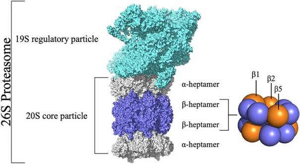
  • Covalent modifications of proteins after translation may include:

    • Proteolysis (cleavage of the protein by proteases)

    • Glycosylation (addition of sugars)

    • Phosphorylation (addition of phosphate groups)

  • Such modifications are often essential to the final functioning of the protein


<ul><li><p>Covalent modifications of proteins after translation may include:</p><ul><li><p>Proteolysis (cleavage of the protein by proteases)</p></li><li><p>Glycosylation (addition of sugars)</p></li><li><p>Phosphorylation (addition of phosphate groups)</p></li></ul></li><li><p>Such modifications are often essential to the final functioning of the protein</p></li></ul><p></p>
97
New cards

Examples of chemical modifications of proteins and their function

knowt flashcard image
98
New cards

Post-translational modifications form a regulatory protein code

  • Can keep the protein inactive until it arrives at the correct site etc.

  • Can have multiple modifications at a time

  • This code regulated the ability, localisation, degradation of the protein (it is the grammar!)
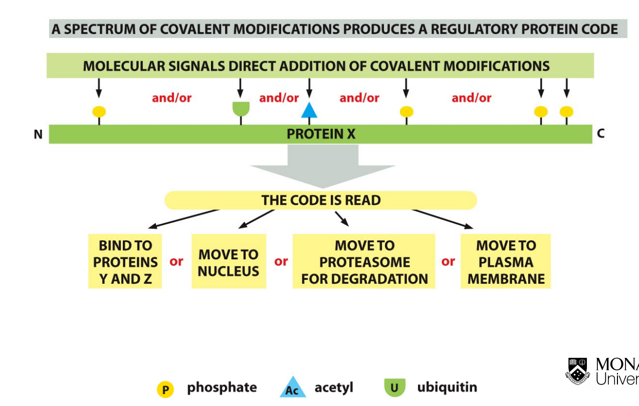

<ul><li><p>Can keep the protein inactive until it arrives at the correct site etc. </p></li><li><p>Can have multiple modifications at a time </p></li><li><p>This code regulated the ability, localisation, degradation of the protein (it is the grammar!) </p></li></ul><p></p>
99
New cards

What are the two ways of activating proteins in a cell signalling cascade?

knowt flashcard image
100
New cards
<p>Post-translational activation: Signalling cascade and cancer</p>

Post-translational activation: Signalling cascade and cancer

  • Oligomerisation

    • The binding of the growth factor onto the membrane and undergoes oligomerisation

  • Auto-phosphorylation of the growth factor

  • Normally small molecule binding:

    • Ras-GTP: on / Ras-GDP: off

    • In cancer mutations in RAS where it continuously energises cells as GTP cannot be hydrolysed into GDP → causing uncontrolled cell division (over-proliferation and onset of cancer)

  • Protein-protein interaction:

    • Ras/Raf

  • Phosphorylation cascade: protein kinases

  • Addressing to specific sub-cellular localisation:

    • Phosphorylation: MAPK => nucleus

    • In nucleus the MAPK can phosphorylate multiple transcriptional factors for cellular division