genetics 3
 Call Kai
 Learn
 Practice Test
 Spaced Repetition
 Match
 Flashcards
 Knowt Play
1/78

There's no tags or description
Looks like no tags are added yet.
79 Terms

chromosome arrangment in humans
each somatic cell has 46 chromosomes
homologous pairs: same length, centromere position, staining pattern, same genes (may or may not be the same form)
autosomes vs sex chromosomes

human life cycle
production of gametes results in haploid cells
upon fertilization, a zygote forms (2n = 46)
mitosis ensures all cells receive 46 chromosomes

meiosis - prophase I
chromosomes partially condense, pair together, and crossing-over occurs
homologous chromosomes are bound together
chromosomes attach fully, spindles attach
nucleolus and nuclear membrane disappear

meiosis - metaphase I
chromosomes align on metaphase plate
each chromosome of the homologous pair is destined for a different pair

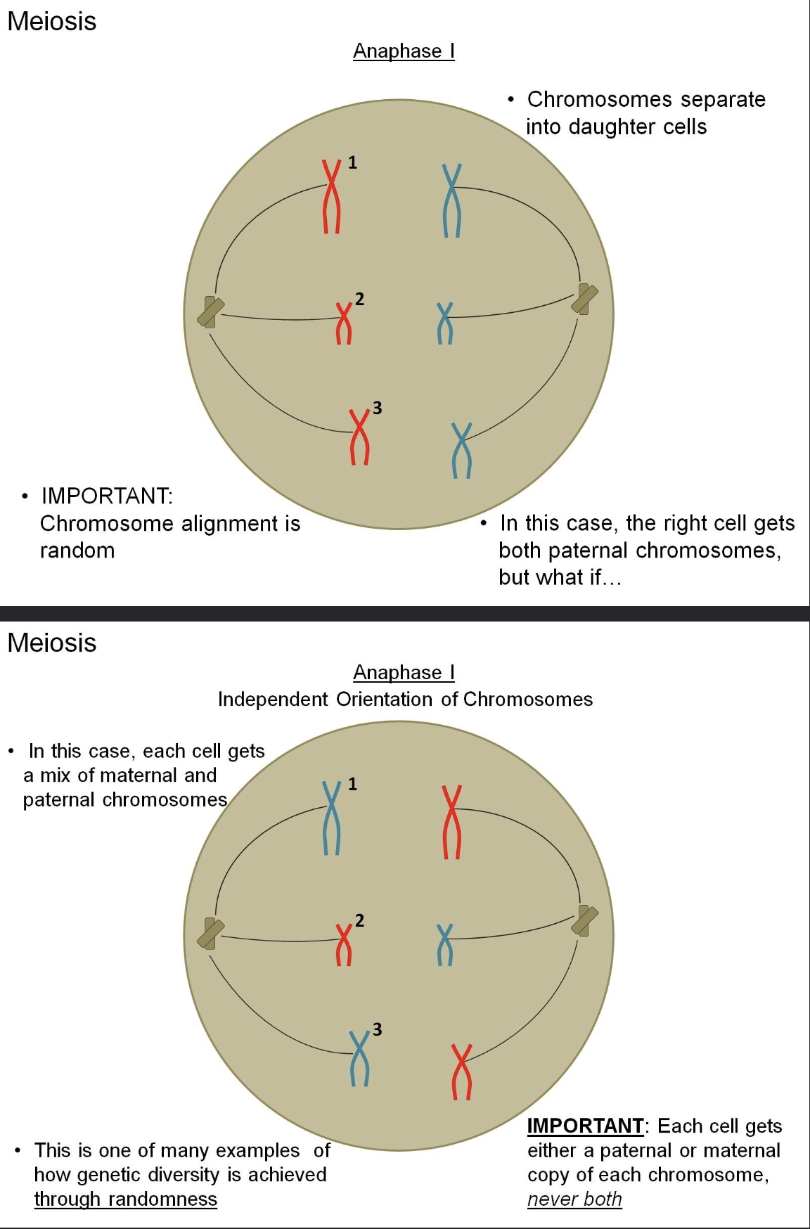
meiosis - anaphase I
chromosomes separate into daughter cells
chromosomes alignment is RANDOM
both paternal chromosomes given to the right cell in example one, a mix of maternal and paternal chromosomes in each cell for example two
genetic diversity achieved through randomness
each cell gets a paternal or maternal copy of each chromosome, but never both

comparing mitosis and meiosis - metaphase
metaphase I
chromosomes are paired so that only the maternal or paternal copy will go into the cell
metaphase (mitosis)
chromosomes are lined up so that each cell with get a maternal AND paternal copy for each pair
comparing mitosis and meiosis - anaphase
anaphase I
daughter cells will be haploid (get either the maternal or paternal chromosome for each pair)
anaphase (mitosis)
daughter cells get the maternal and paternal copy of every chromosome


meiosis - telophase I
telophase I
followed by cytokinesis
meiosis I is called reductional because only one chromosome of each homologous pair is acquired by each daughter cell (although two chromatids are still present)
in other words, this division is where we segregate maternal and paternal chromosomes from each other

meiosis - prophase II and metaphase II
another division occurs immediately after meiosis I without and DNA replication
the steps in this division occur exactly as in mitosis

meiosis - anaphase II and telophase II
ultimately, four haploid gametes are formed, each bearing on chromatid of one chromosome (equational division) = either paternal or maternal
these haploid reproductive cells unite to form a new diploid individual
remember that we’ve shown only one possible combination here (maternal/paternal)

meiosis: from diploid to haploid cells
2n = 6
goal: formation of haploid gametes (n = 3)
interphase
centrosomes (with centriole pairs), centrioles, nuclear envelope, chromatin
prophase I
sites of crossing over, spindle, tetrad, fragments of the nuclear envelope, sister chromatids
metaphase I
spindle microtubules attached to a kinetochore, metaphase plate, centromere with kinetochore
anaphase I
sister chromatids remain attached, homologous chromosomes separate
in telophase I/cytokinesis
chromosomal reduction, cleavage furrow
prophase II
no DNA duplication, n = 3
metaphase II
n/a
anaphase II
sister chromatids separate
telophase II/cytokinesis
haploid daughter cells forming, n = 3

oogenesis

variations in offspring: meiosis and random fertilization
each gamete contains one of several million chromosome combinations
independent orientation of maternal and paternal chromosomes during metaphase I
number of combinations depends on the haploid number
chromosome combinations: total number is 2n, so n = 2 is 22 = 4 combinations. n = 23 in humans, so 223 = 8 million combinations. Factoring in random fertilization: 8 million * 8 million = 64 trillion

meiosis - from the genotype perspective
genotype: AaBbCc
1/8 ABC
1/8 abc
1/8 Abc
1/8 ABc
1/8 aBC
1/8 abC
1/8 aBc
1/8 AbC

variations in offspring: chromosomes carrying different versions of genes

offspring variations: crossing over in prophase I
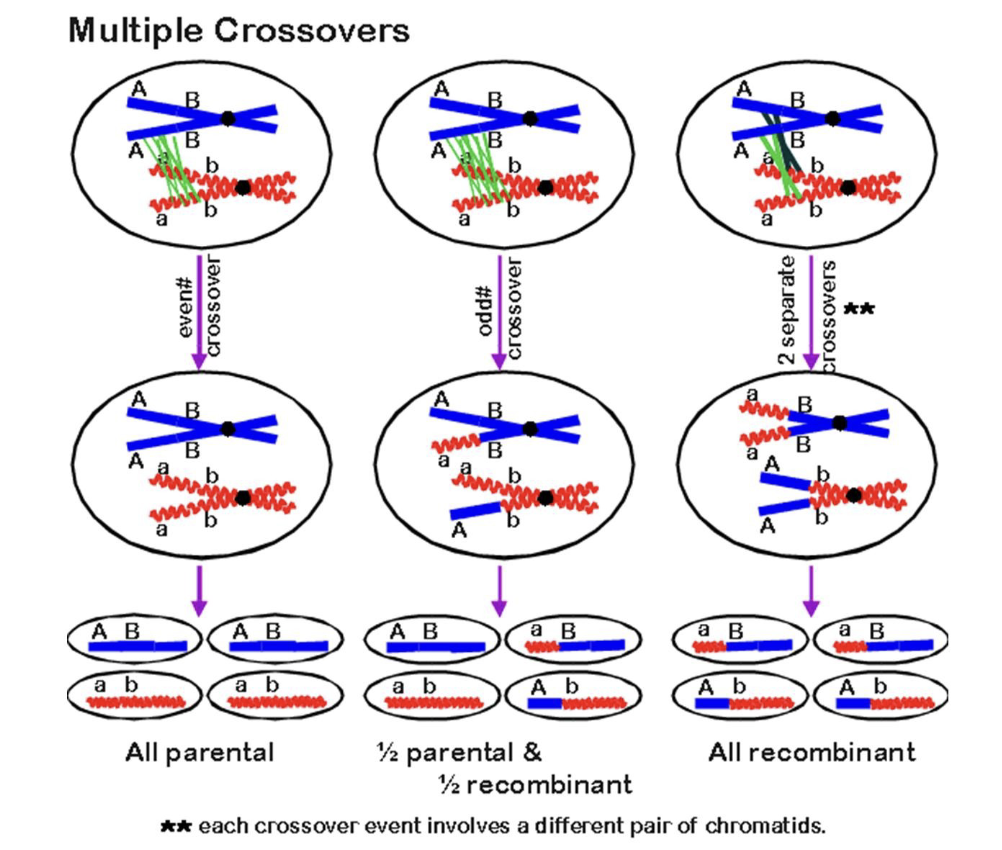
multiple crossovers
different crossovers give different gametes

meiotic abnormalities
nondisjunction: occurs when a chromosome fails to separate properly during meiosis
half the resulting gametes gaining an extra chromosome and half are missing a chromosome
down syndrome is caused by nondisjunction and the inheritance of an extra chromosome 21 (47 chromosomes instead of 46)
meiotic restitution: failure of one of the divisions of meiosis (I or II) fails to occur, producing diploid gametes
nondisjunction and restitution are usually fatal or crippling in animals, but many plants are tolerant of changes in chromosome number
sometimes this is used in agriculture


Gregor Mendel - basic principles of heredity
experimental work with pea plants
easy to cultivate
grow rapidly
produce many offspring
large variety differing in various traits
genetically pure
seven characteristics studied
seed (color and shape)
pod (color and shape)
flower (color and position)
plant height
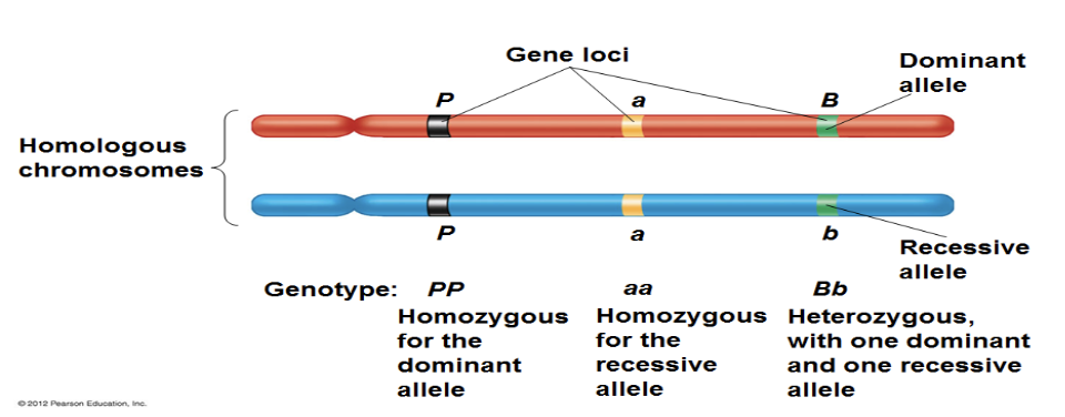
genetics terminology
genotype
an organisms genetic makeup, the heritable information stored in an individuals genes
phenotype
an organisms physical traits represent phenotype
trait
genetic and phenotypic characteristics
gene
molecular unit of hereditary, functional subunit of DNA
locus
specific location of a gene on the chromosome
alleles
variations of genes or alternate form of a gene
dominant
producing dominant phenotype
recessive
producing recessive phenotype
homozygous
containing the same alleles for a gene
heterozygous
containing opposite alleles for a gene

controlled crosses between plants - monohybrid
pea plants are capable of self-fertilization and artificial cross-fertilization
self-fertilization occurs naturally
cross-fertilization involves removing the anthers from a flower and introducing pollen of the desired type with a small brush
established pure-breeding (true-breeding) strains
these are strains that consistently produce the same phenotype
each experiment began with crosses between two pure-breeding parental generation plants (P generation) that produced offspring called F1 (first filial generation)
F1 offspring that are crossed to one another produce the next generation of offspring, the second filial generation F2
pure-breeding parental plants, when crossed, produce F1 that all have the same phenotype
Mendel counted the number of progeny plants of each type and counted many offspring from each cross
he identified patterns in his results, such as the consistent ratios between phenotypes
the ratios he detected are the foundation of his laws of heredity
the trait shown by the F1 offspring was called the dominant phenotype (yellow peas)
the trait that was no apparent in the F1 was called the recessive phenotype (green peas)
when F1 were crossed, 75% of the resulting F2 had the dominant trait, but the recessive trait reappeared in the other 25% [3:1 phenotypic ratio, 1:2:2:1 genotypic ratio]


Mendelian Principles
principle of segregation
the two members of a gene pair segregate away from each other into separate gametes. half the gametes carry one member of the pair and the other half carry the other member of the pair.
in a homozygote, this doesn’t make much difference, but in heterozygotes the two “members” of a pair are different alleles
the basis of this law is the segreation of chromosomes in meiosis I (one copy of the chromosome in each gamete, no homologous chromosomes are present together, unlike in mitosis)
principle of dominance
in a heterozygote, one allele may conceal the presence of another
dominant allele
recessive allele

replicate–, reciprocal–, and test-cross analysis
Mendel made many replicate crosses, producing hundreds or thousands of progeny, by repeating each cross several times (same parent cross)
he performed reciprocal crosses, in which the same genotypes are crossed, but the sexes of the parents are reversed (reverse parent sex, helps determine sex-linked or autosomal)
he also performed test crosses (unknown dominant x homozygous recessive, helps to find the unknown genotype based on offspring) (helpful to determine true-bred individuals)

the dihybrid cross
Mendel’s finding of a 9:3:3:1 fit a mathematical model of two independent pairs of inheritance factors and allowed him to establish his third principle
9/16 WT/WT, 3/16 domA/recB, 3/16 domB/recA, 1/16 double recessive
principle of independent assortment
different gene pairs assort independently into gametes
like with the law of segregation, this is only observable phenotypically in the offspring of a heterozygote
this principle only applies for two genes that are not on the same chromosome
basis for this principle: the random arrangement of chromosomes on either side of the metaphase pate during meiosis I
[when drawing meiosis with two or more traits, make sure to flip the metaphase plate orientation or you will not account for all possible gametes]

genetic notation
the standard or most common allele of a gene is termed the wild-type; note that the wild-type is not necessarily the dominant allele
in general, pick a single letter that is descriptive of the recessive trait; use a lowercase letter to indicate the recessive allele and an uppercase letter to indicate the dominant allele
thus far we have used the Probability Method and Punnet Squares to determine the outcomes of a cross, but this can get messy with crosses involved with three or more genes
the Forked-Line Method is a more rapid method for predicting the phenotype (not genotype) of more complicated crosses

genetic probabilities
AaBbCcDd x AABbCcDd
Aa x AA = ½ Aa, ½ AA
Bb x Bb = ¼ BB, ½ Bb, ¼ bb
Cc x Cc = ¼ CC, ½ Cc, ¼ cc
Dd x Dd = ¼ DD, ½ Dd, ¼ dd
probability of AaBbCcDd?
½ Aa * ½ Bb * ½ Cc * ½ Dd = 1/16 chance
probability of A_B_C_D_?
(½ + ½ ) * (¼ + ½ ) * (¼ + ½ ) * (¼ + ½ ) = 27/64 chance
sample inheritance problem
You have two squirrel strains: red-furred (R) with pointed ears (p) and a grey-furred (r) with rounded ears (P). You cross these two and the F1 population is red with rounded ears. You then randomly mate this population to generate an F2.
a. What are the genotypes of the parents and F1?
parents: RRpp and rrPP
F1: RrPp
b. What proportion of the F2 would you expect to have red-fur and pointed ears?
9:3:3:1 phenotypic ratio → 9/16 R_P_; 3/16 rrP_; 3/16 R_pp; 1/16 rrpp
3/16 will have red fur and pointed ears
c. If you selected the red/pointed F2 individuals and crossed them all back to the
grey/rounded parent stock, what proportion of the resulting population would
still be red/pointed?
parents: R_pp x rrPP → none will be red/pointed because the dominant P will always be given by one parent, overriding the recessive pointy ear gene with a rounded ear gene
sample inheritance problem 2
You have two truebreeding strains of kangaroo rat. One is albino (a) but otherwise
normal (B), and the other has bent ears (b) but otherwise normal (A). You cross these two
and the F1 are all wild-type.
a. What are the genotypes of the parents and F1?
parents: aaBB x AAbb
F1: AaBb (WT/WT)
b. If you mate the entire F1 generation back to the albino parents, what proportion of
the offspring will be wild-type?
aaBB x AaBb → → aa x Aa = ½ Aa, ½ aa → BB x Bb = ½ BB, ½ Bb
WT presented by A_B_ → → ¼ is AaBB, ¼ is AaBb → ½ of backcrossed offspring are WT
c. If you select the wild-type offspring from b) and randomly mate them to each other,
what proportion of the offspring will be albino but have normal ears?
½ AaBB x ½ AaBb
½ AaBB → ¼ AB, ¼ aB
½ AaBb → 1/8 AB, 1/8 Ab, 1/8 aB, 1/8 ab
TOTAL: 3/8 AB, 3/8 aB, 1/8 Ab, 1/8 ab → → 15/64 are WT ear/albino
[punnet squares!!!]
sample inheritance problem 3
The following two genotypes are crossed: AaBbCcddEe × AabbCcDdEe. What
will the proportion of the following genotypes be among the progeny of this
cross?
a. AaBbCcDdEe (completely heterozygous)
½ Aa * ½ Bb * ½ Cc * ½ Dd * ½ Ee = 1/32 offspring
b. AABBCCDDEE
O chance, not possible on B or D gene
The Chi-Square Test
so far we have shown crosses with known genotype, but in genetic experiments we do not know what to expect (hence the experiment)
when we make a cross and examine the progeny, how do we know if our population data fits with a given model or hypothesis?
geneticists use statistics → numbers calculated from data that aid in the analysis of said data
in genetics, one of the most basic and important statistics in the Chi-Square Test, used to compare data with a given hypothesis and determine if the data matches this expectation
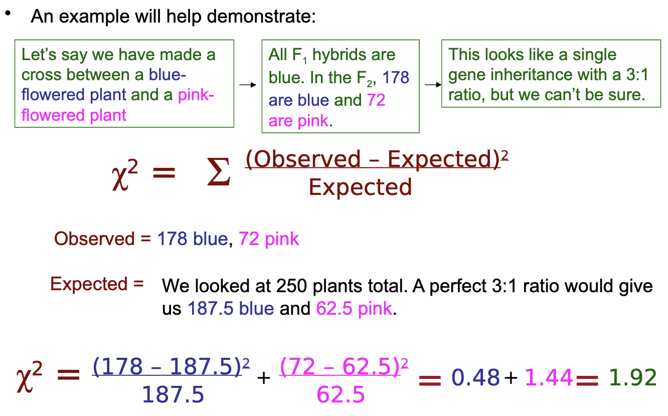
Chi-Square Example
our result was 1.92, to derive meaning from this number we must compare it to chi-square values which are already derived for us
if our chi-square value exceeds the critical value, we reject the hypothesis we tested; if it is lower than the critical value, we do not (the data agrees)
the table shows critical values for a 5% level of confidence; this means that there is only 5% possibility that the test will fail by chance (if hypothesis is mistakenly rejected but is correct)
compare the chi-square value to the critical value for the correct degree of freedom (# of phenotypic classes scored - 1 = DF)
for our data, we have 1 degree of freedom, and our value 1.92 < 3.841, so the data fits the hypothesis
sex-determination
Aristotle proposed that the heat of the male partner during intercourse determined sex (if it overcame the women’s coolness, it would be male)
sex chromosomes discovered in the early 1900s
modes of sex determination - XX/XY
in the XX/XY mode of sex determination, females gametes are all X and male gametes are either X or Y
the y chromosome does not look like a Y, it is just smaller than normal X
zygotes with two X chromosomes become female (homogametous)
zygotes with one X and one Y are male (heterogametous)
occurs in mammals, drosophila, and some plants
modes of sex determination - ZZ/ZW
in ZZ/ZW sex determination, females are the heterogametic sex (ZW) and males are homogametic (ZZ)
W chromosome is usually associated with the development of female characteristics
absence of W leads to ZØ
occurs in birds, butterflies, and some fishes
modes of sex determination - XX/XØ
the XX/XØ mode of sex determination depends on the random distribution of the X chromosome into half of the gametes resulting in males
the presence of two X chromosomes in the zygote results in female offspring
the presence of only one X chromosome results in male offspring
modes of sex determination - haploid/diploid, genic, environmental, dimorphism
haploid-diploid system
some insects have haploid males and diploid females; males produce gametes through mitosis and females through meiosis
genic
genes at one or more loci determine sex, not the presence of a chromosome
environmental
temperature variation controls sex determination in reptiles
in all crocodiles, most turtles, and some lizards, sex determination is achieved according to the incubation temperature of eggs during a critical period of embryonic development
in slugs, the warmer slugs on the bottom of a slug stack will be female
sequential dimorphism
organism can change sex when needed (clownfish)

sex determination in drosophila
the ratio of X chromosomes to sets of autosomes determines the sex of drosophila
Y does not determine sex
sex is determined by the ratio of X chromosomes to autosomes (A)
normal female: AA:XX = 1:1
normal male: AA:XY = 2:1
flies with abnormal chromosome numbers show that: ≥1.0 causes female development, ≤0.5 causes male development, and 0.5–1.0 are intersex
metamales: weak and sterile
metafemales: developmental defects
in contrast to mammals, X chromosome on male is up-regulated so that transcription level equals that of XX female

the Y chromosome and male development
smaller in size compares to the X chromosome
Y chromosome has only 30-50 genes, 900-1400 on X
pseudo-autosomal regions (PARs) at both ends
share homology with regions on the X chromosomes, which allows them to pair together and is critical to segregation of the X and Y chromosomes during male and gametogenesis
has euchromatic and heterochromatic regions
non-recombining region - male specific region of the Y (MSY)
sex-determining region (SRY) located adjacent to PAR

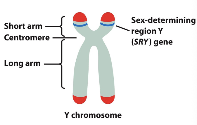
ACTIVE-Y sex determination
in mammals
maleness is caused by a dominant gene (SRY) on the Y-chromosome
proven because XØ mammals are female and XXY are males
at 6-8 weeks of development, the SRY genes become active in XY embryos
produces testis-determining factor (TDF) and Mullerian inhibiting substance
note that other genes (on X and autosomes) are responsible for further male development, but SRY initiates the process
Androgen Insensitivity Syndrome: male has a tfm mutation, testosterone cannot be received, no signal, female secondary sexual characteristics develop, testes remain undescended (can be determined by genetic testing or the development of testicular cancer)


evolution of the Y chromosome
Y chromosome degeneration theory: when sexes evolved, genes that determine that development of one sex over another were developed
additional genes that increased the fitness of the female became linked to the X chromosome
recombination stops between X and Y (don’t crossover to link maternal alleles with SRY, or male fitness decreases and vice versa)
lack of recombination allows deleterious or null alleles to accumulate on Y chromosome (X still recombines with X)
Y becomes cluttered with “junk” DNA that gets deleted

sex-linked genes
males are XY and Y has essentially no genes, males only have one set of alleles for every X-linked gene; termed hemizygous
females have to X chromosomes and sex-linked traits are inherited like normal autosomes, but in males even recessive traits appear dominant
with an X-dominant mutation (XAXa affected mother), half of the children will show the condition, half will not (affected daughter indicates that mother has dominant X)
if the mother is recessive affected (XaXa), only the male offspring will be affected (XaY) because the father’s dominant normal XA masks it in female offspring (XAXa, still affected, but not shown)
recessive genes can go unnoticed until a mother passes it to a son
Queen Victoria carried hemophilia, passed it to 5 kids, daughters were carried and one son died from it

sex-linked genetics problem
XaXa x XAY → → all daughters are carriers (XAXa) and all sons are affected (XaY)
female yellow/WT eye: XyXyPP
male WT/pink eye: XYYpp
XyXyPP x XYYpp
F1: female WT/WT (XYXyPp) and male yellow/WT (XyYPp)
female gametes: XyP, Xyp, XYP, XYp
male gametes: XyP, Xyp, YP, Yp
¼ chance of each gamete, some repeat between the male/female gametes, increasing chance
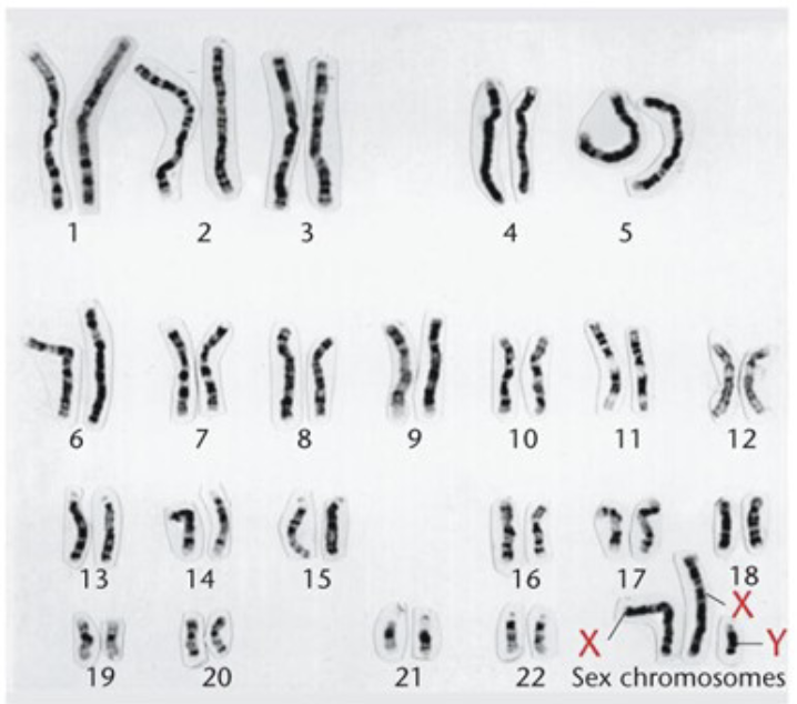
sex-linked chromosomal abnormalities - Klinefelter syndrome
more than one X chromosome (usually 47,XXY; or a 48,XXXY; 49,XXXXY)
tall, with long arms and legs
have underdeveloped testes and prostate gland, no facial hair
phenotypically male, infertile, sometimes with slight gynecomastia
generally normal intelligence, but may be slower to learn
genetically male
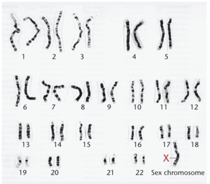
sex-linked chromosomal abnormalities - Turner syndrome
45,XØ karyotype
phenotypically female
occurs in 1/2000 female births
short, may have malformed features
webbed neck
high palate
small jaw
congenital heart
kidney defects
ovarian failure
infertility
generally have normal intelligence, may have learning disabilities
only condition where a missing chromosome results in a viable organism
dosage compensation
in homogametous individuals, there are two X or Z chromosomes, compared to heterogametous individuals
in humans, females have two copies of every X-linked gene and males have one, but their genes are expressed at the same levels
gene expression must be closely regulated; too much would be harmful (as with Down syndrome)
dosage compensation: balances the dose of X chromosome gene expression in females and males
inactivation of one entire X in females (heterochromatinization)
inactivation of some genes on both X chromosomes in females
double expression of male genes (to match normal expression in females)

X chromosome inactivation
dosage compensation in humans is achieved by inactivation of one of the X chromosomes in females
Barr bodies (heterochromatinized X), males do not have any
Lyon Hypothesis
inactivation of X is random
occurs in somatic cells at an early stage of embryonic development and is then passed on to progeny cells by mitosis

mechanism of X inactivation
the X-inactivation center (Xic) is active on the randomly inactivated X (via methylation, histones)
it consists of the X-inactive specific transcript (XIST) gene (RNA produced)
X chromosome encodes X-inactivation center (Xic)
one encodes XIST
expression of Xic leads to inactivation of chromosome expressing the XIST locus

complications in X chromosome inactivation
XØ is not normal, lacks Barr bodies
XY also has no Barr bodies
XX has one
XXY has one (Klinefelter)
XXX has two (triple X)
XXXY has two (Klinefelter)
anomalies in sex determination
hermaphrodites
have both ovaries and testes
external genitalia are ambiguous
generally, true hermaphrodites are sterile, but not always
true hermaphrodites are genetic mosaics — some cells are XX and some are XY
nondisjunction in mitosis, normal females considered mosaic due to the silenced X (which is not the same X in every cell)
pseudo-hermaphrodites
pseudo-hermaphrodites have either testicular or ovarian tissue, but not both
generally the tissue is rudimentary
external genitalia are often ambiguous
some are genetically female, but may look like males
some are genetically male, but may look like females and lead normal female sex lives

Expressivity and Penetrance
penetrance
degree to which individuals in a population express a particular trait for a given genotype
functions as a yes or no outcome, you either have it or you don’t
expressivity
degree to which a given genotype is expressed in the phenotype
varies among individuals

chromosome nondisjunction
chromosome nondisjunction is the failure of homologous chromosomes or sister chromatids to separate as they normally do during cell divison
it can result in abnormalities in chromosome number, cause gain or loss of function (aneuploidy)
in somatic cells, it can result in one daughter cell with an extra chromosome (2n+1) and the other missing one chromosome (2n-1)
the relatively poor survival of these cells normally limits their number in animals
euploidy and aneuploidy
the number of chromosomes in a nucleus and the relative size and shape of each chromosome are species-specific characteristics
the euploid number of chromosomes of an individual is the number of complete sets (n, 2n, 3n, etc…)
euploidy is when there is more than the correct set (2n normally, but showing 3n or 4n)
if cells contain a number of chromosomes that is not euploid, chromosome number is aneuploid (2n+1, 2n-1)
aneuploidy is when there is an extra or missing chromosome

nondisjunction in germ-line cells
the result of nondisjunction in germ-line cells is aneuploid gametes that can produce aneuploid zygotes
nondisjunction in meiosis I results from failure of homologs to separate; the gametes produced are either trisomic n+1 or monosomic n-1 (none are normal)
fusion of these gametes with normal (n) gametes produces trisomic (2n+1) or monosomic (2n-1) offspring
nondisjunction in meiosis II is the failure of sister chromatids to separate normally
among the four gametes produced by meiosis, only two will be affected in this case
two of the gametes will be normal (n); of the remaining two, one will be n+1 and the other will be n-1
when fused with normal gametes, 50% are normal, 50% are not (2n+1 or 2n-1)

gene dosage alteration
Blakeslee and Belling (1913) studied aneuploidy in the jimson weed (2n = 24)
they identified 12 phenotypically distinct lines of trisomic, one for each chromosome
phenotypic effects resulted from changes in gene dosage, the number of copies of a gene; aneuploidy alters dosage of all the genes on the affected chromosomes
depending on extra chromosome/gene, seed shape/size changes (anueploidy)
a euploid of 3n, 4n, etc has the same seed structure, just bigger
gene balance
changes in gene dosage lead to an imbalance of gene products from the affected chromosome relative to the unaffected chromosomes
most animals are highly sensitive to changes in gene dosage
in contrast, plants tolerate gene dosage changes more readily
euploid animals do not exist, but plants do
polyploidy in plants (wheat 6n)

aneuploidy in humans
humans are enormously sensitive to gene dosage changes and aneuploids usually do not survive
only trisomies of chromosomes 13, 18, and 21 are seen in newborn infants, and no autosomal monosomies and observed
trisomy 21 is better tolerated than 13 and 18, there is a lower death risk
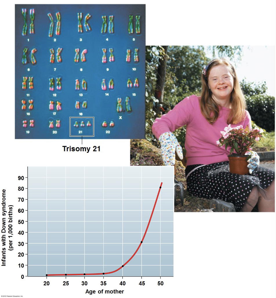
nondisjunction of autosomes: down syndrome (trisomy 21)
individuals have 47 chromosomes (47, +21)
a small number of genes on chromosome 21 are responsible for the cognitive disabilities and heart abnormalities that are principal symptoms
females with down syndrome have a 50% chance of giving birth to an affected individual
research has identified a link between the risk of trisomy 21 and maternal age
age of female’s egg are the same age as she is, so the risk increases as she ages; proteins can lose function as they age (microtubule function compromised, cause nondisjunction)

nondisjunction of autosomes: Patau syndrome (trisomy 13)
individuals have 47 chromosomes (47, +13)
occurs in 1/15,000 newborns
severe mental retardation
cleft palate or lip
causes central nervous system and immune system dysfunction
more than 80% of children with trisomy 13 die in the first year

nondisjunction of autosomes: Edward’s syndrome (trisomy 18)
individuals have 47 chromosomes (47, +18)
half of infants with this condition do not survive beyond the first week of life
some children have survived to the teenage years, but with serious medical and developmental problems
heart defects, kidney problems, delayed growth

nondisjunction of sex chromosomes
multiple forms of sex-chromosome trisomies, and one type of sex chromosome monosomy occurs
nondisjunction of sex chromosomes - Turner syndrome
monosomy X (2n = 44, XØ)
affected individuals are female
the second X may be completely absent (50%) or partially deleted (30%)
tissue mosaicism (mixtures of XØ and XX cells) may also be observed
the most obvious characteristic is very short stature
may also have characteristic facial features and a variety of secondary medical conditions

nondisjunction of sex chromosomes - Klinefelter syndrome
results in males - extra X chromosomes (47, XXY)
occurs in about 1/500-1,000 males
abnormal body proportions, larger breasts, infertility, sexual problems
no mental retardation and most symptoms can be treated by testosterone therapy
greater susceptibility to diseases like breast cancer
in rare cases, females can have an XXY genotype (SRY mutation, or testosterone receptor mutation on the X)
in rarer cases, a male can be XX (because of an SRY translocation to an X)
nondisjunction of sex chromosomes - Jacob and Triple X
Jacob Syndrome (47,XYY)
extra copy of the Y chromosome
Triple X Syndrome (47,XXX)
extra copy of the X chromosome
occurs in about 1/1000 births
individuals are taller than average
no unusual physical features and normal sexual development

reduced fertility in aneuploidy
in trisomics, chromosome separation during meiosis is disturbed because of failure to properly pair and segregate
two patterns of synapsis are possible: a trivalent synapsis or bivalent and univalent arrangement
neither mechanism can segregate three chromosomes equally at anaphase I
in trisomics, meiosis results in two chromosomes moving to one pole and one chromosome moving to the other
thus half the gametes contain two copies of the chromosome; these will produce trisomic offspring that are unlikely to survive
this results in a form of semi-sterility, in which only some of the offspring produced are viable
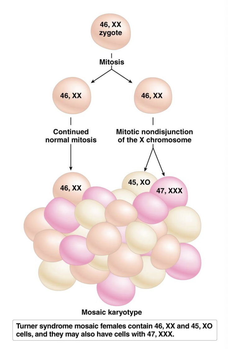
mosaicism
mosaicism can develop as a result of mitotic nondisjunction early in embryogenesis
for example, 25-30% or Turner syndrome cases occur in females that are mosaic, with some 45,XØ cells and some 46,XX
some Turner syndrome individuals carry 47,XXX cells too

gynandromorphs
in some insects, such as butterflies and fruit flies, sex-chromosome mosaicism produces gynandromorphs
these are individuals with some female and some male cells
these occur due to sex-chromosome nondisjunction in early development
uniparental disomy
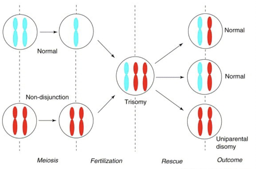
a rare abnormality, uniparental disomy, occurs in humans
this happens when both copies of a homologous chromosome pair arise from the same parent
more often, nondisjunction occurs in one parent, resulting in a gamete with two copies of chromosome (n+1)
this unites with a normal gamete to produce a trisomic zygote
in a process called trisomy rescue, one copy of the chromosome is randomly ejected from the gamete and some of the progeny will have both copies from the parent that underwent nondisjunction

changes in euploidy result in various kinds of polyploidy
polyploidy is the presence of three or more sets of chromosomes in the nucleus of an organism
it can result from duplication of chromosome sets within a species (autopolyploidy)
it can also occur from combining the chromosome sets of different species (allopolyploidy)
many types of polyploidy are possible
not tolerated in humans, only in plants
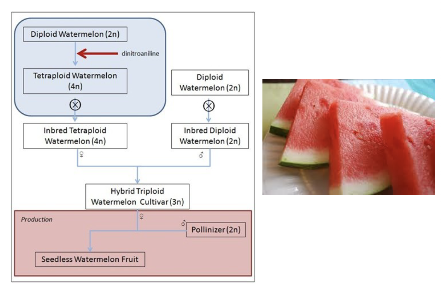
use of odd-ploidy in agriculture
creates seedless watermelon
tetraploid x diploid = triploid seedless

chromosome breakage causes mutation be loss, gain, and rearrangement of chromosomes
mutations that result in loss or gain of chromosome segments can produce severe abnormalities due to gene dosage imbalances
changes may or may not be large enough to detect microscopically
when a chromosome breaks, both DNA strands are severed at a location called a chromosome break point
the broken chromosome ends can adhere to other broken ends or the termini of intact chromosomes
or, since a fragment of the broken chromosome is acentric (lacks a centromere), it may be lost during cell division

larger chromosome deletions
organisms with one normal and one terminally deleted/lost chromosome are called partial deletion heterozygotes
cri-du-chat syndrome (cat’s cry)
a piece of chromosome 5 is missing
occurs in 1/20,000-50,000 newborns
infants have a high-pitched cry, like that of cat
characterized by intellectual disability, delayed development, distinctive facial features, etc

chromosome breakage leads to inversion and translocation of chromosomes
sometimes chromosome breakage leads to reattachment of the wrong broken ends
reattachment in the wrong orientation leads to chromosome inversion; reattachment to a non-homologous chromosome leads to chromosome inversion
two types of chromosome inversion can occur, depending on relative position of the centromere
in a para-centric inversion, the centromere is outside of the inverted region
in a peri-centric inversion, the centromere is within the inverted region
inversion heterozygotes have one normal and one inverted homolog. issues can arise when crossing-over (meiosis) because they are not homologous. codons may change because of this, resulting in different expression of certain genes and a difference in which proteins are created or in the function of the gene

chromosome pairing and recombination - para-centric inversion
alignment of a normal homologous chromosome with its inverted homolog results in the formation of an inversion loop
the crossing-over that occurs outside of the loop proceeds as normal
the crossing-over within the inverted region results in duplications and deletions in the recombinant chromosomes
crossing-over that occurs in para-centric inversions results in a dicentric chromosome and an acentric fragment
the dicentric chromosome is pulled toward both poles of the cell and eventually breaks at a random point; both products of the break are missing genetic materal
the acentric fragment is lost

chromosome pairing and recombination - peri-centric inversion
crossing-over that occurs withing peri-centric inversion results in both duplicated and deleted regions in both of the recombinant products
like para-centric inversions, the recombination event yields two normal gametes (from the non-crossover chromatids) and two abnormal gametes (from the crossover chromatids)
(2) duplication of A/B, deletion of F/G
(3) duplication of F/G, deletion of A/B
50% normal, 50% abnormal gametes
chromosome translocation
translocations occur when broken ends of non-homologous chromosomes are reattached
translocation heterozygous, with one normal copy and no one translocated copy of each chromosome, may be normal in phenotype if not genes are disrupted by the breakage and reattachment events
certain translocation heterozygotes may experience semi-sterility due to segregation abnormalities
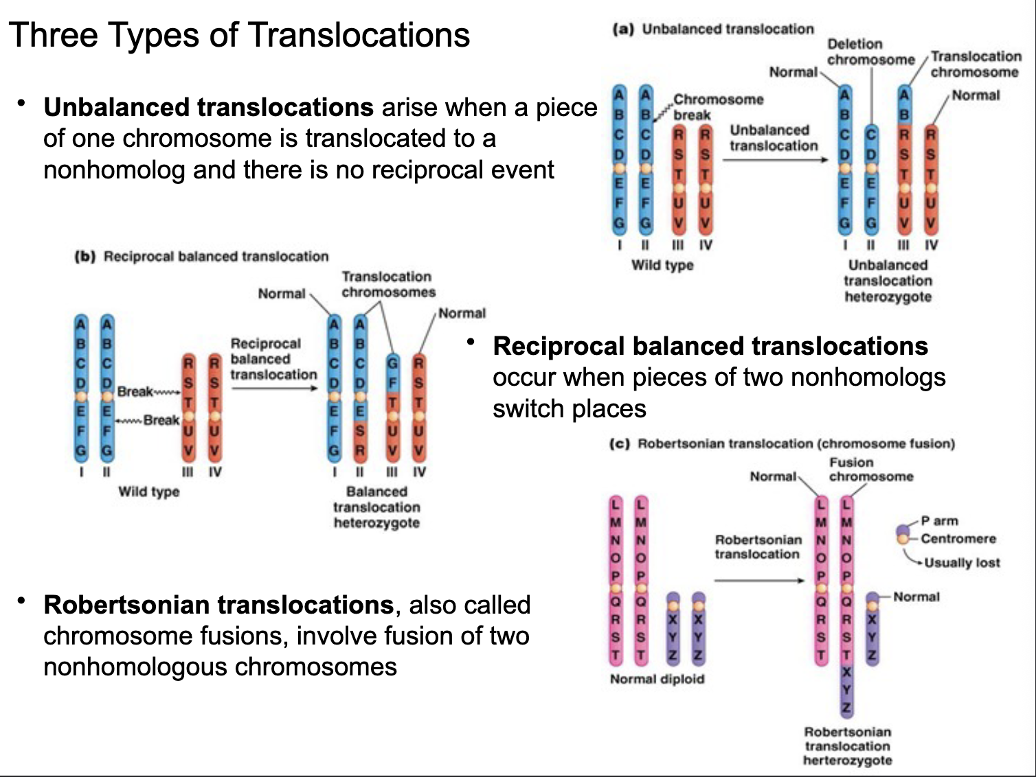
three types of translations
unbalanced translocations
arise when a piece of one chromosome is translocated to a non-homolog and there is no reciprocal event
reciprocal balanced translocations
occurs when pieces of two non-homologs switch places
robertsonian translocation
also called chromosome fusions, involve fusion of q arms of two non-homologous chromosomes
q arms fused, p arm of smaller chromosome is lost
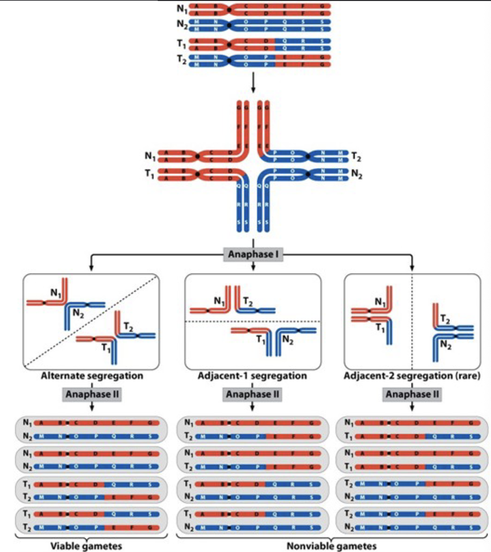
reciprocal balanced translocations
in heterozygotes for reciprocal balanced translocations, none of the four chromosomes has a fully homologous partner
an unusual, cross-like structure is formed at metaphase I of meiosis
two patterns of chromosome segregation are possible
alternate segregation - viable gametes
one set of normal, one translocated (will cause the same issue in future meiosis after pairing with normal chromosomes)
adjacent segregation - non-viable gametes (missing genes/loci from deletions)

translocation: diseases and detections
translocations involving human chromosomes are of great clinical interest because they have been linked to a number of disorders, including mental retardation, infertility, and cancer (somatic cell function compromised)
chromosome abnormalities are usually detected by means of karyotype, chromosome-banding and in situ hybridization
entire individual is affected by germ cell (meiosis) translocation

ring chromosomes
two DNA breaks, one in each arm of the same chromosome, followed by fusion of the proximal broken ends can result in the formation of ring chromosomes
may also be caused by fusion of dysfunctional telomeres
loss of distal material may mimic terminal deletion syndrome
individuals fail to thrive in a manner that is beyond what is expected from the chromosomal imbalance alone
mitotic instability may present normal somatic cell proliferation
also tend to have other anomalies along with the rings (acentric, dicentric)