Sectional Gynecology Part II
 Call Kai
 Learn
 Practice Test
 Spaced Repetition
 Match
 Flashcards
 Knowt Play
1/53

There's no tags or description
Looks like no tags are added yet.
54 Terms
reproductive years
11-13 to 50 years
Ovulation
once a month an ovum, which is the unfertilized egg, is released from one of the two ovaries
Follicle
fluid sac containing a developing ovum within the ovary
Dominant or Graafian Follicle
term for enlarging follicle in which ovulation occurs from. In a normal cycle this is to occur one a month from one ovary. 2CM
Corpus Luteum
cyst producing progesterone at the site of the dominant follicle after it has burst.
Corpus Albicans
scar at the site of a previous corpus luteum
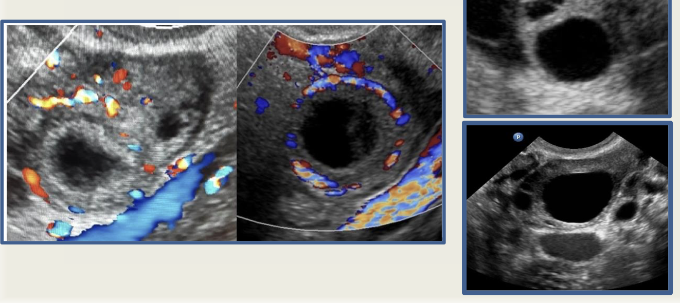
dominant follicle and corpus luteal cysts
1,2,3 Rule
•<1 cm = follicle
•1-2 cm = dominant follicle
•>3 cm = cyst
FSH: Follicle Stimulating Hormone
From the pituitary gland which stimulate follicular growth.
Estrogen
hormone secreted by growing follicles that stimulate the endometrium to regenerate.
LH: Lutenizing Hormone
large surge from pituitary gland causes graafian follicle to rupture
Progesterone
hormone secreted by the corpus luteum that prepares the endometrium to receive the fertilized egg.
Ova
immature oocytes until the onset of menarche
How many oocytes does each female have at the time of birth ?
200,000
With the onset of puberty, the hypothalamus begins the release of…
GONADOTROPIN Releasing hormone (GnRH) : stimulates the anterior pituitary gland to secrete gonadotropins (primary one is FSH).
Ovarian cycle
• Follicular phase (day 1-14): Follicle stimulating hormone (FSH) causes the ovarian follicles to develop which produce estrogen as they grow.
• 5-8 pre-antral follicles develop and one reaches maturity each month called Graafian / Dominant follicle (2cm)
• As estrogen increases in blood, pituitary gland is told to stop producing FSH and secretes the lutenizing hormone (LH).
• LH and some FSH stimulate ovulation on day 14.
• Free fluid in the pelvis can be seen at this time with the rupture of the Corpus Luteum.
• After ovulation, the ovary enters the LUTEAL PHASE
• The corpus luteum secretes progresterone. After around 10 days of ovulation, the CL will degenerate, causing progesterone to decrease.
Mittelschmerz
pain with ovulation
What happens if conception occurs ?
the HCG produced by the zygote causes the Corpus Luteum to persist and to secrete progesterone for 3 months until placenta takes over
Follicles sonography
•Thin, smooth walled
•Anechoic
•Dominant Follicle 2 cm
•Up to 3cm = normal
•Corpus luteum internal echoes and ring of vascularity
Menopause
•Folliculogenesis cease and ovaries decrease in size.
•Ovaries stop making estrogen.
Perimenopause:
FSH increases to get more estrogen.
Estrogen fluctuates
Post menopause:
Estrogen and progesterone are no longer produced.
Physiologic ≤ 3cm cyst
•Dominant Follicle
•Corpus luteum
Functional cyst
•Follicular Cyst
•Hemorrhagic CL
non-neopastic ovarian cysts
•Most <5cm
•Regress 6-8 wks
Follicular & Simple cysts
Grafian follicle → no ovulation → stays active
Hemorrhagic ovarian cysts
•Hemorrhage within CL or functional cyst
•Resolve within 8 weeks
Hemorrhagic Ovarian Cysts sonography
•Cobweb appearance - fibrin strands (?septations)
•Solid appearing area with concave margins / retracting clot
•Circumferential flow

Hemorrhagic Ovarian Cysts (bottom image displays retracting clot)
Endometrial phases
1. Menstrual Phase: days 1-5. Coincide with follicular phase of ovary. Progesterone causes spiral arteries to constrict and decreases blood flow which results in ischemia and shedding of the zona functionalis.
2. Proliferative Phase: day 6-14. As follicles produce estrogen the zona functionalis starts to regenerate. This lasts until ovulation.
3. Secretory Phase: day 15-28. Coincides with luteal phase of ovary
Menstrual Phase begins sonography
• Day 1-4 early menses with hypoechoic cavity represents blood and tissue. Surrounded by hyperechoic endometrial echo.
• Days 3-7 thinner blood has been sloughed.
Proliferative Phase (Days 5-9) sonography
presents as three-line sign:
1. Thin endometrium is in middle
2. The thin endometrium is surrounded on each side by the hypoechoic halo functionalis layer.
3. This hypoechoic halo is surrounded by the thin echogenic basalis layer.
Proliferative phase days 10-14
increase in thickness and echogenicity. Basalis layer increased in thickness therefore echogenicity. Ovulation on day 14.
Secretory phase begins sonography
• Days 15-28
• The endometrium is at its thickest dimension and becomes hyperechoic.

proliferative phase: three line sign
top) basalis layer
middle) endometrial echo
bottom) functionalis layer

secretory phase
Functional layer
glands & stroma that shed menses
HYPOECHOIC during proliferative phase
HYPERECHOIC during secretory phase
Basal layer
glands that regenerate new endometrium

Endometrium days 1-5
menses
fluid and debris

Days 5-14
Early proliferative
Single thin line
Proliferative phase
“3 line sign”

Days 15-28
Secretory Phase
Thick and echogenic
Enhancement

endometrial echo

endometrial/myometrial junction
TECHNIQUE: How to measure ovaries??
•Find the ovary in its longest axis take your length measurement there.
•Go 90 degrees to where you are and take measurement
•3rd measurement (AP) can be taken in either plane. Long axis is easiest as you can see the widest part.
Pelvic Ultrasound Examination uterus
Vagina and uterus are anatomic landmarks.
Document the following:
• Uterine size, shape, orientation
• Endometrium
• Myometrium
• Cervix
Vagina serves as landmark for cervix and lower uterine segment.
Pelvic Ultrasound Examination uterus, cervix
•Length measured in long axis from fundus to cervix
•AP depth measured in long axis from anterior to posterior walls, perpendicular to length
•Width measured from transaxial or coronal view
•Cervical diameters (with pregnancy)
•Endometrium analyzed for thickness and echogenicity
•Myometrium, cervix evaluated for contour changes, echogenicity, and masses
Pelvic Ultrasound Examination Adnexa (Ovaries and Fallopian Tubes)
Ovaries identified anterior to internal iliac vessels
Document the following:
• Size, shape, contour, echogenicity
• Position relative to uterus
Ovarian width measured in transaxial or coronal view
Ovarian volume may be calculated.
Pelvic Ultrasound Examination Cul-de-sac
•Evaluate cul-de-sac for presence of free fluid or mass.
•If mass is detected, document size, position, shape, echographic pattern relationship to ovaries and uterus.
•Differentiate normal loops of bowel from mass.
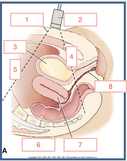
Sagittal scanning plane
1) acoustic beam
2) transducer
3) bladder
4) inferior
5) superior
6) posterior
7) uterus
8) vagina

Sagittal scanning place
1) anterior (approach)
2) bladder
3) superior
4) endometrium
5) myometrium
6) posterior
7) endovaginal canal
8) vagina
9) far field (posterior)
10) inferior
11) near field (anterior)

Sagittal scanning plane
1) anterior
2) cranial
3) posterior
4) caudal

transverse plane
1) anterior
2) left
3) posterior
4) right
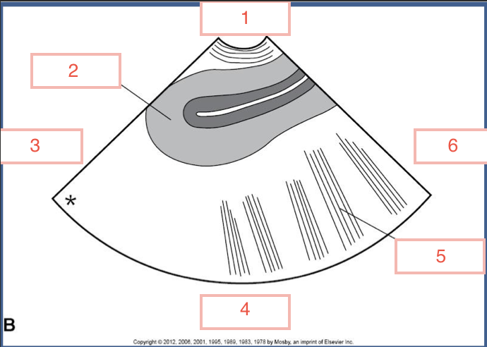
sagittal scanning plane
1) caudal
2) uterus
3) anterior
4) cranial
5) bowel
6) posterior

transverse scanning plane
1) caudal
2) uterus
3) right
4) cranial
5) left