Molds and Fungi
 Call Kai
 Learn
 Practice Test
 Spaced Repetition
 Match
 Flashcards
 Knowt Play
1/21

There's no tags or description
Looks like no tags are added yet.
22 Terms
Structure of trichospororn spp.
Stains live fungi—-
PAS
LPCB stain for fungi
Phenol ___________
Glycerol ___________
Cotton blue ___________
Lactic acid is a fungi ___________
word bank: moisture keeper | preserver | color stains | disinfectant
disinfectant
moisture keeper
color stains
preserver
A negative stain for Cryptococcus neoformans?
Its been replaced by an antigen test
Has limited sensitivity
India Ink (nigrosine stain)
What does KOH in a wet mount do when used on cultures collected for fungi morphology?
a. nothing
b. KOH digest keratin in hair/skin for better visibility
c. KOH digest keratin in hair/nails for better visibility
d. enhances visibility by enlarging fungi
c. KOH digest keratin in hair/nails for better visibility
These are characteristics of:
asexual through spores/conidia
Has hyphae (mass of mycelium)
Macrocondia and microcondia
Multicellular (eukaryotic)
Grows @ 25C-30C (RT)
Mold
These are characteristics of:
Asexual via budding/fission
Unicellular (prokaryotic)
Grows @ 35-37C body temp.
Normal flora BUT shouldn’t be at sterile locations
Yeast

macrocondia(large, asexual multicellular fungal) have tapered ends
Microsporum canis
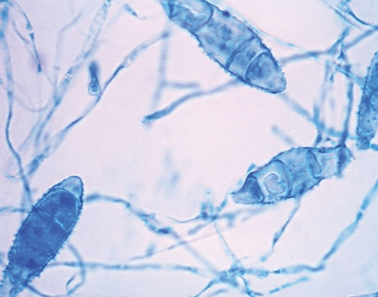
from soil, a macroconidia that has rounded ends; cinnamon color colonies on SDA agar
M. gypseum


small oval microconidia w/ “birds on a wire” hyphae (microscope); white (colonies) with deep red tint
Trichophyton rubrum
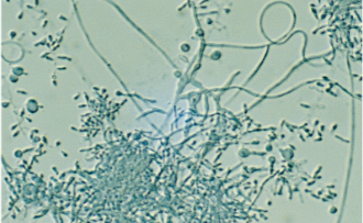
clustered microconidia and spiral hyphae ; white velvet, fluffy (colonies)
Trichophyton mentagrophytes
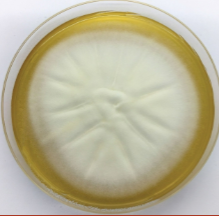

Microscopic: Yeast cells w/ multiple buds “ship’s wheel“
Paracoccidioides brasiliensis
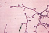
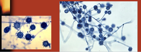
Found in Mississippi River; bird/bat poop soil
“Spelunker’s disease.”
Culture: white/brown fluffy colonies
Microscopic: Tuberculate (spiky) macroconidia
Histoplasmosis capsulatum


Identify species:
Rhinocerebral mucoromycetes Disseminated disease
Structural: Erect sporangiophore, contains rhizoids, Sporangia is intact.
Mucor

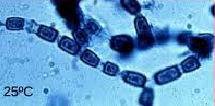
Culture: large spherules
Found in the southwestern US, Central and South America
Microscopic: Barrel shaped arthroconidia w/ empty cells between
Coccidiodes immitis


Identify species:
Budding cells, germ tubes(possible)
Canidia Albicans


Lollipop conidia
Microscopic: round yeast w/ broad based buds
Blastomyces dermatiditis


Oval/cigar shaped yeast cells
Rosette conidia
“Rose handler’s disease“
Sporothrix schenckii
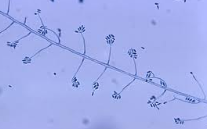

Identify species:
Can be frequently found as the cause of mycotic keratitis
Associated with contact lenses. It has abundant banana shaped macroconidia, singly and in clusters
Fusarium spp.

Identify species:
Rarely causes infection. It has been associated with chronic fungal sinusitis. Growth is demonstrated in shades of blue and green – bread mold
Penicillium spp

Identify species:
Second most common fungal infection. Candida the most common. High mortality rate in immunocompromised patients. Associated with patients having bone marrow transplantation. A. fumigatus grows at 42°C. Long sporangiophore with broad sporangium
Aspergillus Fumigatus

Bread mold
Rhizopus