RED BLOOD CELL MORPHOLOGY SHAPE VARIATION
 Call Kai
 Learn
Learn Practice Test
 Spaced Repetition
 Match
Match Flashcards
 Knowt Play
1/61

There's no tags or description
Looks like no tags are added yet.
Name | Mastery | Learn | Test | Matching | Spaced | Call with Kai | Chat |
|---|
No analytics yet
Send a link to your students to track their progress
62 Terms
What is another name for a target cell?
A. Spherocyte
B. Codocyte
C. Schistocyte
D. Elliptocyte
B. Codocyte
Target cells are primarily a variation in RBC:
A. Size
B. Color
C. Shape
D. Number
C. Shape
What does a target cell look like?
A. Crescent moon
B. Fragment
C. Teardrop
D. Bull's-eye
D. Bull's-eye
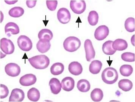
Target cells are described as:
A. Always hyperchromic
B. Always hypochromic
C. Always normochromic
D. Never hypochromic
B. Always hypochromic
What happens to the RBC surface membrane in target cells?
A. It decreases
B. It disappears
C. It increases
D. It becomes rigid
C. It increases
Which substances contribute to the increased RBC membrane in target cells?
A. Iron and calcium
B. Cholesterol and phospholipids
C. Glucose and sodium
D. DNA and RNA
B. Cholesterol and phospholipids
Target cells can result from:
A. Increased cell Hgb
B. Decreased cell Hgb
C. Increased WBCs
D. Increased platelets
B. Decreased cell Hgb
What happens to osmotic fragility in target cells?
A. Increased
B. Decreased
C. No change
D. Completely absent
B. Decreased
Rationale: Target cells have decreased osmotic fragility, meaning they are more resistant to hemolysis in hypotonic solutions.
Which disease is associated with target cells?
•Possible Pathology
•Hgb C disease
•Hgb C trait
•Post-Splenectomy
•Liver disease-flattened target surface
•Iron deficiency anemia
•Hgb abnormality
Which statement BEST describes why target cells form?
A. The RBC has too much Hgb and too little membrane
B. The RBC has increased membrane and decreased Hgb
C. The RBC loses all of its membrane
D. The RBC develops a nucleus
B. The RBC has increased membrane and decreased Hgb
Which combination is correct?
A. Target cell → decreased membrane → increased osmotic fragility
B. Target cell → increased membrane → decreased osmotic fragility
C. Target cell → decreased membrane → decreased osmotic fragility
D. Target cell → increased membrane → increased osmotic fragility
B. Target cell → increased membrane → decreased osmotic fragility
What is the most important appearance of a spherocyte?
A. Large cell with increased central pallor
B. Small cell with no central pallor
C. Crescent-shaped cell
D. Fragmented cell
B. Small cell with no central pallor

Spherocytes have the:
A. Highest surface area-to-volume ratio
B. Lowest surface area-to-volume ratio
C. Same ratio as normal RBCs
D. Highest volume-to-surface ratio only
B. Lowest surface area-to-volume ratio
What is the major defect responsible for the spherocyte shape?
A. Excess hemoglobin
B. Membrane loss
C. DNA increase
D. Increased iron
B. Membrane loss
The shape change of a spherocyte is:
A. Reversible
B. Temporary
C. Irreversible
D. Caused by staining
C. Irreversible
Which RBC membrane protein is decreased in hereditary spherocytosis?
A. Glycogen
B. Spectrin
C. Hemoglobin
D. Albumin
B. Spectrin
what are the possible Pathology for spherocytes
•Immune hemolytic anemia
•Hereditary spherocytosis
•Post-transfusion
Which combination BEST describes a spherocyte?
A. Large + pale + increased central pallor
B. Small + round + no central pallor
C. Fragmented + irregular + pale
D. Crescent + elongated + dark center
B. Small + round + no central pallor
What shape is an ovalocyte?
A. Pencil-shaped
B. Egg-shaped
C. Crescent-shaped
D. Spherical
B. Egg-shaped
What shape is an elliptocyte?
A. Pencil-shaped
B. Egg-shaped
C. Round
D. Fragmented
A. Pencil-shaped

Are ovalocytes and elliptocytes hypochromic according to your notes?
A. Yes, always
B. Yes, sometimes
C. No, they are not hypochromic
D. Only in hereditary elliptocytosis
C. No, they are not hypochromic
Which disease is associated with elliptocytes?
A. Hereditary spherocytosis
B. Hereditary elliptocytosis
C. Hgb C disease
D. Iron deficiency anemia
B. Hereditary elliptocytosis
What type of defect occurs in hereditary elliptocytosis?
A. Platelet defect
B. Membrane defect
C. Nuclear defect
D. Hemoglobin synthesis only
B. Membrane defect
Which protein is specifically mentioned in the membrane defect?
A. Albumin
B. Spectrin
C. Ferritin
D. Transferrin
B. Spectrin
A membrane defect in hereditary elliptocytosis can potentially cause:
A. Hemolysis
B. Leukocytosis
C. Thrombocytosis
D. Increased Hgb production
A. Hemolysis
Some sources consider ovalocytes and elliptocytes to be:
A. Completely unrelated cells
B. The same
C. Opposite cell types
D. WBCs
B. The same
Possible Pathology of ovalocyte
•Myelodysplastic syndrome
•Thalassemic syndromes
•Megaloblastic process
•Macrocyte
Possible Pathology of Elliptocyte
•Iron deficiency anemia
•Hereditary elliptocytosis
•Idiopathic myelofibrosis
What is the key appearance of a stomatocyte?
A. No central pallor
B. Bull's-eye appearance
C. Slit-like central pallor
D. Crescent shape
C. Slit-like central pallor
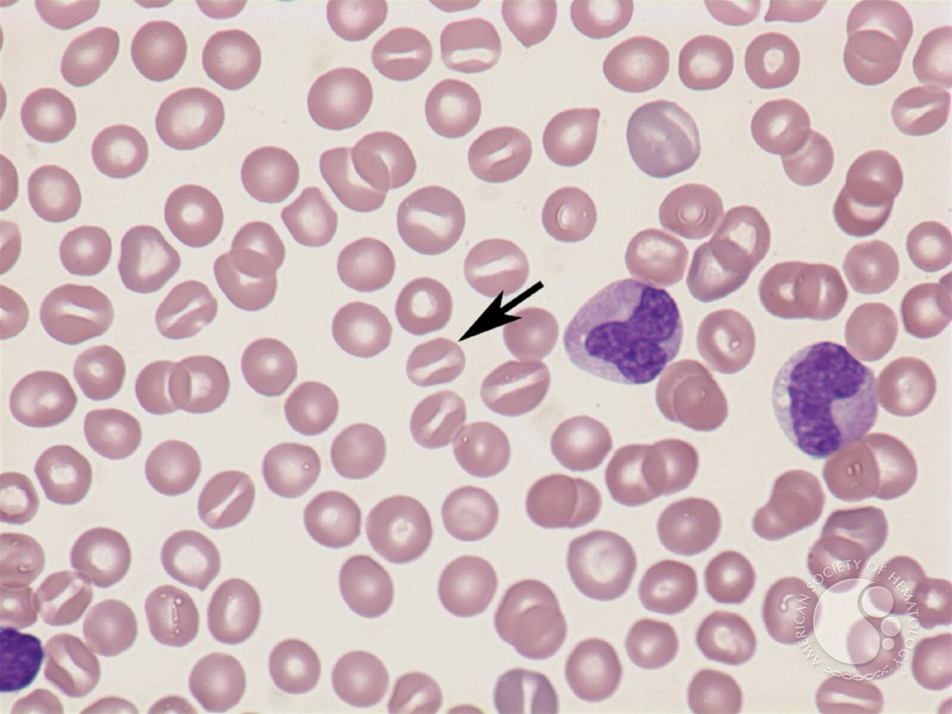
. Stomatocyte shape changes are:
A. Irreversible
B. Reversible
C. Permanent
D. Caused only by genetics
B. Reversible
Which can cause stomatocyte changes?
A. Chemical agents
B. Low platelets
C. Increased WBCs
D. Excess iron
A. Chemical agents
Stomatocytes can sometimes be caused by:
A. An artifact
B. A platelet disorder
C. A neutrophil disorder
D. Bone loss
A. An artifact
Which condition is also listed as an association with a cause of stomatocytosis?
A. Acute alcoholism
B. Iron deficiency
C. Leukemia
D. Thalassemia only
A. Acute alcoholism
Stomatocytosis can be associated with which phenotype?
A. Rh-positive phenotype
B. Rh-null phenotype
C. Hgb S phenotype
D. Hgb C phenotype
B. Rh-null phenotype
What is another name for a sickle cell?
A. Codocyte
B. Drepanocyte
C. Stomatocyte
D. Spherocyte
B. Drepanocyte
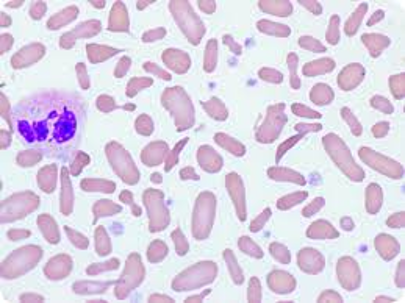
. What happens to an RBC when it sickles?
A. It becomes soft and flexible
B. It becomes rigid and inflexible
C. It becomes larger and round
D. It loses all hemoglobin
B. It becomes rigid and inflexible
What is an important trigger for sickling?
A. High oxygen
B. Low oxygen
C. High glucose
D. Low iron
B. Low oxygen
Rationale: Low O₂ causes the tubules to form and bundle together, deforming the RBC.
What forms inside the RBC during sickling?
A. Crystals
B. Tubules
C. Nuclei
D. Platelets
B. Tubules
Can a sickled RBC return to its normal shape when oxygen is restored?
A. No, never
B. Yes, some can
C. Only in sickle cell trait
D. Only after transfusion
B. Yes, some can
Which statement about sickle cell trait is correct?
A. Sickled cells are always seen on the smear
B. Usually no sickled cells are seen on the smear
C. All RBCs are sickled
D. RBCs become spherocytes
B. Usually no sickled cells are seen on the smear
Which sequence is correct?
A. High O₂ → tubules → normal RBC
B. Low O₂ → tubules form → tubules bundle → RBC deforms
C. Low O₂ → RBC becomes flexible → normal shape
D. High O₂ → tubules bundle → RBC sickles
B. Low O₂ → tubules form → tubules bundle → RBC deforms
What is the typical size of an acanthocyte?
A. Larger than normal
B. Smaller than normal
C. Normal size only
D. Extremely large
B. Smaller than normal
How many spicules can an acanthocyte have?
A. 1–2
B. 3–12
C. 15–20
D. More than 20
B. 3–12
What is the appearance of the acanthocyte's spicules?
A. Smooth and evenly spaced
B. Irregular projections
C. One large projection
D. No projections
B. Irregular projections

What happens to cholesterol and RBC surface area in acanthocytes?
A. It decreases
B. It increases
C. It stays the same
D. It disappears
B. Increases
What happens to lecithin content?
A. Increases
B. Decreases
C. Doubles
D. No change
B. Decreases
What does LCAT stand for?
A. Lecithin-cholesterol acyltransferase
B. Low-cell antigen transfer
C. Lipid-cell antibody transport
D. Lecithin-calcium activation test
A. Lecithin-cholesterol acyltransferase
What are the disease is strongly associated with acanthocytes?
Severe hepatic disease
alcohol intoxication (Zieve's syndrome).
Pyruvate kinase deficiency
Congenital abetalipoproteinemia
vitamin E deficiency.
Post-splenectomy
What causes RBC fragments to form?
A. Increased hemoglobin
B. Loss of RBC membrane
C. Increased platelets
D. Increased oxygen
B. Loss of RBC membrane

RBC fragmentation can occur because of:
A. Altered fluid circulation
B. Increased RBC production only
C. Increased iron
D. Increased oxygen
A. Altered fluid circulation
Rationale: Abnormal fluid/blood circulation can damage RBCs.
What happens to the RBC when it becomes less deformable?
A. It becomes easier to squeeze through vessels
B. It is more likely to fragment
C. It becomes a platelet
D. It gains a nucleus
B. It is more likely to fragment
Rationale: A less flexible RBC is more easily damaged and broken.
What is another name for a helmet cell?
A. Degmacyte
B. Keratocyte
C. Codocyte
D. Drepanocyte
B. Keratocyte
What is another name for a bite cell?
A. Keratocyte
B. Schistocyte
C. Degmacyte
D. Codocyte
C. Degmacyte
What are the an artifact RBC fragments
A. Burr cells
B. Echinocytes
C. Crenated cells
True burr cells seen in what
uremia, dehydration, hypothyroidism, heart disease, stomach cancer, peptic ulcer, post heparin injection, azotemia
Which condition is associated with schistocytes?
•Microangiopathic hemolytic anemia
•Disseminated intravascular coagulation
.burn case
Heart valve surgery
A smear shows many tiny pieces of RBCs. What should you think of first?
A. Schistocytes
B. Target cells
C. Spherocytes
D. Elliptocytes
A. Schistocytes
what are the RBC fragment
•Schistocytes
•Burr cells
•Helmet cells: keratocyte
•Bite cells: degmacyte
What is the characteristic shape of a teardrop cell?
A. Round with no central pallor
B. Bull's-eye
C. Pear-shaped with a tail
D. Pencil-shaped
C. Pear-shaped with a tail

Teardrop cells can be seen in:
A. Thalassemia syndromes
B. Iron deficiency
C. Myelofibrosis
d. inclusion body
What type of teardrops can occur with megaloblastic processes?
A. Micro-teardrops
B. Macro-teardrops
C. Target teardrops
D. No teardrops
B. Macro-teardrops
Which inclusion is associated with drug-induced RBC damage?
A. Howell-Jolly body
B. Heinz body
C. Pappenheimer body
D. Cabot ring
B. Heinz body