Medical Parasitology test 1
 Call Kai
 Learn
Learn Practice Test
 Spaced Repetition
 Match
Match Flashcards
 Knowt Play
1/176

There's no tags or description
Looks like no tags are added yet.
Name | Mastery | Learn | Test | Matching | Spaced | Call with Kai | Chat |
|---|
No analytics yet
Send a link to your students to track their progress
177 Terms
Phylum Nematoda (Roundworms)
one of most abundant types of worms. 12,000 species
live in almost every type of habitat
can detect touch, light, and certain chemicals,
breath across their whole body
parasitize almost every type of plant and animal
5 cm in length
Largest known is 28 ft.
first multicellular organism to have its genome sequenced
nematode C. elegans
Nematodes can live via
free-living or parasitic and live within a host
parasitic nematodes
feed on digestive contents or blood/tissue of their hosts
GI Tract Nematodes
Ascaridida: Ascaris and Pinworms
Strongylida: Hookworms
Trichurida: Trichinella/Trichuris
Filarial Nematodes
Spirurida: Filarial worms
Nematode Basic Anatomy
bilaterally symmetrical
3 cell layers (triploblastic)
known as psuedocoelomates bc they have internal cavity that is not lined w/ mesoderm
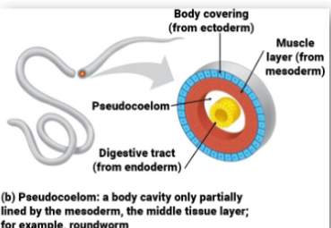
3 cell layers of nematode
Ectoderm: outer tissue layer (epidermis)
Mesoderm: middle tissue layer (muscle)
Endoderm: innermost tissue layer (body cavity)
basic anatomy continued
vermiform structure w/ tapering formed both posteriorly and anteriorly
round body w/ mouth and anus
hypodermis: cellular subcuticular layer that secretes the cuticle
mostly a moving eating tube w/ attached reproductive organs

how is nitrogenous waste excreted vs other material
released through skin while others via the excretory pore
nematode muscles like?
longitudinal and line ventral/dorsal portion
cannot crawl like other wormsd
do nematodes have blood or gas exchange systems
NO
nematodes are most closely related to
anthropods; grow via molting
nematode development
L1: larval nematode develops inside an egg
H: hatched
M1: grows quickly and molts
L2: another rapid growth spurt
M2: transition into a second melting
M3 grows and molts
L4: final larvae stage inside the host
M4: nematodes final molt
Adult: when they become sexually mature
at what stage can nematode become infected
L3
earthworm movement
peristaltic wave; setae anchor
sets own length and girth, anchor, then push
nematode movement
body wall is covered by a thick cuticle which is secreted by the syncytial (multinucleated) epidermis
only longitudinal muscles no circular or diagonal
longitudinal muscles work antagonistically against the outer cuticle like a spring
when muscles contract the restoring force is provided by elastic components of cuticle and hydrostatic force of the pseudocoel
MOVES MORE LIKE A SNAKE
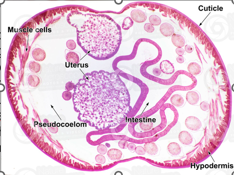
what is cuticle made of
collagen, making it both tough and flexible
this is what is shed as worm grows
nematode mobility is thus generated by
interplay of muscles, pressurized pseudocoel and cuticle

why doesn’t nematode shorten
blocked by geometry
nematode don’t swim they
burrow
they need something to push against to move
nematode eating
have complete (but simple) digestive system w/ mouth at very tip of the animal

nematode eating cont.
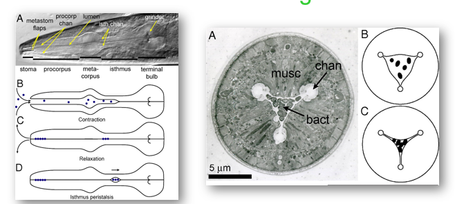
food has to be pumped agianst the pseudocoel pressure by muscular esophagus
Contraction of pharyngeal muscle draws fluid containing suspended food particles (dots) into pharyngeal lumen
Relaxation ejects fluids while trapping particles
(D) Isthmus peristaisis carries food from anterior isthmus to terminal bulb and intestine
food passed rapidly (minutes)
nematode neuroanatomy
brain consists of a single ring of cells around the pharynx
a dorsal and ventral nerve extend back through the length of the worm
these nerves control muscle contraction

why are the brains of a female different form male worm
different behavior wiring and anatomical differences

scientist have labled individual types of neurons of C. elegans with
fluorescent reporters and can track real time neuron activation when exposed to different compoounds
neuroanatomy continued
labial and amphid nerves extend forward to enervate sensory organs
amphids are anterior depressions that contain chemoreceptors
phasmids are posterior chemoreceptors

configurations of neural wiring get established by
natural selection;
behaviors we see are the behaviors which are favorable for survival
Nematode Reproduction
most nematodes are DIOECIOUS (male and female are sep sexes)
exhibit sexual dimorphism - males are smaller
fertilization is internal, and eggs are stored in the uterus
eggs are deposited by the female, embryo develops, juvenile hatches from an egg
male anatomy
during mating, female is held by the male within the bursa
male inserts 2 sclerotized copulatory spicules
sperm duct is muscular and sperm is transferred to female against the pseudocoel pressure
shape and size of spicules and bursa are important features to tell different nematode species apart
males can cement closed the vulva after mating in some species

Female anatomy
most females have 2 ovaries
oogonia are produced at the distal end and mature into oocytes while they move into the growth zone
as oocytes enter the oviduct, they are fertilized by sperm stored in the seminal receptacles

diagram of nematode reproduction

male sperms
not flagellated but have amoeboid movement that isn’t acting based
motility is powered by a dynamic filament system composed of polymers of the 14-kDa major sperm protein (MSP)
after fertilization
eggs are laid
what part of the male anatomy is designed for sensing and attaching to females?
spicules
majority of infections disccused are from
soil transmitted helminths (STHs)
___% of all people in the world are currently infected with GI nematodes
50
Infection generally associated w/ low mortality but can cause considerable morbidity
Ascaris lumbricoides
largest intestinal nematodes parasitizing humans
most common found in humans
most prevalent throughout the tropics, sub-tropics and infects 1.2 billion ppl
can live 1-2 years inside host
single female can lay ~240k eggs per day
eggs are very environmentally resistant (survive up to 15 years in an environment)
Ascaris lumbricoides life cycle
fertilized ascaris egg, still at unicellular stage
passed in stool
Ascaris lumbricoides complete life cycle
beings with ingestion of infectious eggs
eggs become infectious after embryogenesis and molting 2x within the egg
Larvae hatch from egg in small intestine
worms penetrate intestine and go to bloodstream
worms get caught in lung tissue capillaries and enter the alveolar spaces
cough the worm up and swallow it where it goes back into your stomach
one in small intestine again, matures via 1 additional molt and starts laying eggs and feeding on intestinal contents
high worm burdens can lead to bowel obstructions of perforations
time from egg ingestion to egg shedding by mature worms is 60-75 days

Ascaris lumbricoides pathology larval worm phase
larval worm phase:
pneumonitis due to larvae in pulmonary alveoli
migration of larvae through lungs = blood vessels of lungs to hemorrhage
resulting inflammatory response and edema in lungs = ascaris pneumonia which can be fatal
Ascaris lumbricoides pathology adult worm phase
malnutrition and impaired physical growth
mild abdominal discomfort →→small bowel obstruction
wandering ascaris: bile duct obstruction, perforation of small intestine, pancreatitis and liver abscess
Ascaris lumbricoides treatment and prevention
anthelmintic medications such as albendazole and mebendazole
can inhibit microtubule assembly in worm and block glucose uptake by worm cells
infections generally treated for 1-3 days
reducing reinfection based on sanitary disposal of feces
hygienic habits such as cleaning of hands before meals and health education
highlights that poverty itself is a major driver of infection and that access to clean water and sanitation could eliminate vast majority of infection
GI nematode parasites: Pinworms (Enterobius vermicularis)
aka threadworms: cause very few symptoms but are most common worm parasite in US
estimate 30% of children and 16% adults have it
infection occurs via self-innoculation or through exposure to eggs in envi
Pinworms life cycle

GI Nematode Parasites: Hookworms
burrow their hook mouths into the intestines of humans and such blook
can cause anemia in people, delay mental and physical growth in children, and cause physical fatigue
576-740 million of people in world are infected
two main species: Necator americanus and Ancylostoma duodenale
Hookworms life cycle
eggs passed in feces after hatching are free living in the soil
after 5-10 days and 2 molts they become filariform (3rd stage) larvae that are infective
infectious larvae hang out on blades of grass waiting to penetrate the skin of a passing human

Nematode L3 stage potential to become a ____ larva. which is..
Dauer
‘the enduring’
arrested in prepubescent state
can move around but doesn’t eat and is able to survive long-term in the environment
will do this until “conditions improve” and in the case of parasites, this means until they get into their next host
ability to pause is not possible for any of other stages
Hookworm life cycle
eggs passed in feces and after hatching are free living in soil
after 5-10 days and 2 molts they become filariform (3rd stage) larvae that are infective
infectious larvae hang out on blades of grass waiting to penetrate the skin of passing humans

what makes hookworms different
small and life cycle creates potential advantages and disadvantages
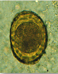
ascaris egg identification and function
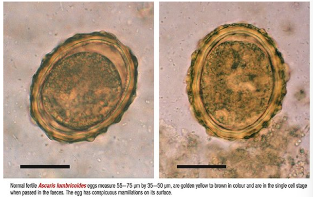
often released as unicellular embryos although they are fertilized
have a thick chitin shell that protects them from the environment
have a heavy, albuminous coating that is characterized as ‘mammilated’
has small protrusions on its surface
hookworm egg identification
Often at some stage of development when released
have a thin shell that is not there to protect them from environmental insults as these larvae hatch and initially grow in the soil

main difference between hookworm and ascaris
ascaris eggs are ingested while hookworms quest on blades of grass and enter bloodstream
what has been suggested about ascarid worms evolution
evolved from previously dixenous (2 host) parasites transmitted by biting insects
others suggested they were originally 2 host parasites w/ one being an intermediate stage in prey animals and a definitive stage in predators
different strategies evolve to maximize dissemination
can get big and blanket the earth in hardy eggs and hope for accidental ingestion
or can get mobile and seek out host directly
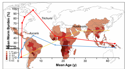
hookworm life cycle facts and world trend
1 year life span (A. duodenale)
2-5 years lifespan (N. americanus)
worm burdens do not decline in adult years
Hookworm Pathology of Infection
skin penetration
secondary bacterial infection = ground itch
ground itch = dry cough, wheezing (1-2 weeks later) in primary infection
pulmonary phase is however usually asymptomatic
Intestinal phase: worms attach to mucosa and feed on blood. worms continuously move to new places exacerbating bleeding
Hookworm treatment and prevention
hookworms were wide spread in south US
Control programs were big part of public health campaigns in South during and right after WWII
1972 12% of school children from rural coastal Georgia tested positive for hookworm infection
Anthelmintic medications (drugs that rid the body of parasitic worms) such as albendazole and mebendazole, drugs of choice for treatment
infections treated for 1-3 days
GI Nematode Trichinella (T. spiralis)
responsible for a potentially fatal disease called trichinosis
worldwide, and an estimated 10k cases occur every year
Females produce live young
Juveniles penetrate skeletal muscles, where they burrow and develop into cysts in muscle tissue called nurse cells
Transmission occurs when undercooked meat from an infected animal is ingested, juvenile worms are liberated into the intestine where they mature
Trichinella life cycle
Encysted larvae are ingested in raw or undercooked meats, released in the stomach
larvae (L1) enter small intestine where they mature and live
mature worms produce live offspring that migrate into the bloodstream
newborn worms (L1) entering into different tissues are the major source of the pathology
juvenile larvae enter skeletal muscle cells where they encyst, waiting to be transmitted to the next host via carnivorism
person infected both a definitive host (where the sexual cycle occurs) and an intermediate host (transmission) at the same time

Trichinella pathology of infection Initial Phase
within 1-2 days after consumption, the person experiences general gastroenteritis
Symptoms include fever, diarrhea, nausea, and vomiting
males have a copulatory pseudobursa at the tail end

Trichinella pathology of infection Late Phase (part 1)
Live (L1) young released in small intestine migrate into the surrounding tissues 2-8 weeks after infection
this is followed by rashes, joint-pain, headache, face swelling, and splinter hemorrhages
symptoms can become more severe but generally subside in 1-2 weeks after beginning
Trichinella pathology of infection Late Phase (Part 2)
Larvae (L1) invade multinucleated muscle cell fibers where they encyst
they use the vasculature of muscle tissue itself to provide nutrients for long term viability
larvae reside in a vascularized collagen capsule waiting to be eaten by a carnivore
fatal infections of trichinosis effects heart, lungs, and central nervous system, giving rise to serious complications including myocarditis, pneumonia, and encephalopathy

Trichinella Treatment and Prevention
prompt treatment w/ antiparasitic drugs can help prevent to progression of trichinosis by killing the adult worms and so preventing further release of larvae
once larvae established in skeletal muscle cells, usually 3-4 weeks post infection, treatment may not completely eliminate the infection and associated symptoms
mebendazole or albendazole
if no initiated within first several days of infection, alternate treatment needed
best way to avoid infection = thoroughly cook meat
Trichuris (whipworm)
infects up to 800 mil ppl globally
comes from ingesting worm eggs passed in feces of infected individuals
live in the colon
adults live in cecum where female produces 20k eggs per day
embryonation occurs in eggs within 3 weeks in soil
children w/ heavy infection = severely anemic and growth-re.
mouth (or front) is at the tin end
Trichuris Life cycle
begins with ingestion of infectious eggs from contaminated dirt or non washed food
larvae hatch in small intestine
worms migrate to colon where they mature, mate and being laying bipolar-plugged eggs
unembryonated eggs pass out into the environment via feces

Trichuris egg

Trichuris pathology of infection
adult worms remain embedded with their thin front end in intestinal mucosa
infections with less than 10 worms are often asymptomatic
moderate to heavy infection = abdominal pain, diarrhea, and weight loss
heavy infected children = profuse bloody diarrhea, cramps, and rectal prolapse
heavy infections = developmental delays
adult worms can live for years
Trichuris Treatment and Prevention
mebendazole or albendazole
reducing reinfection is based on sanitary disposal of feces
hygienic habits such as cleaning of hands before meals and health education help reduce reinfection
availability of clean water, sewage removal and treatment = best chances of reducing overall worm burdens in children
Parasite Biology: obligate relationship
essential for life of one or both (eg tapeworms)
Parasite Biology: facultative relationship
not essential for life or reproduction of one or both associates (free living amoebae)
infection vs disease
infection does not imply malfunctioning process
parasites do not always induce frank disease
dangerous symbionts may cause no disease in some hosts (carriers) (eg polio virus)
types of parasitism
ectoparasite
endoparasite (intracellular (malaria) ; extracellular (tapeworm))
facultative
obligate
accidental parasite
temporary/intermittent
types of hosts
definitive: sexual reproduction, usually vertebrate host
intermediate: asexual, invertebrate (vector)
Platyhelminthes (flat worms) as human parasites
planaria: free-living flatworms that live in quiet bodies of water; bilateral symmetry
soft bodies, no body cavity w/ no specialized circulatory and respiratory organs.
flat shape = oxygen and nutrients to pass through bodies via diffusion
Digenea
body plan variations according to attachment organ position
all important trematodes of humans are distomes
among most common and abundant parasitic worms
development occurs in at least 2 hosts
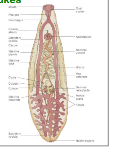
Trematodes or flukes anatomy
digenea: alternate b/w sexual and asexual
possess 2 suckers to attach within the host
oral sucker contains the mouth
muscular pharynx permits worm to pump food into the blind ending gut
most are hermaphrodites
what other organ do trematodes use for food uptake besides the gut
Tegument (skin)
epidermis of trematode is essentially
a singe cell (syncytium) formed by fusion of multiple cells
teguments cell bodies and nuclei unerlie 2 muscle layers under a basal lamina
what are found in many trematode species to help anchor
actin spines
distal cytoplasm is lined by a plasma membrane associated with
layer of glycocalyx
proximal cytoplasm is the cellular region and contains
nuclei, ER, Golgi, mitochondria, and vesicles

trematodes Excretory system
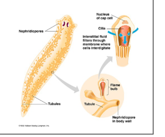
simple protonephridial excretion (kidney) system
protonephridium consists of flame cell and tubule cell
both cells interdigitate forming a micro filter and cilia beating within this cage act as mechanical pump
metabolic wastes are collected from protonephridia through small ducts which merge to central duct, opens to excretion pores
cells can also regulate osmotic pressure of worm and maintain ionic balance
Trematodes reproduction system
produce massive number of offspring
adults most often hermaphrodites
female system complex
oocytes associates with vitelline or yolk cells in ootype which is surrounded by mehlis gland
egg shell forms from secretions of vitelline cells
elongated uterus opens to exterior in genital pore, close to male opening.
sperm travel through uterus to ootype where fertilization occurs

trematode eggs
size and shape distinctive and used for diagnosis

Trematodes Life Cycle
digenic produce enormous number of offspring by combining sexual and asexual
all have at least 2 hosts of which one is a snail
not all stages are found in all species
asexual reproduction (green) occurs in germinal balls. home to omnipotent progenitor cells that can initiate w/o fertilization
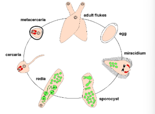
what stages are infective
Miracidia and cercariae
Trematode Life Cycle The Egg
contains embryo rather than an oocyte
must leave body of final host to continue development
mature miracidium in egg uses light, osmolarity, and temperature as clues to when hatching is appropriate
hatching proceeds in most species through preformed ‘dorr’ the operculum
the miracidium
highly motile due to the cilia on surface
may have simple eyes (avoid light) and several chemical and mechanical receptors which use to find intermediate host
penetration glands secrete proteases and other lytic enzymes on contact w/ appropriate host
miracidia of flukes w/ land snails as intermediate host will hatch upon ingestion by snail and penetrate the gut epithelium
the sporocyst
after penetration, miracidum undergoes metamorphosis into sporocyst
this stage = most organ systems reduced to bare minimum and acts as germinal sac
takes up nutrients only over its tegument and germinal mass expands and develops into daughter sporocysts, redia, or cercaria
the redia
sporocyst can produce more sporocysts, cercariae, or redia
have features of adult fluke like oral and ventral sucker, a gut, and ‘birth pore’ to release cercaria
mobile in intermediate host and can prey on sporocysts and redia of same or other species (competition)
the cercaria
stages that leave the intermediate host and infect final host
can be many consecutive waves of ‘shedding’ from snail
already show many anatomical features of adult fluke
metacercaria
cercaria have developed an array of adaptations to acheive successful infection
direct penetration of host skin upon water contact
encystation within muscle of intermediate hosts
encystation on plants
fish an turtle blood flukes life cycle

Dicrocoelium dendriticum
normally found in biliary tract of sheep
bizarre method of transmission
trematodes enhanced transmission
Dicrocoelium dendriticum the lancet fluke
one metacercaria becomes the ‘brain worm’ and lodges into central ganglia of the end
brain worm manipulates behavior of ant, in evening when temp drops, experience spasms of mandibles
Leucochloridium sp.
tiny digenic trematode living in gut of small birds
worm eggs passed with feces and are taken up by amber snails
miracidia hatch, penetrate epithelium and develop into sporocysts within hepatopancreas
cercaria develop which infect birds that eat infected snails
eyes bulge of snail
trematodes of medical importance
Schistosoma, blood flukes
clonorchis & opistorchis, liver flukes with metacercaria in fish
paragonimus, lung flukes with metacercaria in crabs
fasciolopsis, fasciola, dicrocoelium, instestinal and liver flukes w/ metacercaria on plants
Fascioliases
flukes in situ (sheep liver at autopsy)

what is a virulence factor
cathepsin
