U2 med sci group C/D
 Call Kai
 Learn
 Practice Test
 Spaced Repetition
 Match
 Flashcards
 Knowt Play
1/62

There's no tags or description
Looks like no tags are added yet.
63 Terms
functions of skeleton
provide structure/protection movement
structure of synovial joint
end of bones are encased in smooth muscle reducing friction and absorbs shock
what is synovial membrane
layer of connective tissue that lines cavities of joints/tendons and bursae
what is synovial fluid
lubricant in some of your joints reduce friction
actin
protien plays role in muscle contraction
myosin
converts chemical energy into physical form allowing for muscle contraction and movement
troponin
key regulator in muscle contraction and relaxation
slow twitch muscle fibres
contract slowly but made for endurance rely on oxidative phosphorylation
fast twitch muscle fibres
rapid powerful movements rely on anaerobic glycolysis generate greater force but fatigue faster
neuromuscular junction
triggers muscle contraction
role of neurotransmitters
carry messages or signals from one nerve cell to next target cell
the role of tendons in attaching muscles to bones
let bones move as your muscle tighten and relax
osteoarthritis
degenerative joint disease causes joints to become painful and stiff
osteoporosis/rickets
bone disease caused my delayed mineralization of bone
Duchenne muscular dystrophy
causes skeletal and heart muscle weakness its a recessive genetic disorder
Myasthenia gravis
chronic neuromuscular disease causes muscle weakness in voluntary muscles
double circulation
a system where blood passes through the heart twice for every complete circuit of the body.
why is doble circulatory system important
blood can be pumped faster around the body
blood can be pumped around the body at a higher pressure
atria
receive blood returning to the heart
ventricles
pump blood into systemic and pulmonary circulations
myocardium
allow for contraction and relaxation of the heart walls
myogenic muscle
when heart contracts heart betas at a baseline rate without any input from the nervous system
SAN (sino atrial node)
send electrical impulses that start your heart beat
AVN (atrioventricular node)
to send an electrical impulse to the cardiac muscles of the right and left ventricles
atrioventricular valves
Open during atrial systole, allowing blood to flow into the ventricles.
semi lunar valves
Open during ventricular systole, allowing blood to be ejected from the ventricles into the arteries
Close during diastole to prevent blood from flowing back into the ventricles from the arteries.
bundle of his
transmit the cardiac impulse from the atrioventricular node to the ventricles
septum
separates the left and right sides of the heart
purkinje fibre
Specialised, rapid-conducting cells in the heart's ventricular walls.
coronary arteries
supply heart with blood
veins
transport deoxygenated blood back to heart and lungs
stages of cardiac cycle
stage 1- atrial systole
stage 2-ventricular systole
stage 3-diastole
what is stage 1 atrial systole
ventricles relax atria contract
increase in atrial pressure
atrioventricular valves open
blood flows into the ventricles
what is stage 2 ventricular systole
ventricles contract atria relax
ventricular pressure increases
semi-lunar valves open and atrioventricular valves close
blood flows into arteries
what is stage 3 diastole
ventricles and atria relax
semi-lunar valves close
blood flows passively into the atria
arteries
carry oxygenated blood away from heart
thick muscular layer help withstand high pressure
smooth muscle help blood flow reduce friction
arterioles
carry blood from arteries to capillaries
small diameter thin muscle layer and lumen
thin elastic layer bcs blood transport in low pressure
veins
transport deoxygenated blood from body back to the heart
thin muscle and elastic layer bcs blood is transported slowly under low pressure
have valves blood pressure in the veins is slow valves ensure blood dont flow backwards
venules
blood travels from capillaries into venules
venules have thinner walls than arterioles
porous allow fluid and blood to easily flow through their walls
capillaries
exchange substances between blood and body tissues
narrow for increase of diffusion
erythrocytes
bring oxygen to the tissues in your body
flattened biconcave disc no nucleus pale or red color
B cells
B cells is to produce and release antibodies that are specific for unique antigen on the surface of a pathogen.
T cells
T cells are activated when they encounter and bind to their specific antigen
function of a white blood cells
T cells are activated when they encounter and bind to their specific antigen
platelets
Platelets are fragments of cells that are involved in blood clotting and forming scabs.
atherosclerosis
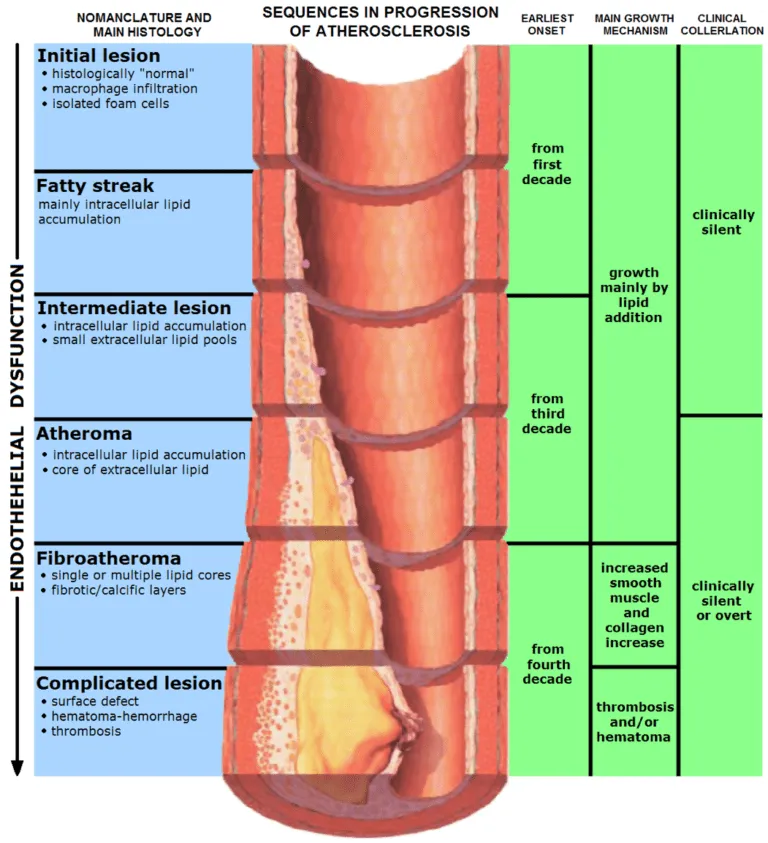
thickening and narrowing of arteries due to the build-up of lipoid plaques in the inner lining of an artery.
arteriosclerosis
blood vessels that carry 02 and nutrients from the heart to rest of body become thick and stiff
squamous epithelium
in the lining of blood vessels and air sacs enables the easy exchange of gases and other molecules between tissues.
surfactant
chemical compound reduce surface tension between diffrent substances such as 2 liquids either gas and a liquid or liquid and a solid
mucous membranes
lubricates and protects these organs and cavities
ciliated epithelium
found in the respiratory tract contain cilia move trapped particulate away from lungs
mucus
lubricates, traps, and expels foreign material such as bacteria, dust, and smoke
macrophages
immune cells that destroy germs, damaged cells and cancer cells. They promote tissue repair and healing
diaphragm
helps you breath sits under lungs separate chest cavity from abdomen
Rib cage
surround and protect vital organs in your chest cavity
intercostal muscles

intercostal muscles work together to expand and shrink the space inside your ribcage
dead space
in the airways no gas exchange takes place
COPD risk factors
tobacco smoke
asbestos
fungal spores
pollution
emphysema risk factors
cigarette smoke
asthma risk factors
cold weather
exercise
stress
pulmonary fibrosis risk factors
radiation treatment
fumes, vapor
chronic bronchitis
smoking
frequent dust /chemical fumes exposure
have asthma