Cytology of tumours
 Call Kai
 Learn
 Practice Test
 Spaced Repetition
 Match
 Flashcards
 Knowt Play
1/51

There's no tags or description
Looks like no tags are added yet.
52 Terms
When is cytology indicated?
Lesion (nodule, mass, plaque) palpable externally or seen on imaging
Organomegaly
Cavitary effusion
Cancer staging (lymph nodes, liver, spleen, BM)
Pyrexia of Unknown Origin
What can cytology tell us and not tell us?
Can tell us:
Inflam present
neoplasia present
If lesion is benign or malig
Type of neoplasia
Test of choice for bone marrrow exam
Cannot tell us:
If hyperplasia or adenoma
If reactive fibroplasia or sarcoma
Type of neoplasia in poorly differetiated tumours —> may need confirmation with histopath
What are the advantages & limitations of cytology?

How do you approach evaluation of a cytological slide?
Macroscopic —> labelling & smear quality
Microscopic —> 10x to assess quality & find sweet spot (thin region w/ intact cells & good cellular detail)
Then ID cell populations on 40-100x
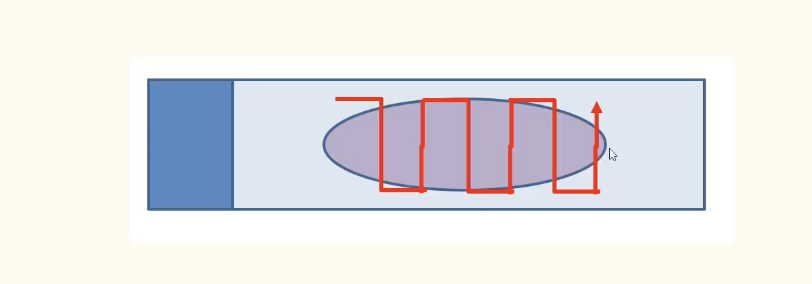
sweet spot = can see nuclei & cytoplasm of cells

List some of the common sample quality issues in cytology
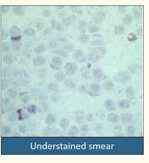
Too thick —> repeat staining & incr staining time
Insufficient staining time
Inadequate drying prior to staining —> should be air-dried promptly
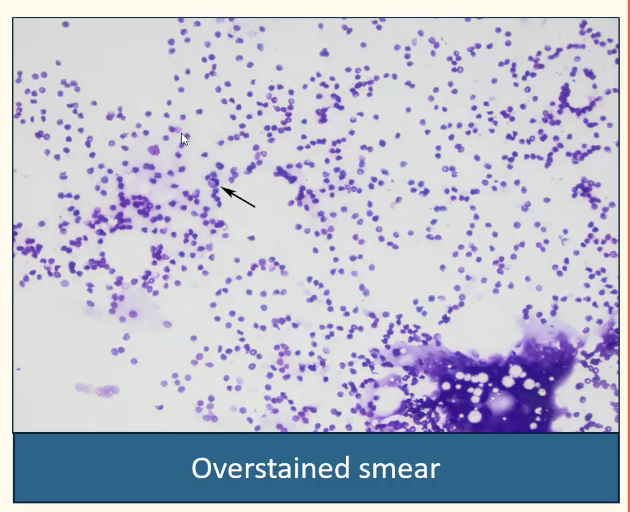
Nuclei no longer “pop out”
Staining time too high —> tailour staining time to sample cellularity (high cellularity, incr time)
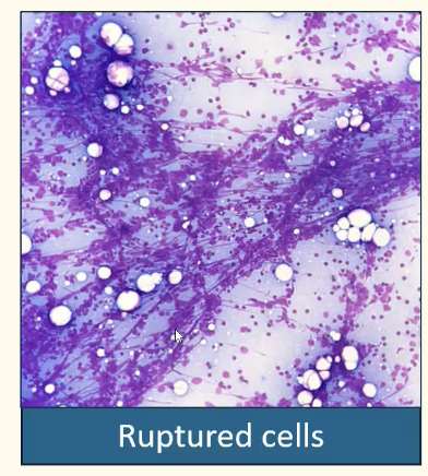
Incr aspiration pressure —> repeat aspiration w/ needle-only technique
Incr pressure during smearing —> repeat applying less pressure
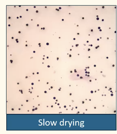
Smear dried slowly
Cells balled up & impossible to ID
should rapidly air dry or use hair drier
List the common cytology artefacts
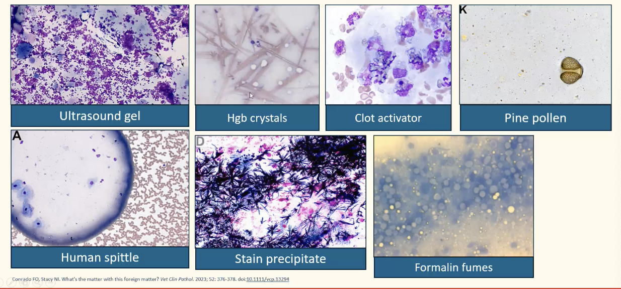
separate biopsy & cytology slides so no formalin artefact on cytology
Compare the general features of inflam to neoplasia on cytology
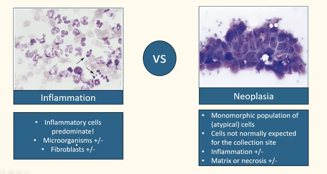
NB. Can often get both —> neoplasias can get inflamed
List some examples of neoplasias that often become inflamed
SCC
Mammary carcinoma
Fibrosarcoma
Mast cell tumour
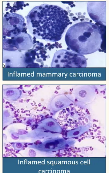
Is this inflammation or neoplasia?
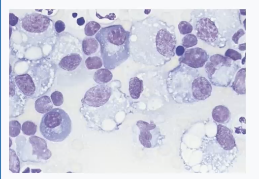
Mixed inflammation
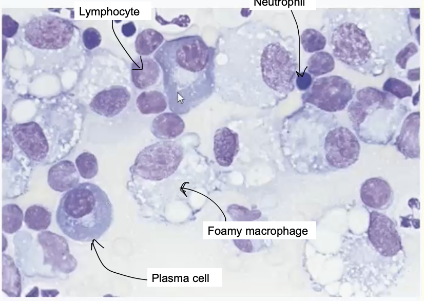
Is this inflammation or neoplasia?
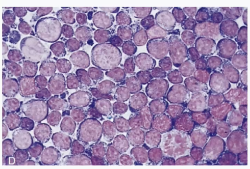
Neoplasia (from a lymphoma)
Monomorphic population of large cells —> can see mitosis
What are the categories of inflam & their poss causes?
Neutrophilic
Immune-mediated conditions, irritants (bile, urine), bacterial, protozoal or fungal infection
Histiocytic/ macrophagic
FB reactions, fungal infection, certain bacterial infections, such as Mycobacterium
Mixed
FB reactions, fungal infection, specific bacteria (e.g. nocardia, actinomyces)
Eosinophilic
Hypersens reactions, parasites, fungi, insect bites, cancer (e.g. mast cell tumours)
Lymphocytic/ lymphoplasmacytic
Antigenic stimulation, viral infections or chronic inflam
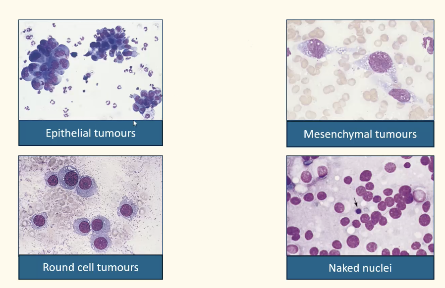
How can you categorise tumours?

What are the types of tumours based on cytomorphology?
How do you differentiate benign and malignant tumours on cytology?

Recall the criteria of malignancy
Pleomorphism
High or variable nuclear:cytoplasmic ratio
Anisokaryosis
Coarse chromatin
Nucleolar changes
Nuclear molding
Multinucleation
Abnormal mitotic figures
At least 3 to call it malignant (nuclear critera = more severe)
What is pleomorphism?
Variation in size, shape or maturation state of cells & nuclei
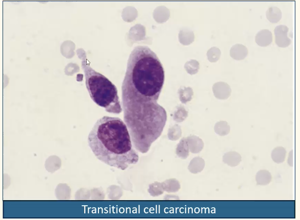
What is anisokaryosis?
Variation in nuclear size between cells of similar origin
High or variable nuclear:cytoplasmic ratio between cells of similar origin
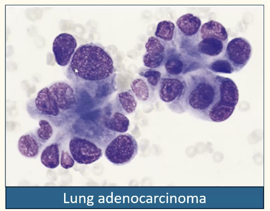
What is coarse chromatin?
Ropy chromatin or clumping of nuclear chromatin is common in immature cells
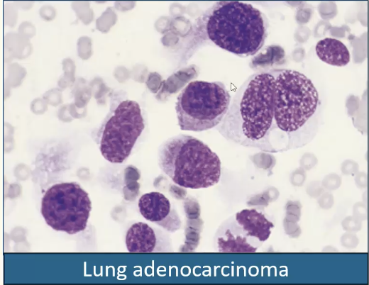
What criteria of malignancy is being shown here?
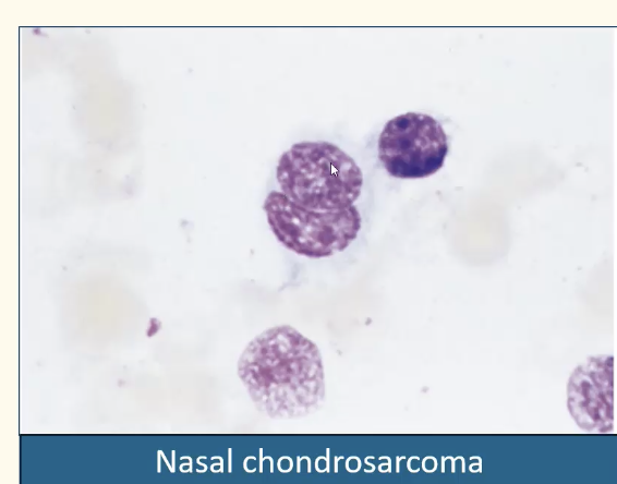
Nuclear moulding —> abnormal nuclear shape related to rapid growth of cells in which tight cell spacing occurs w/p normal crowd inhibition
What is being shown here?
Multinucleation
Two or more nuclei occupy same cell
NB. Binucleation may be found in hyperplasia of some tissues
What criteria of malignancy is being shown here?
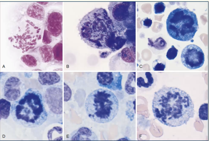
Abnormal mitotic figures
Abnormal chromosomal fragments may appear w/ uneven length & as isolated fragments (lag chromatin)
Incr numbers may be suggestive but not definitive for malignancy
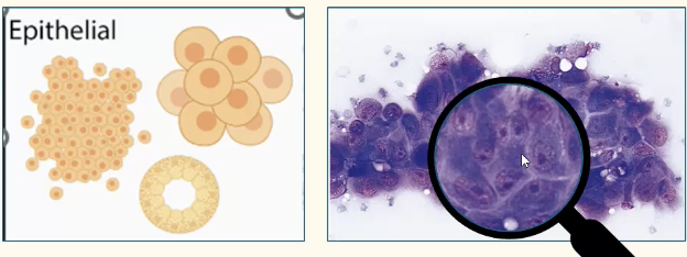
What are the features of an epithelial tumour?
Tight clumps or sheets —> tight junctions (desmosomes)
Large, round to polygonal cells with distinct cytoplasmic borders
Nuclei round to oval
Give some examples of epithelial tumours?
Transitional cell carcinoma
Lung carcinoma
Sebaceous adenoma
What are the features of a sebaceous adenoma:
Clinically
Cytologically?
Clinically —> raised cauliflower alopecic lesion
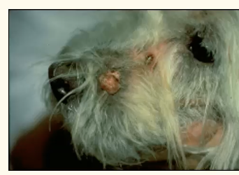
Cytologically —> cluster of sebocytes w/ foamy cytoplasm and small, dense, centrally placed nucleus
NB. cannot differentiate from hyperplasia on cytology
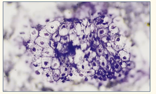
What are the features of perineal (hepatoid) gland tumours?
Ass. with intact male dogs (androgen dependency) but can affect all
Perianal region, also tail, perineum, prepuce, thigh, dorsal or ventral midline
Sheets of cell that look like hepatocytes
large oval cells
abundant, finely granular, pinkish-blue cytoplasm
round nucleus w/ single prominent nucleolus
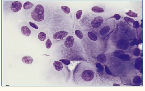
A few 'reserve' cells (Small basophilic cells w/ high N:C ratio
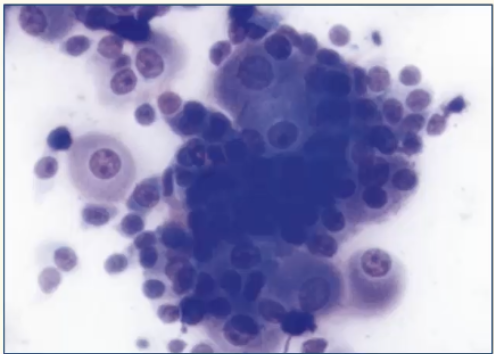
How do SCC present clincially?
Solitary / multiple; proliferative / ulcerative
Limbs —> dog
Pinnae / face —> cats
Locally invasive & may metastasise to regional LNs & digits (highly malignant)
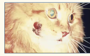
How do SCC present cytologically?
Large polygonal cells w/ variable tendency to cohesion
Typically marked pleomorphism
Perinuclear vacuolation
Often 2° neutrophilic inflm
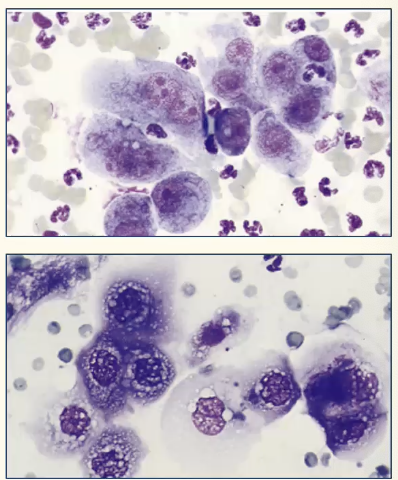
How does a mammary carcinoma present cytologically?
Background w/ blood, basophilic proteinaceous material, lipid and foam cells
Cells arranged in sheets, clusters or acini
Round to polygonal cells w/ round to oval nuclei & basophilic cytoplasm
Mod-marked pleomorphism
Poss incr mitotic activity
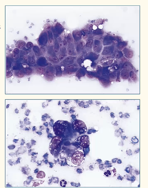
usually require histopath for definitive diag
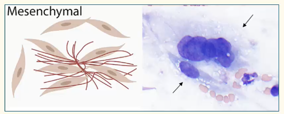
What are the features of mesenchymal tumours?
Cells usually exfoliate individually or in aggregates w/ matrix
Oval, stellate or fusiform often w/ indistinct cytoplasmic borders
Samples often poorly cellular
Nuclei oval to elongate
List some examples of mesenchymal tumours
Soft tissue sarcoma
= Fibrosarcoma, liposarcoma, leimyosarcoma, perivascular tumours, rhabdomyosarcoma etc.
Haemangiosarcoma
Fibroma
What is this showing?
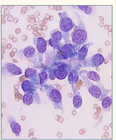
Soft tissue sarcoma
(difficult to distinguish from reactive fibroplasia —> need histopath for grading & ID of specific type)
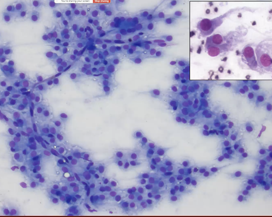
What tumours is being shown here?
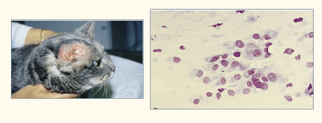
Fibrosarcoma
What mesenchymal tumour is being shown here?
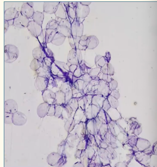
Lipoma (cannot distinguish from normal subcut adipose tissue —> alcohol fixatives may dissolve fat)
What are the features of osteosarcoma?
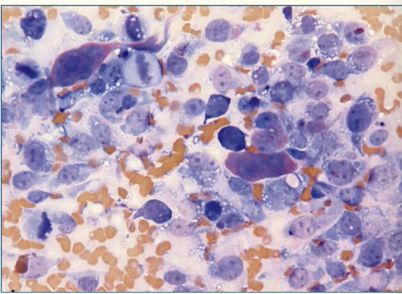
Round to spindle-shaped cells w/ basophilic cytoplasm & eccentrically placed nucleus w/ prominent nucleoli
Osteoclasts (large multinucleated cells) might be present
Pink extracellular matrix (osteoid) produced by cells may be present
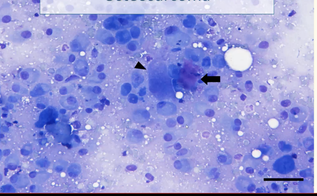
What are the features of round cell tumours?
Cells exfoliate individually
Generally round cells w/ distinct borders
nuceli round to indented
Samples moderately cellular
List some examples of round cell tumours
Transmissible venereal tumour
Mast cell tumour
Lymphoma
Plasmacytoma
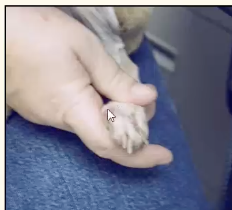
What are the clinical features of a histiocytoma?
Dome-shaped, alopecic, may be ulcertaed
Tends to regress in a few wks
Often (but not only) in young dogs
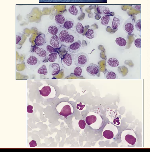
What are the cytological features of histiocytoma?
Oval discrete cells w/ round to oval nuclei & abundant clear to lightly basophilic cytoplasm
Variable numbers of small lymphocytes (common in regressing lesions —> sometimes can predom(^)
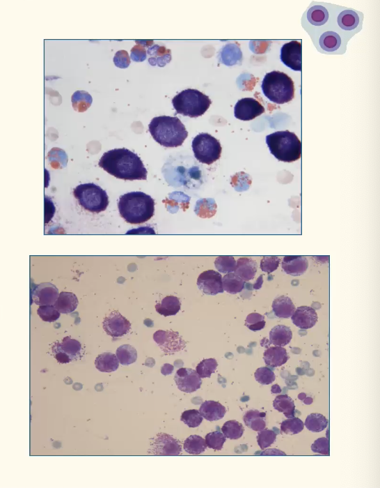
Describe the features of plasmacytomas
Cutaneous plasmacytoma usually benign
Can be well differentiated to pleomorphic
Deep blue cytoplasm, perinuclear clear area, occ. eosinophilic borders
Round, eccentric nucleus

Describe the features of lymphomas
Round cells w/ high nucleus:cytoplasm ration
Large lymphos (larger than neutros) predom
Monomorphic p

opulation
What breeds are predisp to mast cell tumours?
Boxer
Pug
Boston terrier
How do mast cell tumours appear appear cytologically?
Mast cells —> individually or in small groups
Tumour cells vary in degree of granularity and nuclear atypia —> poor granulation can be due to poor differentiation (more aggressive)
Eosinophils usually
How should you stain a mast cell tumour?
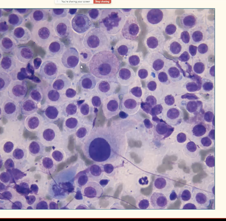
Not with Diff quick (will lose granules)
Toluidine Blue stain
Submit some unstained slides
What are the features of endocrine/ neuroendocrine tumours?
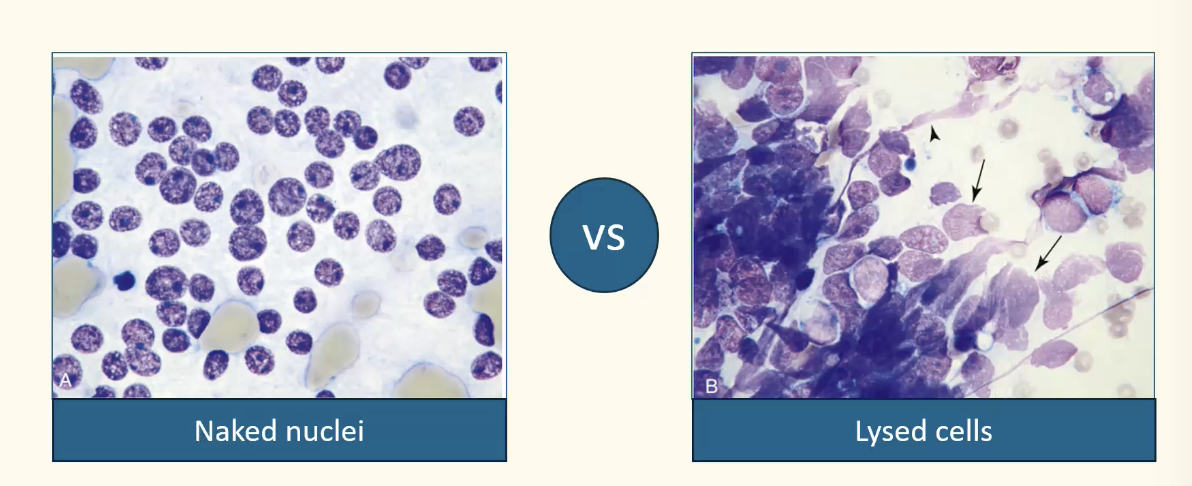
Naked nuclei in lakes of cytoplasm
many free nuclei present & indistinct cytoplasmic borders
Round to polygonal cells
Samples usually highly cellular
Nuclei = round to indented (none to minimal anisokaryosis)
Give some examples of endocrine/ neuroendocrine tumours
Thyroid carcinoma
Phaechromocytoma
What is the difference between these
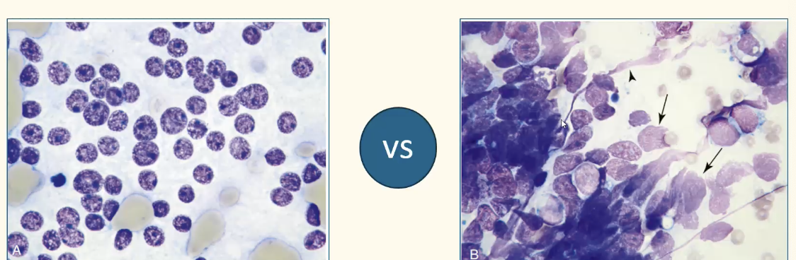
Don't confuse them
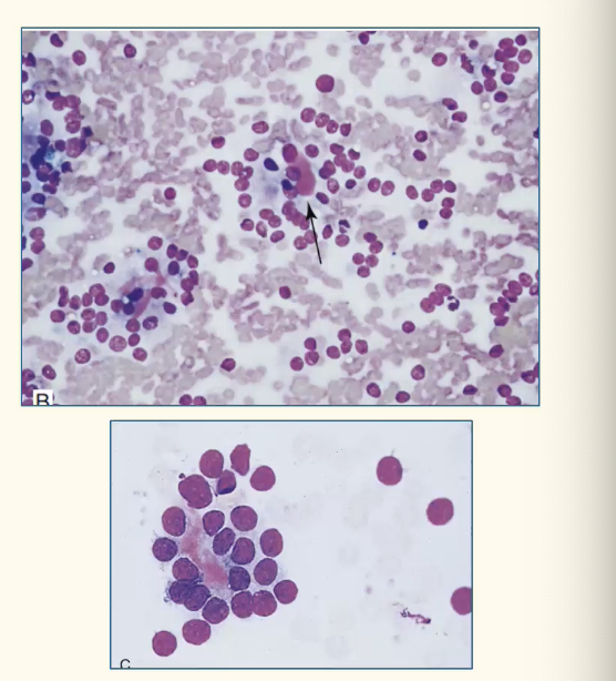
What are the features of a thyroid carcinoma?
Usually blood in background
Cells in clusters mixed w/ variable numbers of naked nuclei in background
Dark blue to black cytoplasmic pigment (tyrosine granules) sometimes intact cells
Extracellular colloid (pink material) may be seen (arrow)
Minimal atypia —> need histopath for degree of malig
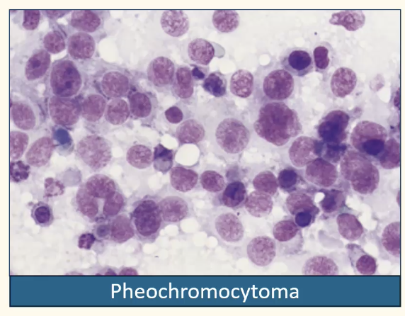
What are the features of pheochromocytoma?
Tumour of chromaffin cells of adrenal medulla
Clin symptoms due to catecholamine release from tumout
Naked nuclei against background of pale basophilic cytoplasma
Both benign & malig poss
Features of malig unreliable in predicting biological behaviour
Summary of tumour types

What neoplasia is being shown here?
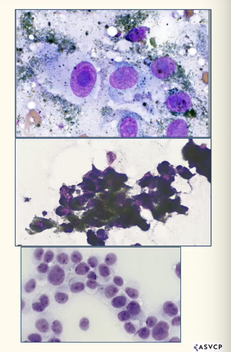
Melanoma
(Dark brown to black, circumscribed, raised, dome-shaped usually hairless mass)
Pleomorphic (epitheliod to fusiform to round)
Fine black-green granules masking nucleus, large single nucleolus
May have sparse granulation
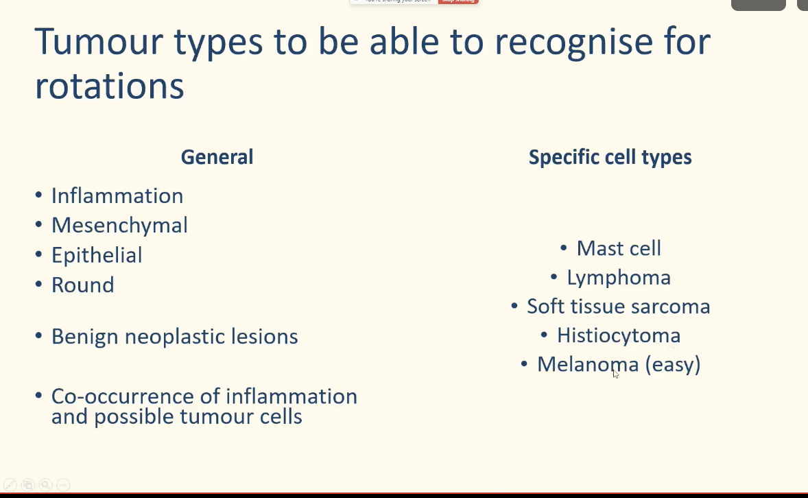
What are the general vs specific cell tumour types?
What is this type of neoplasia?
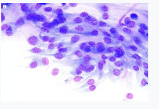
Mesenchymal