13. CAN. DR. HOLLAND. SPEAK. ANY FASTER?? - Congenital heart disease
 Call Kai
 Learn
Learn Practice Test
 Spaced Repetition
 Match
Match Flashcards
 Knowt Play
1/51

There's no tags or description
Looks like no tags are added yet.
Name | Mastery | Learn | Test | Matching | Spaced | Call with Kai | Chat |
|---|
No analytics yet
Send a link to your students to track their progress
52 Terms
congenital heart disease
_________: most common cause of cardiac disease in animals under 1 year
patent ductus arteriosus (PDA), pulmonic stenosis (PS), subvalvular aortic stenosis (SAS), ventricular septal defects (VSD), mitral and tricuspid dysplasia, peritoneopericardial diaphragmatic hernia, tetralogy of Fallot
List off the common congenital defects!!!!
echocardiography
Thoracic radiographs will evaluate the cardiac size and shape, but ___________ is needed for a definitive diagnosis
enlargement, dilation, hypertrophy
________, _________ and __________ of the heart help determine the significance of the lesion
doppler
_________: noninvasive determination of the severity of the lesion
patent ductus arteriosus
_______________: the ductus fails to close after birth causing abnormal communication between the aorta and pulmonary artery
both
Does shunting of blood between the aorta and the pulmonary arteries with a PDA happen in systole or diastole?
Left
Does a PDA enlarge the right or left heart?
ductus bump
segmental enlargement of the proximal descending aorta due to turbulent blood flow in the PDA is called a ________________
trachea mildly dorsally deviated, straightening of caudal cardiac waist, prominent right cranial lobar artery and vein
What are the abnormalities?

ductus bump
What is highlighted here as the KEY to a diagnosis of a PDA?

ductus bump, MPA, left auricle enlarged, enlarged artery and vein
This is a TEXTBOOK case. What are the abnormalities?
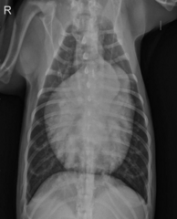
cool
AAAAAnd heres that same case with my crappy annotations.
(translation top to bottom: Ductus bump, MPA, Left auricle, enlarged artery and vein)
Cool?

ductus bump
Whats the abnormality at A?

double opacity, enlarged LA
Whats the abnormality at B?

pulmonic stenosis
________: obstruction of blood flow from the right ventricle to the main pulmonary artery
pulmonic valves
The most common form of pulmonic stenosis is at the level of the _________
coronary artery
Bulldogs may have an aberrant _____________ that encircles the pulmonary annulus causing a pulmonic stenosis
right heart wall thickens
So what changes in the heart will there be with pulmonic stenosis?
enlarged right ventricle/reverse D, prominent main pulmonary artery, decrease of at least one set of pulmonary arteries/veins, maybe enlarged CVC and hepatomegally
What are the radiographic signs of pulmonic stenosis
hat/cap sign,
What sign of pulmonic stenosis is shown at the white arrow here?

enlarged main pulmonary artery, reverse D, small caudal pulmonary vasculature
What abnormalities are here? (starting with the white arrow)

dorsal trachea deviation, enlarged CVC compared to aorta, decreased cranial pulmonary vasculature visualization, liver mildly enlarged (shifted gastric line)
What are the abnormalities?

no
Are we in failure?

left to right
Ventricular septal defects allow for what type of shunting?
there will be left heart disease but no ductus bump
What is the difference in ventricular septal defects radiographically when compared to PDAs?
true
true/false: in cases with small ventricular septal defects, the radiographs can look normal. Larger defects cause left heart disease signs
enlarged cardiac silhouette, malformed sternum (incidental), prominant liver
What are the abnormalities?

enlarged right caudal lobar artery and vein, rounding of the left ventricular margin
What are the abnormalities

dorsally deviate trachea, backpack sign, compressed left caudal mainstem bronchus, enlarged cranial lobar vessels, CVC is larger than aorta, deviated gastric axis, loss of abdominal detail (fluid)
what are the abnormalities?

cool
She said you;re not responsible for recognizing aortic stenosis on radiographs, but know its an outflow obstruction that leads to increased LV systolic pressure. AND that it predisposes animals for endocarditis and myocardial infarctions
Look at other stuff on your own if you want. Cool?
large breed dogs
Who most typically gets tricuspid dysplasia?
tricuspid/mitral dysplasia
_________: any combination of malformed valve leaflets or cusps, abnormal chordae tendinae, and abnormal papillary muscles
cardiac enlargement (severe), severe right atrial enlargement, right ventricular enlargement, enlarged CVC, hepatomegaly, failure
What are signs of tricuspid dysplassia radiographically?
doral deviated trachea, CVC larger than aorta
What tricuspid dyplasia signs are seen here?

no, no
Do we have hepatomegaly? Do we have heart failure?

greatly enlarged right atrium
This animal has tricuspid dysplasia. This dramatic reverse D sign is due mostly to the _________________

yes
Are we in heart failure?

holosystolic
Mitral dysplasia in GSDs, bull terriors and great danes is often acompanied by what type of murmur?
variable left atrial and ventricular enlargement, left heart failure, pulmonary venous enlargement, pulmonary edema
What are radiographic signs we can have from mitral dysplasia?
yes
Is this cat in heart failure?

dorsal deviation of trachea, elongated left ventricular border (tall heart)
This is that same case after aggressive treatment for pulmonary edema. What abnormalities are still present?

enlarged left atrium
The widened heart base on the VD image of a mitral stenosis is due to what abnormality?
mitral valve regurgitation
Mitral dysplasia looks similar to older dogs who have ____________________.
peritonealpericardial diaphragmatic hernia
_________________: most common diaphragmatic hernia in cats and dogs, resulting from an embryonic fusion defect in the ventral diaphragm leading to communication of the pericardium and peritoneal cavity
liver
The _________ is the most commonly herniated organ in a PPDH
trick question, most commonly incidental finding and the clinical signs are dependent on the organ herniated
What clinical signs do PPDH dogs commonly present with?
moderate to severe cardiac silhouette enlargement without any evidence of decompensation, dorsally displaced trachea and CVC, heterogenous opacity over cardiac silhouette, cranially displaced organs
What are radiographic signs of PPDH?
PPDH, (cant see the dophragm borders, and look at how far back the is because the liver is herniated into the pericardium)
Yo, whats happening here?

enlarged cardiac silhouette, indistinc diaphragm borders, loss of normal liver volume
Abnormalities?

mesothelial remnant sign
What is the abnormality in this PPDH cat?

cool
She said shes not making us ID tetrology of fallot either, so you can go look at it if you want. Cool?