Human Anatomy Exam 2.1
 Call Kai
 Learn
Learn Practice Test
 Spaced Repetition
 Match
Match Flashcards
 Knowt Play
1/347

There's no tags or description
Looks like no tags are added yet.
Name | Mastery | Learn | Test | Matching | Spaced | Call with Kai | Chat |
|---|
No analytics yet
Send a link to your students to track their progress
348 Terms
# of bones in body
206
# of bones in Axial skeleton
80
# of bones in Appendicular skeleton
126
Spongy bone characteristics
- Internal layer
- Protects Medullary Cavity
- Yellow & Red bone marrow
- Trabeculae
Spongy bone arrangement and reason
- Parallel thick branching plates/bars called trabeculae
- Reduce weight of skeleton, easier bone movement
Compact bone characteristics
- External wall
- Osteon functional unit
Large bone: Epiphysis
Superior and Inferior sections (2)
Large bone: Metaphysis
Small sections between Epiphysis and Diaphysis (2)
Large bone: Diaphysis
Middle section of the bone, the shaft (1)
Large bone: Head
- Round
- Separated from shaft by neck
- Forms part of joint
Bone functions
- Support
- Mineral storage
- Blood cell production
- Protection
- Movement
- Production of osteocalcin (fat storage & insulin production)
4 bone specialized cells
- Osteoprogenitor
- Osteoclast
- Osteoblast
- Osteocyte
Osteoprogenitor cell
- Stem cell
- Divides to produce osteoblasts
Osteoclast cell
- Multi-nucleate cell
- Secretes acids and enzymes to dissolve bone matrix
Osteoblast cell
- Immature bone cell
- Secretes osteoid (organic bone matrix)
Osteocyte cell
- Mature bone cell
- Maintains bone matrix
Osteogenesis & cells
Bone formation
- Osteocyte
- Osteoblast
Osteolysis & cells
Bone destroying
- Osteoprogenitor
- Osteoclast
Bone blood supply: Nutrient artery & vein
Through nutrient foramen in diaphysis
Bone blood supply: Epiphyseal vessels
- Through foramina of epiphysis
- To medullary cavities of epiphysis
Bone blood supply: Metaphyseal vessels
- Through artery & vein of metaphysis
- To diaphyseal surface
Bone blood supply: Periosteal vessels
- Through periosteum
- To superficial osteons (compact bone)
Periosteum
Thin membrane of dense irregular connective tissue that covers bone outer surface
Periosteum functions
- Isolate and protect bone
- Provide route and place of attachment for insertion of ligaments and tendons
- Bone growth and repair
Endosteum
Thin membrane of connective tissue that lines bone inner surface
Endosteum functions
Bone growth and repair
7 categories of bone shape
- Sutural
- Pneumatized
- Short
- Irregular
- Flat
- Long
- Sesamoid
Sutural bone characteristics
- Small
- Flat
- Oddly shaped
- Along sutures
Sutural bone example
Inca bone (suture where parietal & occipital bones meet)
Pneumatized bone characteristics
- Hollow
- Air pockets
Pneumatized bone example
Ethmoid bone
Short bone characteristics
- Boxlike
- Exterior compact & interior spongy
Short bone example
Carpal bones
Irregular bone characteristics
- Complex shape
- Short, flat, notched, or ridged surfaces
Irregular bone example
Vertebrae of spinal column
Flat bone characteristics
- Thin, roughly parallel surfaces of compact bone
- Spongy bone sandwich
Flat bone example
Ribs
Long bone characteristics
- Long
- Slender
- 2 epiphysis, 2 metaphysis, diaphysis, marrow cavity
Long bone example
Humerus
Sesamoid bone characteristics
- Small
- Round
- Flat
Sesamoid bone example
Patella
Depressions and openings
- Foramen
- Groove
- Notch
- Fissure
- Fossa
- Meatus
- Sinus
Bone marking: Foramen
Round or oval opening through a bone
Bone marking: Groove
Furrow
Bone marking: Fissure
Narrow, slit-like opening
Bone marking: Notch
Indentation at edge of structure
Bone marking: Fossa
Shallow basin-like depression, often serving as articular surface
Bone marking: Meatus
Canal-like passageway
Bone marking: Sinus
Cavity in a bone, filled with air and mucus
Surfaces that form joints
- Head
- Facet
- Condyle
Bone marking: Head
Bony expansion carried on narrow neck
Bone marking: Facet
Smooth, nearly flat articular surface
Bone marking: Condyle
Rounded articular projection
Projections that are sites of muscle or ligament attachment
- Tuberosity
- Crest
- Trochanter
- Line
- Tubercle
- Epicondyle
- Spine
Bone marking: Tuberosity
Large rounded projection, may be roughened
Bone marking: Crest
Narrow ridge of bone, usually prominent
Bone marking: Trochanter
Very large, blunt, irregularly shaped process (only example is femur)
Bone marking: Line
Narrow ridge of bone, less prominent than crest
Bone marking: Tubercle
Small rounded projection or process
Bone marking: Epicondyle
Raised area on or above a condyle
Bone marking: Spine
Sharp, slender, often pointed projection
Elevations and projections
- Process
- Ramus
Bone marking: Process
Any projection or bump
Bone marking: Ramus
Extension of a bone that forms an angle with the rest of the structure
Bone fracture
Break or crack in a bone
Fracture: Comminuted
- Bone fragments in multiple pieces
- Common in aged
Fracture: Compression
- Bone is crushed
- Common in porous bones subjected to trauma (fall)
Fracture: Spiral
- Ragged break with excessive twisting forces
- Common in sports
Fracture: Epiphyseal
- Epiphysis separates from diaphysis along epiphyseal plate
- Common where cartilage cells are dying and calcification of matrix is occurring
Fracture: Depressed
- Broken portion is pressed forward
- Common in skull
Fracture: Greenstick
- Bone breaks incompletely, one side of shaft breaks & other bends
- Common in children
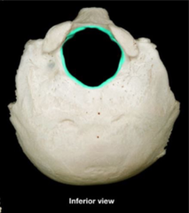
Foramen magnum
Occipital bone
- Large hole (inferior view)
Occipital condyles
Occipital bone
- Bumps next to foramen magnum
- Make joints with 1st vertebrae of column

Hypoglossal canal
Occipital bone
- Small holes next to occipital condyles
- Room for blood vessels & nerves

Nuchal lines
Occipital bone
- Attachment of nuchal ligament that maintains straight forward position
- 2 lines: Superior & Inferior

External occipital protuberance
Occipital bone
- Small ridge, attachment for ligaments or muscle

External occipital crest
Occipital bone
- Small ridge inferior of protuberance

Superior border
Parietal bone
- Border of sagittal suture

Parietal eminence
Parietal bone
- Lump in the middle
Parietal bone lines
- Superior & Inferior
- Horizontal
- Indicate attachment of temporalis muscle

Inferior border
Parietal bone
- Border of squamous suture
- Connects to neck

Frontal bone shape
Shell
Frontal bone: Squamous part
Anterior surface (forehead)
Metopic suture
Frontal bone
- Indicates frontal bone used to be 2 parts, then bind into 1
- Only in some skulls

Supra-orbital margin & notch
Frontal bone
- Where eyes are

Supra-orbital foramen
Frontal bone
- Move eyes via cranial nerves

Superciliary arch
Frontal bone
- Where eyebrows are

Lacrimal fossa
Frontal bone
- Holds lacrimal gland that makes us cry

Frontal air cells
Ethmoid bone
- Lots of little holes connected to sinuses

Orbital part
Frontal bone
- Forms roof of each orbit
- Protects eyes

Superior margin
Frontal bone
- Posterior section
- Margin of coronal suture

Frontal crest
Frontal bone
- Posterior section
- Attach membranes that protect brain

Frontal bone notch
- Posterior section
- Notch for ethmoid

Temporal bone articulations
- Zygomatic
- Parietal
- Occipital
- Sphenoid
- Mandible
Temporal bone 3 parts
- Squamous
- Tympanic
- Petrous
Temporal bone squamous shape
Scale-like
Temporal bone squamous location
- Right superior
- Above zygomatic process
Zygomatic process
Temporal bone
- Ends squamous part
- Long stem that sticks out
- Cheek arch

Temporal bone tympanic part
- Internal ear organs
- Right below external acoustic meatus (middle inferior)
Mastoid process
Temporal bone
- Attachment site for muscles that rotate head
