Long bone/ Nerves/ Arteries/ Epiphyseal Plate
 Call Kai
 Learn
Learn Practice Test
 Spaced Repetition
 Match
Match Flashcards
 Knowt Play
1/32

Earn XP
Description and Tags
Name | Mastery | Learn | Test | Matching | Spaced | Call with Kai | Chat |
|---|
No analytics yet
Send a link to your students to track their progress
33 Terms
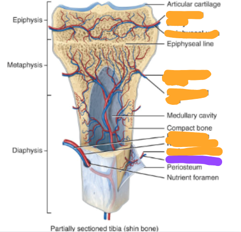
Diaphysis
bone shaft

2 Epipyses
both ends of the bone at the joints
proximal and distal

2 Metaphyses
region between diaphysis and epiphysis

Articular cartilage covering both epiphyses

Periosteum
connective tissue surrounding the diaphysis

Medullary cavity
Hollow space within diaphysis
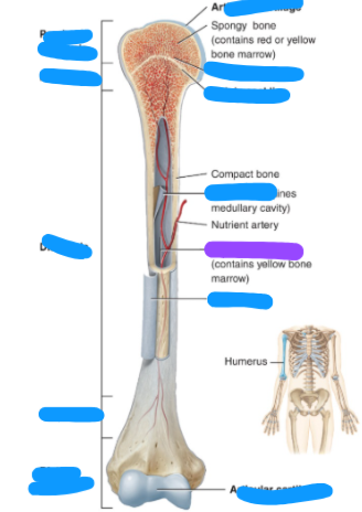
Endosteum
thin membrane lining the medullary cavity

Epiphyseal line

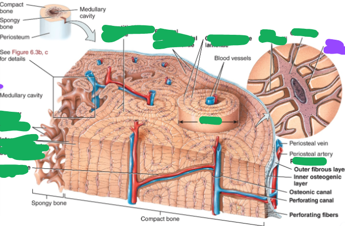
compact bone and spongy bone

spongy bone trabeculae

internal circumferential bone lamellae
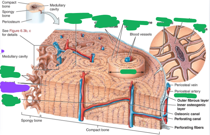
transverse canal

interstitial bone lamellae

external circumferential bone lamelae

concentric bone lamellae

osteon

periosteum

Bone canaliculi
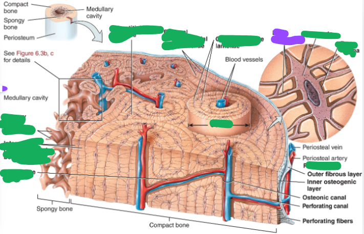
osteocyte
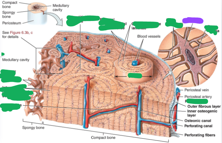
Bone lacuna
epipyseal artery

epiphyseal vein

metaphyseal artery

metaphyseal vein

nutrient vein

nutrient artery

perosteal artery

periosteal vein
resting cartilage zone
Area of cartilage on epiphyseal side of epiphyseal plate
inactive

proliferating cartilage zone
Area of cartilage on diaphysis side of epiphyseal plate that is rapidly dividing
New cells formed move upward, causing lengthening

hypertrophic cartilage zone
Area with older chondrocytes closer to diaphysis
Cartilage lacunae enlarge and erode

calcification cartilage zone
Surrounding cartilage matrix calcifies
chondrocytesharden and die

ossification zone
Osteoclasts erode calcified cartilage spicules, and osteoblasts build new bone over the remaining spicules
bone forms