lecture 9 blood & circulatory system review
 Call Kai
 Learn
Learn Practice Test
 Spaced Repetition
 Match
Match Flashcards
 Knowt Play
1/114

There's no tags or description
Looks like no tags are added yet.
Name | Mastery | Learn | Test | Matching | Spaced | Call with Kai | Chat |
|---|
No analytics yet
Send a link to your students to track their progress
115 Terms
blood
Fluid connective tissue (cells in ECM)
Cells:
Erythrocytes
Leukocytes
Thrombocytes
Extracellular matrix:
Plasma (watery substance)
Dissolved proteins (ex/ antibodies, hormones)
blood separation and composition
Red blood cells ~44% of blood volume
Buffy coat ~1%
Leukocytes and platelets
Plasma ~54%
transportation
Respiratory gases
Nutrients and wastes
Hormones
function of blood
regulation
Body fluid distribution (leak plasma)
Body temperature
function of blood
protection
Immune cells
Platelets
Inflammatory response
function of blood
plasma
Composition
~ 92% water
Ions (Na+ and Cl-) (Ca+)
Nutrients (Sugars, amino acids, etc.)
Proteins (ex/ antibodies)
Plasma leaks out of capillaries to form interstitial fluid
Plasma proteins stay in blood vessels
erythrocytes (RBCs)
No nuclei → biconcave disk
30% more surface area (flat in middle)
No organelles
No mitochondria – do not consume O2
Life span ~120 days
97% composed of hemoglobin
Transport O2 and CO2
New cells made in the red marrow (spongy bone)
Life span ~120 days
Old cells removed in liver or spleen
hemoglobin
280 million molecules of hemoglobin per RBC
1 molecule holds up to 4 O2 molecules and 4 CO2 molecules
O2 is bound by iron atoms in molecule
Oxygenated blood is bright red
Deoxygenated blood is dark/deep red
blood typing
describes antigens on the surface of erythrocytes.
Two factors determine blood type:
ABO blood group
Rh factor
Blood type determines what antibodies the immune system will produce:
Anti-A, Anti-B, and Anti-Rh
Important for blood transfusions, organ transplants, health risks, etc.
ABO blood group
presence or absence of A and B antigens
Rh factor
presence or absence of Rh surface antigen (aka: D antigen, have +)
A+ blood type
antigens: A, D
antibodies: anti-B
A- blood type
antigens: A
antibodies: anti-B, anti-D
B+ blood type
antigens: B, D
antibodies: anti-A
B- blood type
antigens: B
antibodies: anti-A, anti-D
AB+ blood type
antigens: A, B, D
antibodies: none
universal acceptor
AB- blood type
antigens: A, B
antibodies: anti-D
O+ blood type
antigens: D
antibodies: anti-A, anti-B
O- blood type
antigens: none
antibodies: anti-A, anti-B, anti-D
universal donor
anemia
low erythrocyte level or hemoglobin concentration
Erythrocyte Volume Disorders
sickle cell disease
erythrocytes distort into a sickle shape
Hereditary – mutation in DNA results in defective hemoglobin molecule
Insufficient O2 and CO2 binding
Erythrocyte Volume Disorders
polycythemia
abnormal excess of erythrocytes
Thicker, more viscous blood
Tremendous strain on the heart
Erythrocyte Volume Disorders
leukocytes
Part of the immune system - defend against pathogens
Originate in red bone marrow
Mainly function outside the bloodstream in loose CT
Diapedesis: circulating leukocytes leave the capillaries
granulocytes
have granules filled with chemicals
Neutrophils, Eosinophils, Basophils
leukocytes
agranulocytes
no granules
Lymphocytes, Monocytes
leukocytes
leukocytes cells to most to least
Neutrophils (60%)
Lymphocytes (30%)
Monocytes (6%)
Eosinophils (3%)
Basophils (1%)
Never Let Monkeys Eat Bananas
neutrophils
Phagocytize and destroy pathogens (harmful cells)
Granules of various digestive enzymes
First responder to infection
granulocytes
eosinophils
Fights parasitic worm infections
Granules of parasite-killing proteins
Also involved in allergies
granulocytes
basophils
Granules of histamine and heparin
Secrete during inflammatory response and allergic reactions
granulocytes
lymphocytes
Involved in adaptive immune response --> fight specific foreign molecules (antigens)
T-Lymphocytes
B-Lymphocyte
Natural Killer Cells: Attack abnormal and infected cells
agranulocytes
T-lymphocytes
Attack intracellular pathogens (viruses and cancer cells)
Attack cells directly
B-lymphocyte
Attack extracellular pathogens (bacteria, fungi, etc.)
Produce antibodies
monocytes
Leave circulation to enter tissues and differentiate into macrophages (not in blood, in tissues)
Macrophages perform phagocytosis of pathogens, cellular debris, etc.
agranulocytes
leukemia
cancer in leukocyte-forming cells
Abnormal development and proliferation of leukocytes
Poorly differentiated cells – don’t function properly
Interfere with maturation of normal blood cells
Poor resistance to infection
platelets
Platelets are cell fragments that break off from megakaryocytes
Function in blood clotting
hemopoiesis
Occurs in the red marrow
Red marrow transplant for certain blood disorders
lumen
Central blood-filled space of vessel
endothelium
simple squamous ET
Smooth surface to prevent RBC rupture
tunica intima
subendothelium
areolar CT
Nourish endothelium
tunica intima
tunica media
Circular sheets of smooth muscle
Vasoconstriction (contract)
Vasodilation (relax)
Elastic fibers help with recoil
tunica adventitia/externa
Dense irregular CT that loosens
Anchor neighboring tissues
Vaso vasorum in larger vessels to nourish tissue
arteries
thicker walls
higher blood pressure
tunica media thickest layer
veins
thinner walls
lower blood pressure
tunica adventitia thickest
valves present
skeletal muscle pumps propel blood towards heart
elastic arteries
Largest arteries. Located closest to the heart.
Ex Aorta, pulmonary trunk, brachiocephalic trunk.
Unique feature: Abundant elastin fibers throughout tunics (prevent stretch) for stretch and recoil to pump blood (systole)
muscular arteries
Distal to elastic arteries (middle size)
Regulate diameter to control blood supply to organs
Unique feature: Internal and external elastic laminae separate tunics – provide stretch and recoil
arterioles
Smallest arteries
Can close off diameter completely to shunt blood away from capillary bed
Metarterioles just upstream of capillary bed
Thoroughfare channel in the center of capillary bed
capillaries
Smallest blood vessels
Lumen is ~ diameter of RBCs
Endothelium (and small subendothelium) only
Leak plasma to supply interstitial fluid
Gas exchange (O2 and CO2)
Nutrient/waste exchange
continuous capillaries
Intercellular clefts for diapedesis and to leak plasma
in most tissues
fenestrated capillaries
Have small windows for extra plasma leakage
in kidney
in synovial membrane
sinusoid capillaries
Have large holes where RBC’s can enter/exit
red marrow (new RBC can enter)
liver/spleen (old RBC can leave)
venules
smallest veins (microscopic)
Venules merge to form veins
valves
prevent backflow of blood
Cusps forms from tunica intima
veins
varicose veins
occurs when valves prolapse
lots of standing
old age (loss of elastic fibers)
deep vein thrombosis
a blood clot forms in a deep vein, usually in the leg or pelvis.
Clot can break loose, travel to the lungs and block blood flow (pulmonary embolism)
arteriosclerosis
hardening of arteries
atherosclerosis
fatty deposits (plaques) build up in arterial walls.
Narrows arteries over time
Can damage lining of blood vessel (rupture RBC, blood clot from tear)
hypertensive arteriosclerosis
hardening and thickening of arteries and arterioles caused by chronic high blood pressure
aneurysm
“bulge” or “ballooning” in the wall of an artery due to weak areas in the blood vessel wall
Most commonly seen in the brain and aorta
Ruptured aneurysm is a medical emergency
Arteriovenous Malformation
abnormal, tangled network of blood vessels where arteries and veins connect without a bed of capillaries
Can lead to tissue damage, hemorrhagic stroke, or rupture
aorta

brachiocephalic trunk (right side only)
splits into Common carotid a. and Subclavian a

subclavian artery (to arm)

common carotid artery (brain & face)

external carotid artery (to face)

internal carotid artery (to brain)

subclavian artery (under clavian)

axillary artery (armpit)

brachial artery (arm)

ulnar artery (pinky)

radial artery (thumb)

thoracic aorta

abdominal aorta artery (at the diaphragm)

celiac trunk
(splits into branches that go to the stomach, spleen, and liver)

superior mesenteric artery (to various intestines)
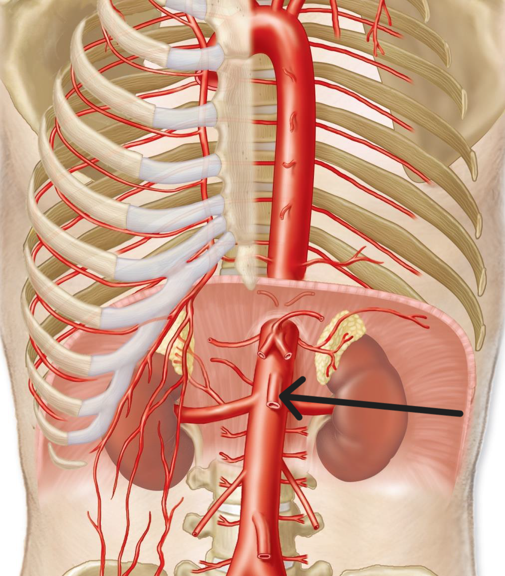
renal artery (2) (to kidneys)
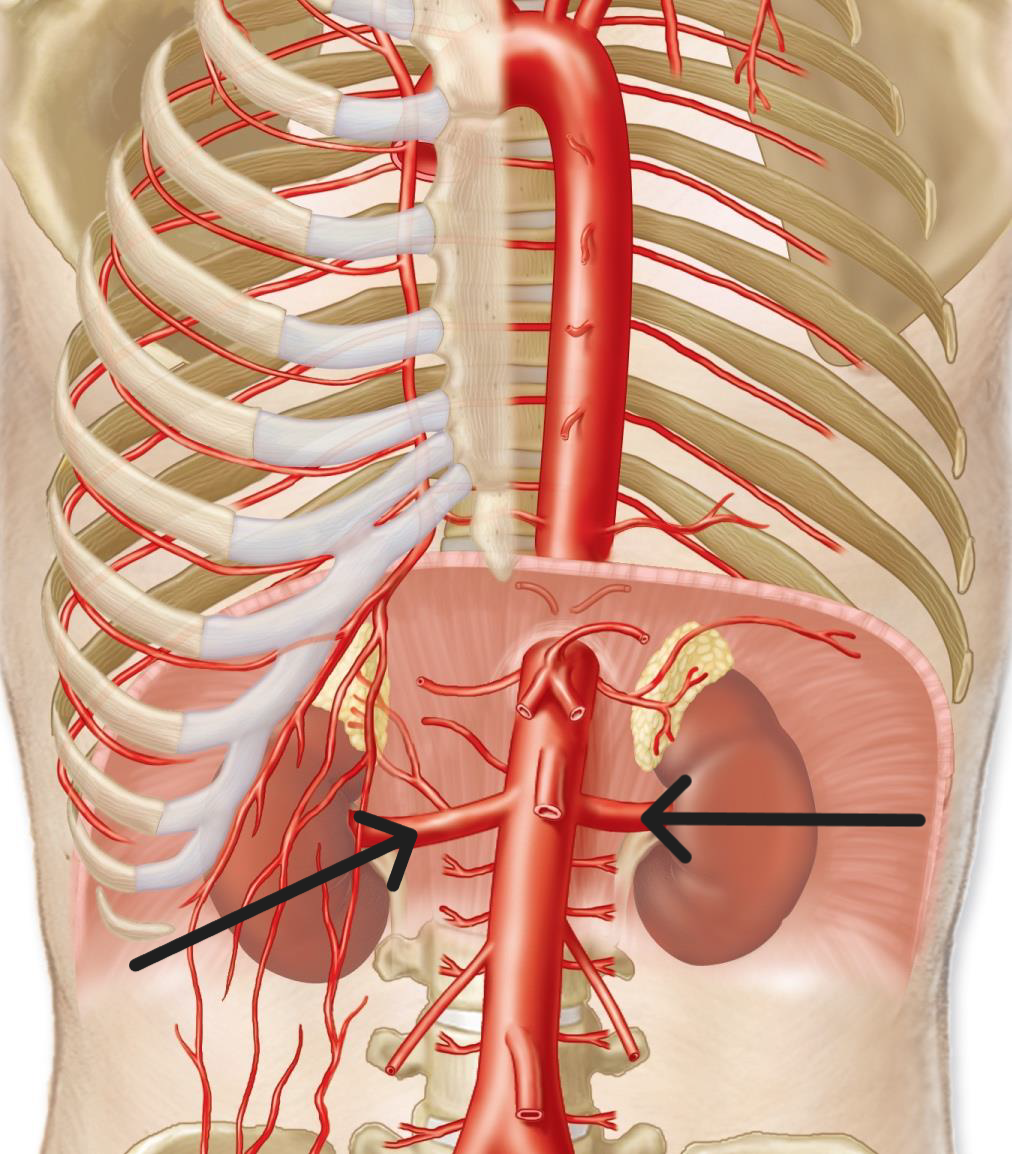
gonadal artery (2) (to gonads)

inferior mesenteric artery (to intestines)

abdominal aorta artery (until pelvic area)
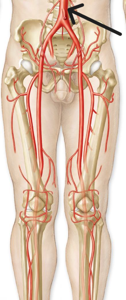
common iliac artery

external iliac artery (leg)

internal iliac artery (to pelvic organs)

femoral artery (to thigh)

anterior tibial artery

superior vena cava vein (brachiocephalic veins merge)

brachiocephalic veins (2)
internal jugular vein and subclavian vein merge
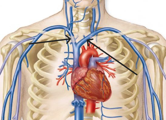
internal jugular vein (from brain)
drains into brachiocephalic vein

external jugular vein (from face)
drains into subclavian vein

brachiocephalic vein

subclavian vein

axillary vein

brachial vein
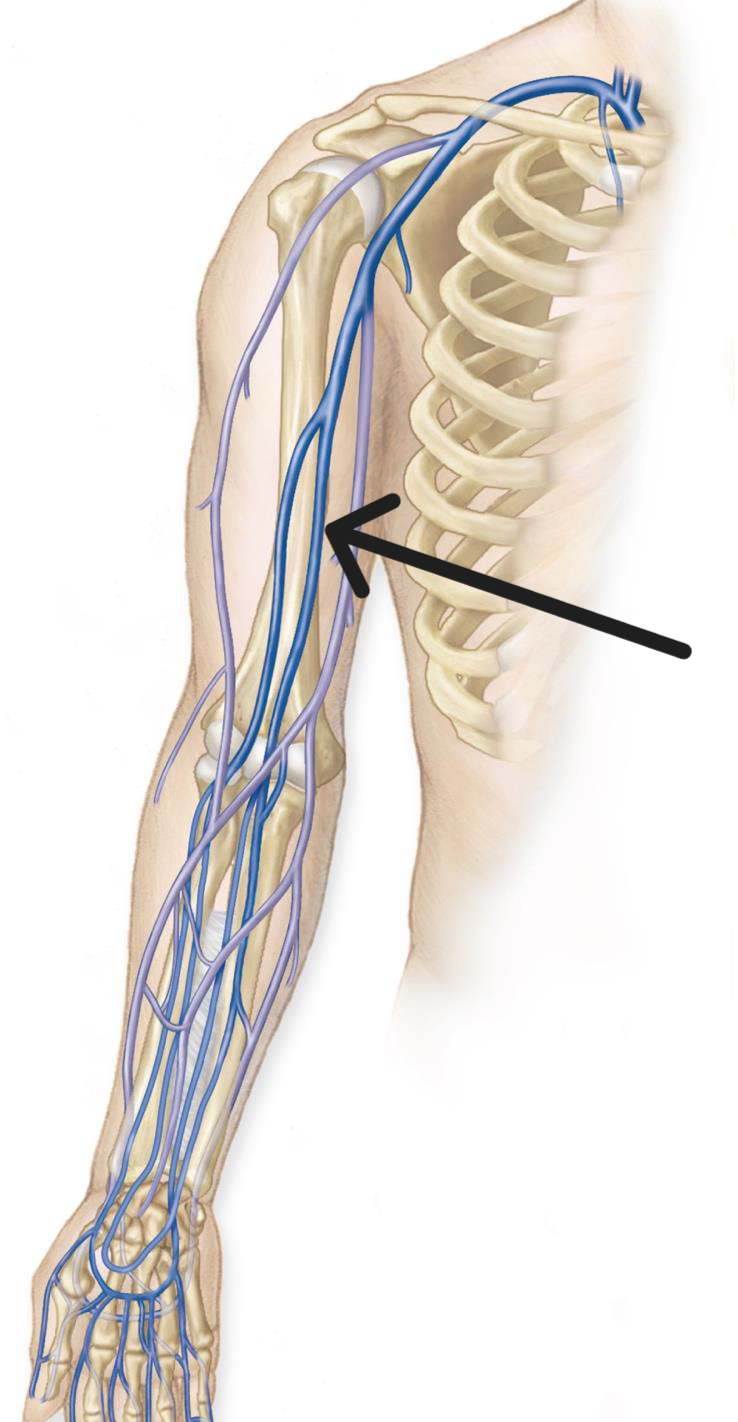
ulnar vein

radial vein

renal vein

gonadal vein

inferior vena cava (common iliac veins merge)

common iliac vein

external iliac vein

internal iliac vein

femoral vein
