Midterm
 Call Kai
 Learn
Learn Practice Test
 Spaced Repetition
 Match
Match Flashcards
 Knowt Play
1/176

There's no tags or description
Looks like no tags are added yet.
Name | Mastery | Learn | Test | Matching | Spaced | Call with Kai | Chat |
|---|
No analytics yet
Send a link to your students to track their progress
177 Terms
Quality Assurance
implementing strategies or procedures to ensure the reliability, accuracy, and consistent of lab results
System put in place to prevent errors or contamination
Proactive, preventative system focused on establishing processes and procedures like SOPs and training
Quality Assurance means
building quality in
preventative
Quality control
reactive
reactive processes that use routine checks to identify, measure, and correct deficiencies and confirm that the quality system is working effectively
day to day procedures to make sure QA is being maintained
Quality control means
checking quality out
reactive
What factors determine the correctness and reliability of tests? - QA
Human Factors
Organization Structure
Physical facilities and environmental conditions
test methods and method validation
equipment
handling of test items and standards
measurement standards
technical records and reporting
SOPS
Human Factors - QA Factor
Staff needs to be suitably qualified and experienced for the activities they are required to perform. Includes
appropriate training and competency tests
regular monitoring of staff performance (periodic proficiency testing or evaluation of QC data produced by them)
records kept that detail and explain everything
Organization Structure - QA Factor
People need to be trained to have a background for their job and there must be a hierarchy of job roles
should have supervisors with analysts under them
everyone is laid out having different jobs and letting everyone know who is who, so they know who to report to
Physical Facilities and Environmental Conditions - QA factor
lab and office accommodations must be suitable in construction and size to perform the range of activities undertaken
Environmental conditions must be appropriate (temperature, humidity, lighting, dust free, clean and sterile lab, etc.)
space should allow for separation of noncompatible activities
ex. DNA extraction separated from DNA amplification, or firearms testing separated from GSR analysis area
DNA labs mostly have one direction of movement to ensure excess contamination does not occur
space must be secured with controlled access
Test Methods and Method Validation - QA factor
It must be possible to demonstrate, through validation or verification records, that all analytical methods used are fit-for-purpose
precision and accuracy of test methods
LOD and LOQ
specificity, linearity, range
repeatability and reproducibility
life cucye of method validation
Precision vs Accuracy of test results
precision - all runs give same results
accurate - results are correct
repeatability vs reproducibility
repeatability - if I do the procedure 10 times it will be the same all 10 times
reproducibility - if multiple people do the procedure, the results are the same
Equipment - QA factor
all equipment and instrumentation needs to meet specifications consistent with intended purpose, be suitably maintained to ensure satisfactory performance, and be calibrated against recognized standards where relevant
maintenance
safety
calibration checks
records maintained and monitored
handling of test items and standards - QA factor
COC and retention of retesting - must be operation procedures in place to determine how long we are keeping evidence
lab must have and apply procedures to protect samples from loss, contamination and degradation
Measurement Standards - QA factor
chemical or physical standards used for calibration or validation purposes
documented use and storage of these standards to preserve integrity and should be certified by an external body
Technical Records and Reporting - QA factor
Should include everything that has to do with evidence
may include
handwritten notes
worksheets
instrument print outs
photographs or digital images and administrative paperwork
SOPs - QA Factor
Must be detailed, step-by-step instructions for routine tasks to ensure consistency and accuracy
Should be
detailed and specific
consistency
comprehensive
accessible
compliance
Internal QC Procedures
Analysis of reagents or subtract blanks - make sure reagent itself isn’t giving wrong detection
analysis of previously analyzed samples
replicate analysis of samples
analysis of interlaboratory “blind” samples
5% of samples analyzed should be a QC sample - control or blanks
External QC
interlaboratory collaborative trials or samples exchanged to see if everyone gets the same results
Proficiency tests
“blind” test submitted to an analyst for processing and the analyst does not know the expected outcome
serves as QC for individual and the lab for validity of test methods, equiptments, reagents, etc
Horizontal Internal Audits
cover a single aspect of a lab’s quality system to check on compliance against documented procedures
vertical internal audits
follow the progress of a case from receipt through examination, sample analysis, interpretation of results, reporting and case finalization
external audits
audit undertaken by an accreditation agency or by representatives from another lab
ISO 17025
international std for competence of testing and calibration labs setting general requirements for quality management system
ISO 17020
international std designed to ensure that "inspection bodies" carry out competent, independent, and impartial inspections to provide their clients with info in terms of conformity with regulations, stds, or other specifications
What could happen without a robust QA/QC system?
evidence may be contaminated
reports may be inaccurate
findings may lack credibility
Direct Evidence
evidence that if accepted would prove a fact without any need of an inference to be drawn from it
Circumstantial or Presumptive Evidence
suggests a fact by implication or inference, requires reasoning to connect it to a conclusion
Evidence must be…
relevant, probative, and admissible
Physical Evidence
any tangible object that can connect a suspect to a CS
Ex) Dirt, gun, bullets, fingerprint, fibers, sand, soil, tool marks, ammunition, firearms, glass, footprint, documents
Biological Evidence
comes from living organisms and contains DNA or other organic material
Ex) blood, semen, saliva, urine, sweat, skin cells, hair, organs, bone
Biological Evidence Processing Steps
Identification - determining type of body fluid and if from human or animal
individualization - linking the biological evidence to a specific individual
reconstruction - using biological evidence to understand the sequence of events at a CS
Control Samples
samples, swabs or cuttings from material alone the biological evidence is on
used to confirm the results of the test performed
reference samples
Samples collected from all individuals who may be linked to the CS to act as eliminatory or comparative evidence
What must biological evidence packing be labeled with?
evidence description
evidence source - the origin, where it was, when collected
COC
unique identifier number
can indicate what processing is needed, health hazards, required or preferred storage conditions
When should biological evidence be wet or dry?
Blood-draw samples or some contents of a SA kit are the only evidence that remain in liquid form, everything else should be dried properly to be stored and then tested in the future.
limits microbial growth that will impede DNA
Packaging Types
Evidence Bags
More bulky evidence such as clothing and bedding
Boxes
sharps box - knives, or gun box
Evidence envelopes
small items of evidence - swabs, sm fibers/trace evidence, cigarette butts
Short-Term Storage Conditions for biological evidence
Holds evidence up to 72-hours
liquid blood - refrigerated
Urine - Frozen
Dry Biological Stained Item - Temperature controlled
Wet Bloody Item (cannot be dried) - frozen
Bones - can be frozen, temp controlled, or room temp
Hair - temp controlled
swabs with biological material - wet: refrigerated, dry: temp controlled
Vaginal Smears - Temp controlled
feces - frozen
buccal swabs - temp controlled

Long-Term Storage Conditions for biological evidence
Hold evidence more than 72 hours
liquid blood - refrigerated
Urine - Frozen
Dry Biological Stained Item - Temperature controlled
Bones - temperature controlled
Hair - temp controlled
swabs with biological material - dry: temp controlled
Vaginal Smears - Temp controlled
feces - frozen
buccal swabs - temp controlled
DNA extracts - liquid: Frozen

COC Documentation Should Include
Description of the evidence
Unique case identifier (case #)
Where the evidence was collected
Where the evidence was stored
Who was in possession of the evidence and for what purpose
What was done to the evidence (analysis or re-packaging)
Numerical identifiers for cases and evidence items
Case # and evidence # always associated
Case # always the same, evidence # tends to change
Biological Evidence Disposal
Incineration - burning, destroys and contaminates evidence
Thermal Treatment - heat to destroy pathogens
Chemical Treatment - chlorine in form of sodium hypochlorite solution or bleach or chlorine dioxide gas
contaminating the evidence
SAFE - Sexual Assault Medical Forensic Examination
An examination conducted by a clinician on a patient who has experienced SA done by a SANE
Need to be coordinated approach, victim-centered care, informed consent, confidentiality, reporting to law enforcement (up to victim), payment waved by VAWA
SANE - Sexual assault nurse examiner
Qualification for forensic nurses who have received special training to conduct SA evidentiary exams for rape victims in the US
SAK - Sexual assault kit
Contains
Kit container
Instruction sheet
Forms
Materials to collect and preserve evidence
Patients clothing/material dislodged from clothing
Foreign material on patients body
Fingernail scrapings/cuttings
Hairs
Vaginal/cervical swabs
Penile swabs
Anal/perianal swabs
Oral swabs
Body swabs
all skin swabs collected with light touch, in circular motion for strangulation, oral contact (kissing, licking, biting, sucking), ejaculation on skin, touch DNA by holding skin
Known blood/saliva sample, or buccal swab for DNA analysis and comparison
Must be very careful while processing SAKs - need to wear proper PPE, don’t sneeze or breathe too closely to kit
Serology
The study of serum - the watery liquid part of blood that holds the water, proteins, hormones, lipids, antibodies and antigens NOT RBC, WBC or platelets
Forensic Serology
Detection and Identification of serum or serological fluids in a legal context
identification of body fluids in a forensic context (not only analyzing blood, other bodily fluids)
Ways to Identify a potential Biological Stain
1st step - oblique and ALS
2nd step - Presumptive Testing
color tests
3rd Step - Confirmatory Testing
immunologic testing
Alternate Light Source
Can make a stain or substrate fluoresce if the correct wavelength is used.
Mainly used for locating and enhancing suspected stains.
Semen, Saliva, urine will glow under certain light because they will absorb the original light and reemit the light at a lower energy, lower wavelength EM radiation.
Blood absorbs light making it look darker
Presumptive Testing
Make use of a target chemical to establish the possibility that a specific body tissue or fluid is present to narrow down possibilities and help decide what tests to do next.
Presumptive tests INDICATE the presence of a bodily fluid
Should be quick, simple and safe
most rely on color changes
Confirmatory Testing
Used to IDENTIFY the specific biological material, which can then be typed.
Always after a presumptive test, to conclusively identify a substance because it costs more, requires additional equipment, takes longer, has less false positives.
Most rely on interactions of antibodies and antigens
Confirmatory tests IDENTIFY the presence of a bodily fluid
Color Tests
Most presumptive tests
Either the chemical structure changes through an electron transfer losing a hydrogen atom, changing the structure and allowing the substance to absorb and reflect light differently
OR the chemical reaction releases a colored molecule from binding interactions with the substrate’s molecular structure and relies on fluid-specific enzymes.
Immunologic Testing
Often used for confirmatory testing
Lab techniques that detect antigens or antibodies using specific antigen-antibody binding.
Antigen: Protein/polysaccharide structure recognized by the immune system.
Antibody: Immune protein that binds specifically to an antigen.
Confirmatory test: Detects an antigen unique to one body fluid.
Could you have a negative for a presumptive and a positive for a confirmatory?
Yes, because there are false negatives and confirmatory tests are more sensitive.
3 major components in blood
RBC - no nucleus
possess the pigment molecule of hemoglobin to give red coloration
transport O2 and CO2
WBC - have a nucleus
responsible for immune defense - secrete antibodies that react to foreign components in the body
Platelets - no nucleus
responsible for clotting mechanism
Plasma
Majority of the composition of blood - 55%
45% = RBC and 5% = WBC and platelets
ABO Blood system
RBC and WBC have antigens on surface recognized by antibodies
Key rule: You produce antibodies against the A/B antigen you do NOT have.
Type A: A antigens + anti-B antibodies
Type B: B antigens + anti-A antibodies
Type AB: A + B antigens + no antibodies
Type O: No A/B antigens + anti-A + anti-B antibodies
Blood typing test: Add a known antibody to the blood. If the matching antigen is present, agglutination (clumping) occurs.
Presumptive Blood Detection Reaction
Presumptive blood tests detect hemoglobin by using the peroxidase-like activity of heme iron to oxidize a colorless dye, producing a color or light.
Reaction:
Blood (heme/Fe) + H₂O₂ + colorless dye → oxidized colored dye
How it works:
Heme contains iron (Fe).
Iron reacts with H₂O₂, becoming activated.
Activated heme removes electrons from the dye → oxidation.
Oxidized dye changes structure → visible color/light.
Remember:
Blood → hemoglobin → heme → iron → reacts with H₂O₂ → oxidizes dye → color/light
Kastle-Meyer Test
Presumptive Color Test for Blood
all steps done directly on the swab
Heme catalyzes H₂O₂ oxidation of phenolphthalin → pink phenolphthalein = positive result
Kastle-Meyer Steps
Apply phenolphthalin to the suspected stain/swab.
Add H₂O₂.
Heme acts as a peroxidase-like catalyst.
Heme helps break down H₂O₂ → reactive oxygen species.
Reactive species oxidize phenolphthalin → phenolphthalein.
Pink = positive.
Key: Heme + H₂O₂ → oxidation → pink.
Leucomalachite Green Test
Presumptive Color Test for blood
Heme catalyzes oxidation of leucomalachite green → green malachite green = positive result
Leucomalachite Green Steps
Apply leucomalachite green to the suspected stain.
Add H₂O₂.
Heme iron acts as a peroxidase-like catalyst.
Heme catalyzes oxidation of leucomalachite green.
Leucomalachite green → malachite green.
Green = positive.
Key: Heme + H₂O₂ → oxidation → green.
Luminol Test
Presumptive Color test for Blood
Heme catalyzes H₂O₂ reactions that oxidize luminol, producing a blue glow if positive
Luminol Steps
Mix luminol + NaOH + H₂O₂.
Heme iron catalyzes the breakdown of H₂O₂.
OH⁻ reacts with luminol → luminol dianion.
Dianion reacts with oxygen.
Oxidation produces blue light.
Blue glow = positive.
Key: Heme + luminol + H₂O₂ → blue chemiluminescence.
Hematrace Test
Confirmatory Test for Blood
Antibodies on a test strip bind human hemoglobin antigens, producing a positive result.
Hematrace Steps
Apply the suspected blood sample to the test strip.
Sample encounters antibodies specific to human hemoglobin.
Human hemoglobin binds to the antibodies.
Antigen-antibody reaction produces colored lines.
Positive = human hemoglobin detected.
Key: Antibody + human hemoglobin → positive test.
Takayama Test
Confirmatory Test for Blood
Blood forms pink, feathery hemochromogen crystals visible under a microscope.
Takayama Steps
Add pyridine, glucose, and NaOH to the suspected blood.
Hemoglobin is reduced to hemochromogen.
Hemochromogen forms crystals.
Examine crystals under a microscope.
Pink, feathery crystals = positive.
Key: Takayama → pink, feathery hemochromogen crystals.
Teichmann Test
Confirmatory Test for Blood
Blood forms brownish-black, rhomboid hemin crystals under a microscope.
Teichmann Steps
Add acetic acid + NaCl to the suspected blood.
Gently heat the sample.
Iron in hemoglobin reacts with chloride → hemin.
Allow the solution to cool.
Examine under a microscope.
Brownish-black, rhomboid crystals = positive.
Key: Teichmann → brown/black rhomboid hemin crystals
What is the major limitation of Kastle-Meyer and Leucomalachite Green?
They detect hemoglobin, but do not confirm that it is human blood.
Major advantage for luminol
It is highly sensitive, non-destructive, and can detect old or washed blood.
Saliva
Produce 1-1.5L/day
most common biological evidence found next to blood
identification of saliva largely based on detecting the presence of amylases
salivary alpha-amylases
beta-amylase
gamma-amylase
Starch-Iodine Test
Presumptive color test for saliva
Salivary amylase breaks down starch, preventing the starch-iodine complex from forming.
yellow/brown color = saliva present, blue = saliva absent
Starch-Iodine Steps
Add starch to the suspected sample.
Add iodine.
If starch is present, starch + iodine → blue.
If saliva is present, α-amylase breaks down starch.
Starch is no longer available to react with iodine.
Yellow/brown = positive for saliva.
Key: Amylase breaks down starch → no blue color.
Phadebas Test
Presumptive Color test for saliva
α-amylase digests starch microspheres, releasing a water-soluble blue dye. it works by salivary α-amylase breaks down starch microspheres, releasing the blue dye.
Phadebas Steps
Add the sample to Phadebas reagent.
Reagent contains starch microspheres with blue dye.
Salivary α-amylase digests the starch.
Digestion releases the water-soluble blue dye.
Dye diffuses into the solution.
Blue = positive for saliva.
Key: Amylase digests starch microspheres → blue dye released
SALIgAE
Presumptive Color Test for Saliva
A yellow color indicates the presence of saliva.
Urine/feces can cause cross-reactivity, and blood can interfere with interpretation.
SALIgAE Steps
Add the suspected sample to the SALIgAE reagent.
Reagent reacts with components associated with saliva.
Allow the reaction to develop.
Observe the resulting color change.
Yellow = positive for saliva.
Key: Yellow color = positive.
RSID-Saliva
Confirmatory Antibody Test for Saliva
Uses antibodies specific to saliva to detect saliva antigens.
RSID-Saliva Steps
Apply the suspected sample to the RSID-Saliva test strip.
Sample encounters saliva-specific antibodies.
Saliva antigen binds to the antibodies.
Antigen-antibody complex produces a colored test line.
Positive line = saliva detected.
Key: Saliva antigen + specific antibody → positive test.
What enzyme is primarily used to detect saliva?
α-Amylase, which breaks down starch.
Semen
Thick, viscous liquid
Male ejaculation is a mixture of 5-10% sperm and 90-95% seminal plasma
Healthy human male can release 2-6 mL per ejaculation
Acid Phosphatase Test
Presumptive Color Test for Semen
Apply AP substrate to the stain.
AP breaks down the substrate.
Product reacts with Fast Blue B color developer.
Blue/purple = positive for AP.
Key: Semen contains high concentrations of acid phosphatase.Test for Semen
Sperm Search
Confirmatory microscopic test for semen
Prepare the suspected semen stain.
Examine the sample under a microscope.
Look for spermatozoa.
Sperm present = confirmation of semen.
Key: Provides direct visual identification of sperm morphology.
P-30 PSA
Confirmatory Antibody Test for Semen
Apply the suspected stain to the ABA card/test.
Add antibodies specific to PSA/P-30.
PSA binds to the antibodies.
Antigen-antibody reaction produces a positive result.
Key: PSA/P-30 = prostate-specific antigen → semen.
Role of Forensic Serology
to aid in the corroboration of the scenario or refute the scenario based on what we find
Reports follow KISS
Keep It Simple, Stupid
The criminalistics value of hair
Can get information on how, when, who
how the hair was cut
when in the life cycle was the hair lost
who - if have root, can do DNA analysis
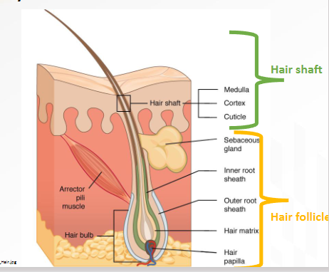
Hair Follicle
is the living, tunnel-shaped structure in the skin that grows hair, includes the root
A downgrowth or invagination of the epidermis into the dermis
Part located inside the skin and where the hair grows
Hair follicle has
Hair bulb which holds the
Root

Hair Shaft
visible, nonliving strand that emerges above the skin
Part of hair we see
Hair Cycle Phases
Anagen
Catagen
Telogen
stages in alphabetical order

Anagen Stage
1st Stage
Cell division
Formation of keratin - main proteins in hair that gives hair structure
Phase lasts 2-7 years
Determines hair length
Growth phase
Catagen Stage
2nd stage
Cell division slows - bulb part isnt as big as in anagen stage
Follicle shrinks and starts to detach from bulb
Lasts about 2-3 weeks
Telogen Stage
Final stage
Hair growth stops and old hair eventually falls out
Lasts around 3 months before cycle restarts
What stages can you get DNA from the hair?
Anagen and Catagen bc still have the root and living cells in the hair
90% of human head hairs in anagen or catagen growth phase
What gives hair color?
melanocytes in the hair follicle are responsible for synthesizing melanin
with age, production of melanin stops - gray hair appears
vitiligo or albinism involve melanocyte dysfunction or absence
What stage hair do you usually find at crime scenes?
Telogen
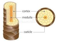
3 layers of the hair shaft
cuticle - outer layer of overlapping cells
cortex - provides strength and color, majority of the hair
medulla - core, sometimes absent
can be absent, fragmented, continuous
Cuticle → cortex → medulla
Level 1 Hair examination
recognition and separation of human and animals hair
based on shaft-profile
Looking at
Hair type
hair length
root shape
color
medulla
cuticle
Hair Type - Level 1 exam
Animals have 2 types of hair - outer hair or under coat hair present in different shapes, don’t see in human hair
Format of hair can see differences in shape