BISC205 Topic 2
 Call Kai
 Learn
Learn Practice Test
 Spaced Repetition
 Match
Match Flashcards
 Knowt Play
1/53

There's no tags or description
Looks like no tags are added yet.
Name | Mastery | Learn | Test | Matching | Spaced | Call with Kai | Chat |
|---|
No analytics yet
Send a link to your students to track their progress
54 Terms
What is “amphipathic”? Why are phospholipids amphipathic?
Amphipathic: A chemical compound or molecule possessing both a hydrophilic (water-loving) and hydrophobic (water-hating) region.
Why are phospholipids amphipathic: it allows them to self-rearrange into a bilayer when placed in water, which prevents water from entering the cell and cause it to swell/burst.

Why are membranes described as fluid mosaic models?
Fluid means “lots of movement” and mosaic means “mixture”. A membrane is a fluid structure, in which is can move. It also has various proteins embedded in it, which makes it appear as a mixture/mosaic

Two classes of phospholipids in animal cell membranes
1) Phosphoglycerides (MOST ABUNDANT IN ANIMAL CELL MEMBRANES)
2) sphingolipids

Phosphoglyceride structure
phosphate groups linked to a polar head group, such as serine, choline, ethanolamine, or inositol. It has glycerol (3 carbons) as the backbone. It has two fatty acid chains bound to glycerol through ester bonds.
Four types of phosphoglycerides
1) phosphatidylethanolamine
2) phosphatidylcholine
3) phosphatidylserine
4) phosphatidylinositol

Sphingolipid structure
Has sphingosine as backbone (18-carbon amino alcohol with an unsaturated hydrocarbon chain). It has one fatty acid chain bound to sphingosine through amide bonds. Highly enriched in the cell membranes of brain and nervous tissue.

Three main types of sphingolipids
1) ceramides
2) sphingomyelin
3) glycosphingolipids

Ceramides
The simplest type of sphingolipid, in which one fatty acid chain is attached to sphingosine with an amide bond. They form as base structures of the other two types of sphingolipids.

Sphingomyelin
Ceramide bound with either phosphocholine or phosphoethanolamine.

Glycosphingolipids
Ceramides with one or more sugar residues. There are two common types of glycosphingolipids:
1) cerebrosides: single glucose or galactose as sugar residue.
2) gangliosides: at least three sugars as residues (but one sugar must be sialic acid)

Factors that can increase membrane fluidity
1). Temperature increase: membrane can move from a solid (gel) state to a more fluid state
2) shorter fatty acid chains: allow membranes to become more fluid than longer fatty acid chains. Shorter fatty acid chains means less surface area for van der waals’s or hydrophobic interactions to occur between phospholipids.
3) membranes rich in unsaturated fatty acids: kinks (or bends) in unsaturated fatty acids prevents tight packing between phospholipids. Each bend indicates a double bond. This makes it easier for them to drift laterally or rotate

Factors that can decrease membrane fluidity
1) temperature decreases: membrane moves from a fluid state to a solid (gel) state
2) longer fatty acid chains: more surface area which allow for more stabilizing van der waal’s or hydrophobic interactions to occur between phospholipids
3) membranes rich in saturated fatty acids: makes phospholipids very straight with NO bends. This makes it harder for them to drift laterally or rotate

How cholesterol decreases membrane fluidity
At warm temperatures, cholesterol restrains movement of phospholipids, since too much movement can sacrifice the structural integrity of the membrane. The rigid steroid ring of cholesterol inserts between the flexible hydrocarbon chains of phospholipids, limiting their movement and interactions.

how cholesterol increases membrane fluidity
At cool temperatures, cholesterol prevents tight packing between phospholipids (cholesterol increases membrane fluidity). It wedges themselves in between the tails of phospholipids to prevent their orderly-like packing

Lipid raft
A spatial variation of the membrane that is thicker with a higher concentration of molecules. They are enriched with cholesterol, sphingomyelin, gangliosides, and phosphoglycerides with saturated fatty acid chains.
Why do lipid rafts have reduced membrane fluidity, and how do they compensate for it?
Reduces fluidity: Their high concentration of molecules are tightly packed together, reducing fluidity within the lipid raft.
Compensates: the surrounding membrane around rafts have phosphoglycerides with unsaturated fatty acid chains. This means the surrounding membrane is more fluid and allows rafts to move along membrane with combine with other rafts.

Functions of lipid rafts
1) serving as organizing centers for the assembly of signalling molecules
2) recruiting actin cytoskeleton (actin filaments interact with actin binding proteins). This allows for further stabilization of lipid raft.
3) facilitating formation of transport vesicles (ex/ lipid raft mediated endocytosis)

Six major functions of membrane proteins
1) transport
2) enzymatic activity
3) signal transduction
4) cell-cell recognition
5) intercellular joining
6) attachment to the cytoskeleton and extracellular matrix

Plasma membrane asymmetry: primary location (inner or outer leaflet (or monolayer)) of glycolipids
Located in the outer monolayer of the plasma membrane
Membrane carbohydrates (glucose or galactose) may be covalently bonded to lipids (forming glycolipids) and carbohydrates are on the outer leaflet to participate in cell-cell recognition. This means that glycolipids need to be on the outer monolayer of the plasma membrane if carbohydrates are
Plasma membrane asymmetry: primary location (inner or outer leaflet (or monolayer)) of phosphoglycerides (PE, PC, PI, and PS)
Outer layer
phosphatidylcholine (PC)
Inner layer
Phosphatidylserine (PS)
Phosphatidylethanolamine (PE)
Phosphatidylinosital (PI)
The two phosphoglycerides that create the surface potential difference across the membrane
PS and PI have a net negative charge.
This creates a more negative charge in the inner layer

Plasma membrane asymmetry: primary location (inner or outer leaflet (or monolayer)) of Sphingomyelin
Sphingomyelin is in the outer leaflet/monolayer

Important reasons for maintaining plasma membrane asymmetry
1) to create a surface potential difference across the membrane: negatively charged inner layer allows to interact with positively charged amino acid residues and peripheral membrane proteins
2) to create curvature within membrane: cylindrical phospholipids creates bilayer, while cone or conical shaped phospholipids create membrane curvature
3) preservation of cell viability: PS on the outer leaflet is an indication of a loss of membrane integrity. This is because it indicates cell apoptosis and targets the cell for engulfment by macrophages

Cylindrical shaped vs conical chapes phospholipids
Cylindrical phospholipids
PC
PS
PI
Sphingomyelin
Conical phospholipids
PE
what can and cannot pass through the plasma membrane
Can pass
hydrophobic molecules can dissolve in the lipid bilayer and pass through
Cannot pass
Hydrophilic molecules can pass through transport proteins

Concentration gradient definition
Differential concentrations of a substance across a space or a membrane. Cells have a higher concentration of potassium (K+) and a lower concentration of sodium (Na+). Concentration gradients cause ions to move from high to low concentrations. An example of concentration gradient could be the concentration gradient of K+ drives it into the cell.

Electrical gradient
A difference of charge across the plasma membrane. The interior of the living cell is electrically negative due to ammonia protein and chlorine ions. Electrical gradients cause ions to move to opposite charged sides. An example of electrical gradient could be Na+ entering the negatively charged interior of the cell.

Electrochemical gradient
The combined gradients of concentration and electrical charge that affects an ion. For example, the electrical gradient of K+ drives it into the cell, but the concentration gradient of K+ drives it out of the cell.

Passive transport
Diffusion of a substance across of a biological membrane that requires no energy,
Two types of passive transport
1) simple diffusion: where no transport proteins are needed. Small, uncharged polar molecules like urea, water, and ethanol cross the membrane by simple diffusion.
2) facilitated diffusion: transport proteins speed passive movement of molecules across the plasma membrane. This is ideal for polar molecules and charged ions
(List) Three main types of proteins that carry our facilitated transport
1) porins
2) permeases
3) ion channels

Porins
Large, non-selective barrel-shaped transport proteins that move hydrophilic molecules across the membrane based on size rather than charge. Aquaporins transport water, whereas aquaglyceroporins transport non-water molecules such as glycerol.

Permeases
facilitates the transport of specific molecules across a membrane by binding to the molecule and undergoing conformational changes. Unlike porins and ion channels, permeases can become saturated when all their binding sites are occupied by molecules

Ion channels and the five types of ion channels
form smaller pores through only which specific ions may pass based on size and charge. They are typically gated. Channels are specific to one or sometimes two ions.
There are five types
1) ligand-gated channels
2) voltage-gated channels
3) mechanogated channels
4) signal gated channels
5) Leak channels

Ligand gated channels
opens when specific regulatory proteins bind. They also open or close in response to specific extracellular molecules

Voltage gated channel
opens or closes in response to different membrane potentials

Mechanogated channels
regulated through interactions with subcellular proteins that make up cytoskeleton. Changes in cell shape, such as cell swelling, alter arrangement of cytoskeleton. Upon sensing changes in cytoskeleton, mechanogated channels open or close

Signal gated channels
opens or closes in response to specific intracellular molecules

Leak channels
non-gated channels, which means no trigger is required for their opening and closing. It has an intrinsic rate of switching between open and closed states

active transport definition and the types of active transport
Active transport: when substances diffuse against their electrochemical gradient and move from area of high concentration to low concentration. It requires energy in the form of ATP from the cell.
Two types of active transport
1) primary active transport
2) secondary active transport

Primary active transport vs secondary active transport
Primary active transport: Involves permease carrier proteins that use an exergonic reaction (ie. ATP hydrolysis) to provide the energy to transport a molecule
Secondary active transport: involves permeases carrier proteins that move one molecule down its electrochemical gradient to help move another molecule against its concentration gradient

Four primary active transporters that use ATP hydrolysis and some examples
1) P-type ATPases: pump specific ions across membranes. This includes Na+ and K+ ATPases and Ca2+ ATPases
2) V-Type and F-Type ATPases: structurally related in that they both exclusively pump H+ ions across a membrane. Vacuolar ATPases is an example of V-Type whereas mitochondrial ATP synthase is an example of F-type (can also create an H+ gradient using energy gained from ATP hydrolysis)
3) ABC (ATP Binding Cassette) transporters: pump larger organic molecules like drugs and cellular metabolites. They include multi-drug resistance proteins like P-glycoproteins

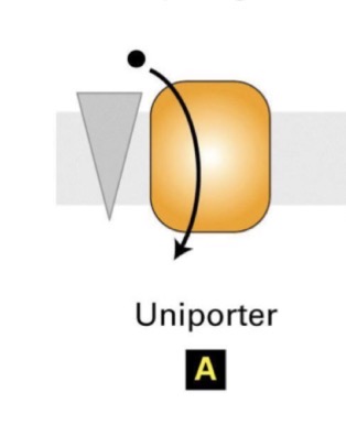
Uniporter (primary active transport)
Involves moving one specific ion or molecule at a time. They can be channel proteins too, but with active transport, only the carriers are used.
Antiporter or exchanger (secondary active transport) with example
Involves moving two different ions or molecules in opposite directions

Symporter or co-transporter (secondary active transport) with example
Involves moving two different ions or molecules in the same direction

Osmosis definition
The passive transport of water across a selectively permeable membrane that separates two solutions differing in osmolarity. Water diffuses across a membrane from a region of lower total solute concentration to the region of higher total solute concentration. This also means that water moves from high H2O concentration to low H2O concentration

Osmolarity definition
The number of particles in a solution. It is a measure of solute concentration (Osm/L or OsM). It takes into consideration the TOTAL number of solutes (permeable and impermeable). Osmolarity involves terms like iso-osmotic, hypo-osmotic, and hyper-osmotic.
Hyper-osmotic, hypo-osmotic, and iso-osmotic
Hyper-osmotic: osmolarity solution is greater than that of another solution
Hypo-osmotic: osmolarity solution is lower than that of another solution
ISO-osmotic: solutions are equal in osmolarity, and therefore no net osmosis occurs between them if they are separated by a selectively permeable membrane.

How to calculate osmolarity

How to determine n (dissociation coefficient of a particular solute in solution)
It is the number of particles that result from dissociation of a molecule in water. A molecule that can dissociate into two particles has n=2. A molecule that can dissociate into three particles has n=3.

Tonicity definition
Describes what happens to a cell when placed in solution. It takes into account concentration only relating to impermeable solutes only. This is because the penetrating solutes reach equilibrium and not affect tonicity
Hypertonic, hypotonic, and isotonic
Hypertonic: solution with higher concentration of non-penetrating solutes than that found in cells. Result: water rushes out of cell and cell shrinks.
hypotonic: solution with lower concentration of non-penetrating solutes than that found in cells. Result: water rushes into cell, causing it to swell and burst.
Isotonic: solution with the same concentration of non-penetrating solutes as that found in cells. Result: nothing happens.

Penetrating vs non-penetrating solutes
Penetrating solutes: solutes that are permeable to the cell. They are NOT accounted for when considering tonicity.
Non-penetrating solutes: solutes that are impermeable to the cell. They are accounted for when considering tonicity.

Osmolarity vs tonicity
A solution’s osmolarity is based solely on its total solute concentration. However, its tonicity is determined by how it affects cell volume. Osmolarity considers both non-penetrating and penetrating solute concentrations, whereas tonicity only considers non-penetrating solutes.
