Anatomy 1 Practical 1- Bones
 Call Kai
 Learn
Learn Practice Test
 Spaced Repetition
 Match
Match Flashcards
 Knowt Play
1/164

There's no tags or description
Looks like no tags are added yet.
Name | Mastery | Learn | Test | Matching | Spaced | Call with Kai | Chat |
|---|
No analytics yet
Send a link to your students to track their progress
165 Terms
frontal bone
Your forehead, covers top bit of orbit. Articulates with parietal, sphenoid, zygomatic, lacrimal, maxilla, and nasal bones. Can be seen by the yellow part
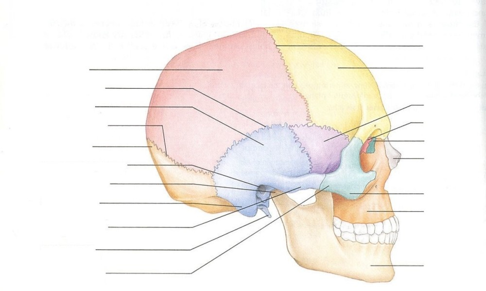
coronal suture
suture on the top of the head. exists between frontal bone and parietal bone.

parietal bone
2 bones (left and right), that make up the top part of the skull- labeled as red

sagittal suture
runs between the 2 parietal bones, separating the 2 bones

occipital bone
very back of skull, forms the base of skull. most posterior part of cranium. labeled as orange on diagram
sort of stretches anteriorly and wraps around the underside of the skull. there's a section that is on the inferior side of the skull, and is most anterior, where it has a flat edge and points upward, that’s where the sphenoid bone will connect

lambdoid suture
suture between parietal and occipital bone

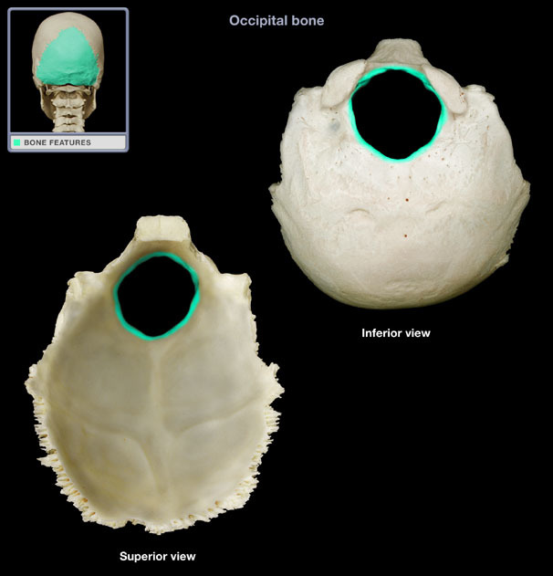
foramen magnum
big hole at the bottom of occipital bone. where the brain stem is going to go through to connect with the spinal cord
temporal bone
on the side of head, by both the ears, is the bluish bone

zygomatic process (of the temporal bone)
a thin projection that sticks out going anteriorly, and will connect with the zygomatic bone. kinda separated from the temporal bone itself, like will have a space in between each section they’re not just right on top of each other

styloid process
pointy part of the temporal bone that sticks out inferiorly. tilts a little bit towards the front
muscles that connect there help with tongue movement

external auditory meatus
forms the auditory canal, where sound enters the ear
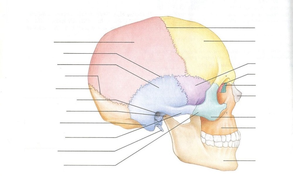
mandibular fossa (of the temporal bone)
on the squamous portion of the temporal bone, kinda underish of the temporal bone
hollow of bone
is where the mandible articulates with the temporal bone, TMJ joint is there
the condyle of the mandible fits into the fossa, allowing the jaw (only freely moving bone in skull) to move up and down

mastoid process
at the bottom (inferior) of the temporal bone. Is a little bump/projection that attaches to the neck.


zygomatic arch
the zygomatic process of the temporal bone and the temporal process of the zygomatic bone (which is kinda thicker compared to the zygomatic process) fuse together and form this
squamous portion of the temporal bone
kinda a superior & anterior portion of the temporal bone. Where mandibular fossa and zygomatic process are

sphenoid bone lateral view
behind eye/ zygomatic bone

spehnoid anterior view of skull
can view it inside the orbital, and on the sides of your face (orange)

optic canal/foramen anterior view
2 holes on the sphenoid bone, inside of the orbitals, where the optic nerves will go through

optic foramen superior view (inside of skull)
those very top holes, right by the lesser wings

lesser wing & greater wing of sphenoid
flaps on the top, right by the optic foramen

pterygoid process & plates
those 4 inferior-pointing projections, 2 on each side. the process itself is just like the 2 plates together on each side. what is in yellow
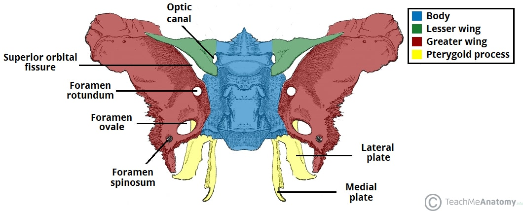
sella turcica
on the back of the sphenoid bone, there’s a saddle shape. that’s where the pituitary gland sits

how can you generally describe the shape of the sphenoid bone on its own, at least looking at it from the front?
a butterfly-looking shape that expands the width of the skull
zygomatic bone
next to sphenoid bone. forms a bit of the lower and more lateral side of the orbit

maxilla/ maxillary bones
1 on each side, they meet in the middle.
forms a large part of middle of the face and even the sides of the nose and connect to the bone
known as the keystone of the face, bc it articulates with all other face bones

mandible
jawbone
connects with teeth and moves at the TMJ joint
ramus is that flat part that goes up
mandibular condyle is that round thing that will be posterior on both sides and will be what connects at the TMJ joint with the mandibular fossa on the temporal bone
coronoid process/mandibular process: the spikey part up top, will fall under the zygomatic bone. More anterior compared to the condyloid process. like the ridge of a crown

mental foramen
hole of mandible located inferior to the teeth

mandibular foramen
holes that are behind your teeth that are responsible for the feeling of your teeth. kinda on the opposite side of the ramus

nasal bone
kinda js self explanatory, where your nose is. 2 that meet up together and form the bridge of the nose

lacrimal bone
where your tear ducts are, form the inner eye part that’s more towards the side of the nose

ethmoid lateral view
can kinda see it sticking into the back of the orbit, where that red is

ethmoid anterior view
can see it in the gap where the nasal region is since it forms part of the septum is the blue color

ethmoid bone landmarks
the perpendicular plate makes up a lot of the nasal septum. is the inferior part of the bone, and runs from the back to the front in a straight line. is a very thing, plate-like part
on the top side of the bone, there will be a bunch of little holes. that is where the cribriform plate is, and through those holes will be where the olfactory bulbs go thorugh
the spike on top of the bone is the crista galli
to help kinda tell the difference of front vs back, know the cribiform plate is on the anterior side

inferior nasal conchae
in light blue
their own 2 separate bones, unlike the superior and middle conchae.
right under ethmoid bone
can be seen on lateral side of the hole in the nose
are kinda thin, like vomer, but instead of how it flattens out to connect to the sphenoid bone, it flaps at the end kinda

vomer
ok i know it’s a little hard to see in the anterior view but it forms the bottom half of the nasal septum.
it’s a long skinny bone, similar in shape to the perpendicular plate of the ethmoid bone, that runs straight back and forth.
it will spread out and flatten to connect to the sphenoid bone
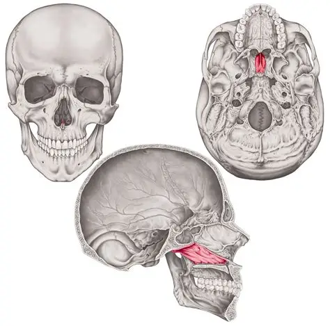
occipital condyles
roundish projections that are on lateral side of the foramen magnum, will articulate with the C1 vertebrae to allow you to nod.

palatine process of the maxilla
a lot of the mouth’s hard palate is made up by the maxilla itself, the anterior portion of the mouth at least. is the dark gray

palatine bones
a left and right, meet in the middle
makes up the posterior part of the hard palate
also makes up the back of the nasal cavity
IMPORTANT: know what the bones look like on their own!
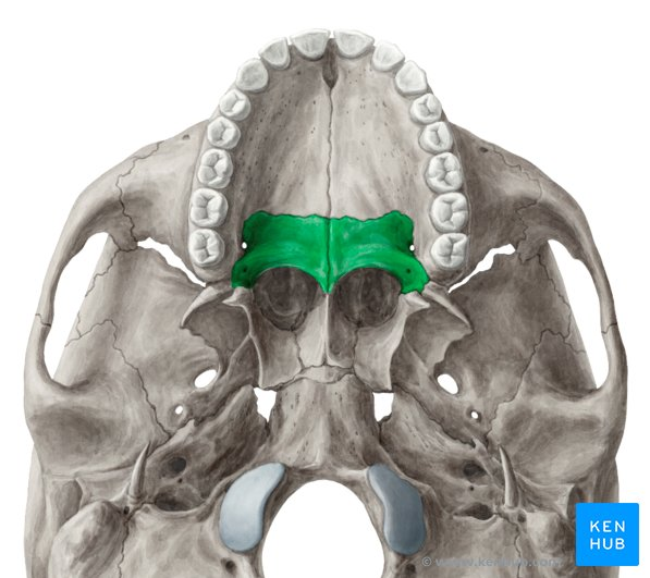
supraorbital foramen
2 holes above each eye

infraorbital foramen
foramen on the anterior surface of the maxillary, under the eye

incisive foramen of the maxilla
holes on the underside of the mouth, on the hard palate, towards the front

greater palatine foramen
2 holes on the sides of the palatine (not the 2 small ones actually on it) that are kinda next to the maxilla

petrous portion of temporal bone
can be seen from a superior view, is where the middle ear activities take place

external occipital proturbence
bump on back of head

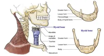
hyoid bone
doesn’t articulate with any bone, js kinda suspended
U-shaped
more in the throat area

foramen ovale (inferior view)
kinda one of those most anterior holes. located on the sphenoid bone. right by the sella turcica

foramen ovale (superior view)
same oval shape hole, on the sphenoid bone. highlighted in green (only the hole tho). is lateral to the foramen lacerum


stylomastoid foramen
small hole right in between the styloid and mastoid processes. on the temporal bone


foramen lacerum (inferior view)
kinda in between to the part of the occipital bone that extends to the sphenoid and the temporal bone

foramen lacerum (superior view)
hole be close to the sella turcica


jugular foramen
medial to the carotid canal and closer to the neck for the jugular vein to go through. highlighted in green will be big and kinda peanut shaped.


carotid canal
anterior to the jugular vein. on the temporal bone. highlighted in green

anterior/frontal fontanelle
the very part on top of baby’s skull that is soft

anterior lateral fontanelle/ sphenoid fontanelle
fontanelle that’s just above the sphenoid bone

mastoid fontanelle/ posterior lateral fontanelle
fontanelle on the side of the skull, towards the back

cervical vertebrae general
vertebrae towards the top of the neck, there’s 7
thoracic vertebrae general
vertebrae located where your chest is, there’s 12
lumbar vertebrae general
there’s 5 of these vertebrae, located on your lower back
sacrum
at the very bottom of spine, connects your tailbone

coccyx
your tailbone, connected to sacrum

body of vertebrae
anterior, cylinders that stack together (with discs in between). the lower you go, the larger these become because that part of the spine holds more weight. the atlas/c1 vertebrae does not have this. in the light pink

vertebral arch
formed by the lamina and the pedicles. is on the posterior portion of the vertebrae

lamina
forms the point of the vertebral arch. in between the transverse and spinous processes (easiest to see that on thoracic vertebrae)

pedicles
the part of the vertebral arch that’s closer to the body of the vertebrae. 2 sort of cylinders. on the thoracic and lumbar vertebrae they go straight back and forth from the body, but on C2-C7 they go out diagonally. C1 has no pedicles

vertebral foramen
the hole that is in the middle of every vertebrae so that the spinal cord can go through. once all the vertebrae are stacked, these form the vertebral canal

spinous process
highlighted in yellow. points straight back. what you feel on someone’s back. C1 doesn’t have this. in most cervical vertebrae, this is bifid- 2 flaps.

transverse process
points straight out to the side. in green
in the thoracic vertebrae, it’s really obvious. this and the spinous process form a distinguishable W shape going posteriorly.
in lumbar vertebrae (which is shown in the picture), it’s a little less of an angle and straighter out to the side, but still easy to see. not as easy to see the W shape tho bc it has a facet in between the 2 processes
cervical vertebrae also have transverse processes but are hard to see because they are short compared to those 2. just know they’re next to the transverse foramen

transverse foramen
highlighted in red. 2 holes on each side of the cervical vertebrae. what distinguishes them from the other types of vertebrae. arteries go through

atlas/C1
no body- instead there’s an anterior and posterior arch
rotates on dens of axis (where that face is towards the anterior side)
side of transverse foramen are toward the front/anterior side
no pedicles
those big indents u can see medial to the transverse foramen are the superior articular surface and is where the occipital condyles articulate

axis/ C2 dens process
the superior facing projection that articulates with C1

costal facets (of thoracic spine)
are highlighted, where the ribs articulate with the thoracic spine. amts of facets on each vertebrae may differ, but these are the typical ones
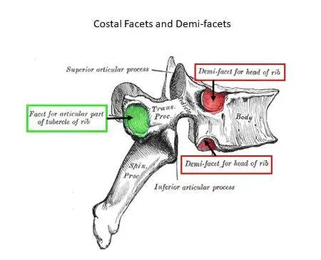
sacral promontory
on top and in the middle of the sacrum, forms a flat-ish line

true ribs
in purple. pairs 1-7, have direct connections to sternum with the cartilage they attach to. rib-cartilage-sternum

false ribs
3 pairs (green), but also include the 2 bottom ribs (blue) because they have no direct rib-cartilage-sternum connection.
the cartilage off the ribs in green connect to the cartilage of the rib above it, so there’s no direct connection to the sternum

floating ribs
2 pairs, 11 and 12, highlighted in blue. they can be classified as also false ribs bc their cartilage coming off the rib doesn’t have a direct connection to the sternum, but they’re on their own bc unlike false ribs, they don’t have connection of cartilage to the one above it. they are the only ribs that are only connected to the vertebrae

head of ribs
indent, on the side that’s more curved and connects to the vertebrae

neck of rib
the part of the rib that’s in between the head and that other facet

tubercule of rib
on posterior side, a bony bump that’s close to the head of the head, but closer to the other fact

costal groove
on the bottom and interior surface of the rib, there is a ridge and an inferior border. in between them is the costal groove

body
part of the sternum in red

manubrium
part of the sternum in green, the top part

xyphoid process
the little point on the bottom of the sternum, in purple

suprasternal notch
little notch on top of body of manubrium

right scapula
the glenoid cavity will be most lateral, and the opposite side, kinda the most simplest, will be towards the middle/ closest to the spine

left scapula
you know it’s on this side of the body bc of where the glenoid cavity connect to the humerus and where the medial border is

suprascapular notch (superior view)
a notch/indent on the superior part of the scapula. where number 4 is located. right by the spine of the scapula

suprascapular notch (anterior view)
indent on the top of the scapula. right by coracoid process
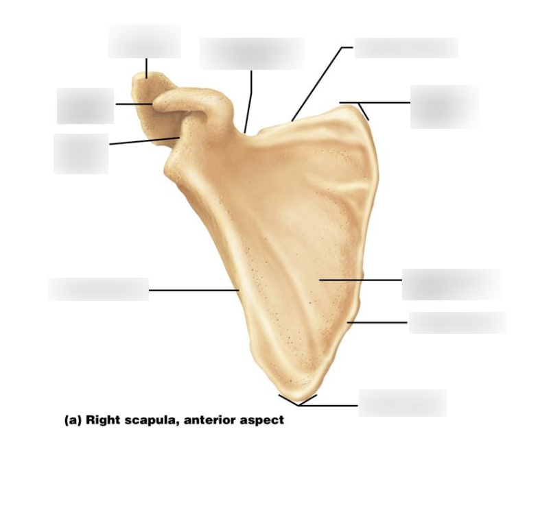
superior angle
the top point of the scapula

inferior angle
bottom point of the scapula

subscapular fossa
the flat side, on the anterior side so that your scapula can glide over your ribs.

medial border
the side of the scapula that runs down toward the middle of the body

lateral border
the side of the scapula that runs toward the outer part of the body, close to the glenoid cavity. will be closer to where humerus connects

glenoid cavity
the big indent of the scapula where the head of the humerus will connect to. in this picture, is indicated where the pointer without a color is.

spine of scapula
on the back part of the scapula, will stick out and travel horizontally

acromion
forms the very top of the shoulder, and branches off the spine of the scapula (on lateral side). will come from around the back and go towards the front of the body to connect with the clavicle

supraspinous fossa
flat part above the spine of the scapula

infraspinous fossa
flat part of scapula below the spine

coracoid process
on anterior side of scapula. a part that kind of looks like a horn and projects out to the side. close to the acromion

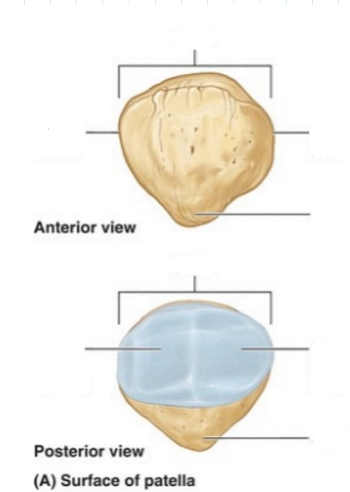
how to differentiate between the front and back of the patella
the front is smooth, and the back has the 2 facets with the ridge in the middle to articulate with the femur and fit into it. apex is the point of bottom, base is the top
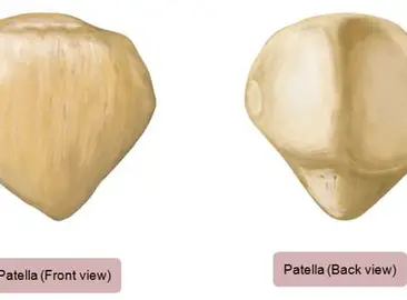
medial facet of patella
on the posterior side of the patella, the indent that sort of bulges out to the side. will be the smaller facet of the 2. is the surface that articulates with the medial condyle of the femur