BSCI331 Exam 3 - L10 Post-Translational Modifications to Proteins
 Call Kai
 Learn
 Practice Test
 Spaced Repetition
 Match
 Flashcards
 Knowt Play
1/63

There's no tags or description
Looks like no tags are added yet.
64 Terms
What are ways that proteins are modified post-translation? Post-translational modifications of proteins
Covalent modification of amino acid side chains of proteins to control protein function
Protein function can also be regulated by ligand binding (e.g. non-covalent interactions)
covalent modification
a chemical change to a molecule, most commonly a protein, that happens by forming or breaking a covalent bond
How do we alter amino acid side chains to change protein function?
Covalent modification of an amino acid side chain changes their structure, which changes their chemical properties
What are the ways we can covalently modify amino acid side chains of proteins to change their chemical properties?
Proteolytic cleavage
Phosphorylation
Ubiquitylation
proteolytic cleavage
Hydrolysis (addition of water) to break down the peptide bonds between amino acids in a protein by protease enzyme; in other words, after a protein is made and translated by the ribosomes in the cytoplasm, certain amino acids may be removed
why is proteolytic cleavage considered a covalent modification?
Because it involves the hydrolyzing/breaking of peptide bonds (strong covalent bonds) in the primary structure of the protein backbone
a given protein may have…
multiple potential modification sites, not all of which may be used at the same time - certain covalent modifications occur in response to certain signals, such as changes in pH and temperature; the cell responds to certain conditions in whatever combination by covalent modifications to proteins
phosphorylation
addition of a negatively CHARGED phosphate group to the hydroxyl (OH) of the R-groups serine, threonine, tyrosine (eukaryotes) by the class of enzymes called protein kinases
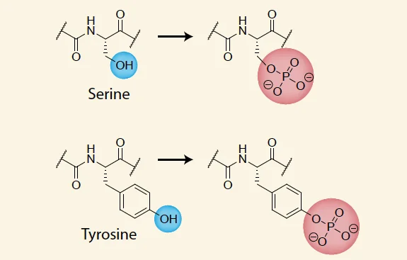
protein kinases
enzyme that transfers phosphate from ATP to the amino acid residue
In bacterial cells, what amino acid R groups can be phosphorylated?
serine, threonine, tyrosine, AND histidine (transient - used to transfer the phosphate to somewhere else; histidine acts as a phosphate carrier)
kinase domain
the region of a protein kinase responsible for adding phosphate groups to other proteins.
Serine/threonine kinase domain: Recognizes serine or threonine residues and phosphorylates them
Tyrosine kinase domain: Recognizes tyrosine residues and phosphorylates them
Why do the amino acids require different kinases?
The amino acids serine and threonine can be phosphorylated by the same type of kinase (with a serine/threonine domain), whereas tyrosine requires a different type of kinase (with a tyrosine domain)
This is because serine and threonine share similar chemical structures (Serine has a –CH₂OH side chain; Threonine has a –CH(OH)CH₃ side chain)
Tyrosine is chemically different because it while it contains an –OH group, its side chain is much larger with an aromatic ring - requires different set of kinase
For serine and tyrosine to both be phosphorylated, what must be true?
If the protein has two separate kinase domains (serine-threonine kinase domain and tyrosine-kinase domain)
Serine vs. phosphoserine
Serine: R= -CH2-OH
Phosphoserine (phosphorylated serine by kinase): R = -CH2-O-PO32-
How is the phosphate added to proteins?
Addition of the phosphate is a non-spontaneous reaction that requires energy that comes from the hydrolysis of ATP
Hydrolysis of ATP forms the phosphorylated amino acid residue and ADP
Every phosphate molecule added to a protein = costs 1 ATP
amino acid residue
The word for an individual amino acid in a sequence. For example, in the sequence UGPDEQLMFRH, the third residue is a proline.
Why is it called a residue? Because when it's in a polypeptide chain, it's no longer technically an amino acid. When you react amino acids: NH2-CHR1-COOH + NH2-CHR2-COOH -> NH2-CHR1-CO-NH-CHR2-COOH + H2O
The individual amino acids in the chain have lost the hydroxy-terminus from their carboxylic acid, so it wouldn't be accurate to call them amino acids anymore. Therefore, they're referred to as amino acid residues
Is phosphorylation reversible?
Yes, the phosphorylation is reversible
However, the reaction that leads to phosphorylation is not reversible - if you take phosphate off of phosphoserine, you don’t have enough energy to put the phosphate back onto ADP and reform ATP (loss of energy, ATP still used)
What catalyzes the removal of a phosphate from a phosphorylated residue/protein?
Protein phosphatases catalyze the removal of a phosphate
Kinase and phosphatases always…
come in pairs; a kinase that adds a phosphate to a residue is coupled with a phosphatase that can remove said phosphate from the same residue
For every kinase, there must always be a phosphatase that reverses the reaction
However, the phosphatase does not put the phosphate back onto ADP - it just releases the phosphate to be free
Therefore, every phosphorylation and phosphate removal costs 1 ATP
What is the most common covalent modification in the cell?
Phosphorylation - many changes in protein structure and activity are driven by phosphorylation
Why is phosphorylation the most common covalent modification in the cell?
Because:
Each phosphate group adds two negative charges to the protein
This can drive major structural changes, activity changes, or changes in protein solubility (ex. serine + phosphate = negatively charged serine can repel a neighboring negatively charged aspartic acid, changing the shape of the protein; or attracted to nearby positively charged residue)
Addition of a phosphate renders that amino acid different from the other versions of the amino acid in other parts of the protein - therefore, the added phosphate group may create a new recognition site that allows other proteins to recognize and bind to the phosphorylated protein (especially true for tyrosines - phosphorylated tyrosines generate a binding site for a “SH2 domain” of another protein that specifically recognizes the phosphorylated tyrosine but not the normal, nonphosphorylated tyrosine) = modifies function of protein by changing what proteins it can interact with
The SH2 domain is a specialized region of another protein that recognizes and binds to a phosphorylated tyrosine residue on a target protein
“SH2 domain” is a…
phosphotyrosine-binding motif - the SH2 domain is a specialized region of a protein that recognizes and binds to phosphorylated tyrosine residues on other proteins
Motif: A specific structural feature/amino acid sequence associated with a particular function. Here, the SH2 domain's ability of the protein to bind to phosphotyrosine
You are studying a protein that is turned on by phosphorylation at a specific serine residue. What would you expect would happen if the serine is mutated to aspartic acid?
A. It would be impossible for the protein to be turned on
B. The protein will always be turned on
C. I have no idea
A & B - Both answers - it depends. If the purpose of the phosphate is to generate a negative charge, then the protein will always be turned on because the aspartic acid itself under physiological conditions is also negatively charged.
However, if the purpose of the phosphate is to generate a specific recognition/binding site, then the aspartic acid would not suffice and it will be impossible for the protein to be turned on
A - Suppose a protein must bind to the phosphorylated serine to activate it
Phosphorylated serine has a specific chemical structure recognized by the binding protein
Aspartic acid has a negative charge but does not have the same structure as phosphorylated serine.
Therefore, the binding protein may not recognize aspartic acid.
Without this interaction, the protein cannot become activated through that mechanism.
Ubiquitylation
the covalent addition of ubiquitin to the side chain of a lysine residue in a protein; reversible
ubiquitin
a small cytosolic protein (76 amino acids) - cytosolic means it floats freely within the cytosol that can be covalently attached to side chain of a lysine residue
what is the purpose of ubiquitylation?
compared to the addition of a phosphate, the addition of ubiquitin is much bigger (it’s a whole protein), so it can serve as a tag that can be recognized
what functions does the ubiquitin “tag” have?
Ubiquitin serves as a tag that can either:
Mark proteins for selective degradation (in response to a specific stimulus) by adding a string of ubiquitins to a lysine of a protein (polyubiquitination) OR
Direct proteins to specific locations in the cell by adding ONE ubiquitin (monoubiquitination)
polyubiquitination
Within its structure, the ubiquitin protein contains two lysine residues (lysine 48 and 63). The first ubiquitin attaches to the target protein by a covalent bond with the lysine residue on the target protein. Then, additional ubiquitins attach to the first ubiquitin via the lysine 48 and 63 residues (the C-terminus Gly-76 of another ubiquitin can form a covalent bond with one of these lysines). This forms a chain of ubiquitins covalently bonded to the target protein.
You can also make branched chains of ubiquitin, where you can add a ubiquitin to lysine 63 and lysine 48

monoubiquitination
Covalently bonds one ubiquitin to the lysine residue of the target protein; serves as a tag that directs proteins to specific locations in the cell
The TARGET protein that gets directed to specific locations in the cell, not the ubiquitin itself.
When ubiquitin is covalently attached to a lysine residue on a target protein, it acts as a molecular tag that tells the cell what should happen to that target protein.
How does this work?
1. Ubiquitin attaches to the target protein through a covalent bond.
2. Other proteins in the cell recognize the ubiquitin tag.
3. These proteins help direct the ubiquitinated target protein to a particular cellular location or pathway.
Per ubiquitin, how much ATP is used?
2 ATP is used to add each ubiquitin
So a chain of 20 ubiquitins = cost of 40 ATP + cost of making ubiquitin
However, the ubiquitin is reusable (does not have to spend lots of energy to make it over and over again)
Hydrolysis of a peptide bond is..
a spontaneous reaction - requires 0 added energy because making the peptide bond costs ATP; therefore, breaking the bond releases energy
Ubiquitylation is..
the only covalent modification that is an actual protein (76 amino acid protein modification)
Noncovalent interactions between proteins and other molecules
All proteins bind to other molecules - these molecules are called ligands of the particular protein in question
Ex. a structural protein interacts with other structural proteins to build a structure
ligand
molecules or ions that bind reversibly to a target protein and regulate the protein’s function
Why is ligand binding reversible?
Because ligand binding is noncovalent, so it is REVERSIBLE (equilibrium chemistry event that can bind and nonbind)
What is the strength of ligand binding?
Covalent interactions are strong - you form a chemical bond that has to be broken in order to reverse the interaction
However, ligand binding are weaker - noncovalent interaction can be reversed because molecules are in constant motion in aqueous solution, bumping into other nearby molecules
In order for ligand binding to be “strong” enough where the ligand is actually considered bound to the protein, the protein binding must be STRONG enough to withstand the jolting of molecular motions (thermal motion)
Aka the constant motion of both the ligand and the target protein, as well as the surrounding water molecules. This is called thermal motion, which occurs because molecules possess kinetic energy.
How is ligand binding strength achieved?
Ligand binds strongly to the protein by:
3D complementary of binding - ligand fits nicely into the shape of the protein that matches its shape
Formation of several noncovalent bonds - additional bonds = greater contact; one noncovalent bond is comparatively weaker - strength in numbers
The more the shape of the protein corresponds to the ligand, the stronger the ligand binding is going to be
Ligand binding sites are..
3 dimensional; they result from the three-dimensional folding of the tertiary or quartenary structure of the protein, generating a 3 dimensional binding site for the ligand
The amino acids of a protein that contribute to binding a ligand (form the 3D binding site for the ligand) are..
often quite far apart on a protein’s primary sequence but come together when the protein folds

How can we characterize the strength of ligand-protein binding?
A + B ←→ AB
A = protein
B = ligand (regulates the function of the protein)
AB = protein-ligand complex
Reversible and dynamic equilibrium; AB can be formed or broken down into A and B
We can quantify the rates for the forward and reverse reactions by the kon (rate constant - how rapidly do the protein and ligands combine to form the complex) and koff (rate constant - how rapidly does the protein-ligand complex dissociate into protein and ligand)

Does equilibrium mean the protein and ligand stop binding and unbinding?
No - The kon and koff reactions are occurring simultaneously; for every chemical reaction, both the forward and the reverse reactions are happening at the same time
kON and kOFF are the…
kON - the rate of forward (association) reaction that creates the protein-ligand complex (A + B —> AB)
kOFF - the rate of backward (dissociation) reaction that breakdown the protein-ligand complex (AB —> A + B)
Ka
The association constant that measures the strength of binding (ligand-protein binding)
Ka = kON/kOFF
If Ka is large, then..
kON is larger, and there is strong ligand-protein binding
Conversely, if Ka is small, then there is weak ligand-protein binding because KOFF is bigger (dissociation of protein-ligand complex)
Kd
The dissociation constant
Kd = 1/Ka
A large Kd means…
weak ligand-protein binding; more dissociation of protein-ligand complex into the protein and ligand
Relationship between Ka and Kd
Inverse; Greater Ka means greater association of protein and ligand into the protein-ligand complex and smaller Kd which means less dissociation of the protein-ligand complex into protein and ligand
Why is the dissociation constant used more frequently than the association constant?
Because the dissociation constant quantifies the concentration of ligand needed for 50% of protein A to be bound to the ligand and 50% of protein A to be unbound (concentration of ligand at 50% binding)
A (protein) + B (ligand) ← AB (protein-ligand complex)
Kd = ([A][B])/[AB]
At 50% binding, the concentration of free protein [A] equals the concentration of protein-ligand complex [AB], so they cancel out
Kd = [B] (ligand concentration)
So therefore, Kd tells you the concentration of the ligand at which 50% of binding occurs between protein and ligand
Therefore, Kd tells you how much ligand you need for 50% of protein A to be bound to the ligand and 50% of protein A to be unbound from the ligand
What does 50% binding mean?
50% binding means 50% of the total protein is bound to ligand (protein-ligand complex), while the other 50% is free (unbound) protein.
For example, imagine you have 100 protein molecules:
50 proteins are bound to ligands, forming protein-ligand complexes (AB).
50 proteins are unbound, existing as free protein (A).
Free ligands (B) are also present in the solution, but their number is not necessarily 50.
A - protein; B - ligand
At 50% binding: [A]=[AB]
Therefore: Kd = [A][B] / [AB]
Since [A] = [AB], they cancel, and Kd = [B]
This means the dissociation constant, Kd, equals the concentration of free ligand needed for 50% of the proteins to be bound at equilibrium
Therefore, small Kd indicates…
Strong protein-ligand binding; small concentration of ligand [B] needed for 50% protein-ligand binding - if a ligand binds strongly to a protein, you don't need much ligand to keep 50% of the proteins occupied
Small concentration of ligand [B] needed for 50% protein-ligand binding (50% of protein bound to ligand, 50% free protein) - so the ligand must be bound very strongly
Large Kd indicates…
[B] is large; you need lots of free ligand for 50% protein-ligand binding; indicates that the ligand doesn’t bind for very long - so you need lots of ligand around so that 50% of the protein is bound to the ligand
Lower dissociation rates = [blank] Kd values = [blank] binding
Lower dissociation rates = lower Kd values = stronger binding
Which of the following values represents the stronger protein-ligand interaction?
A. Antibody binding to bacterial toxin with a Kd of 10^(-12)M
B. Calmodulin binding to calcium with a Kd of 10^(-6)M
A. because lower Kd means less free ligand is needed for 50% of protein-ligand binding to occur = stronger protein-ligand
What are examples of protein-ligand interactions?
Protein regulation by direct phosphorylation
Protein regulation by binding to phosphorylated/dephosphorylated forms of another nucleotide
Ligand regulates protein function by direct phosphorylation
A protein is regulated by the addition or removal of a phosphate group directly on the protein (protein-ligand interaction: ligand = ATP, protein = protein kinase)
Ligand ATP binds to protein kinase in its active site
The protein kinase hydrolyzes the ATP and transfers the terminal phosphate groups to another protein (covalent modification) on the serine, threonine, or tyrosine, basically using ATP to alter protein function

In direct phosphorylation, the protein that is being regulated either..
has a phosphate bound or no phosphate bound
Ligand regulates protein function by binding to phosphorylated/dephosphorylated forms of another nucleotide
GTP-binding proteins act as molecular switches for OTHER proteins that turn ON and OFF depending on whether the GTP-binding proteins are bound to GTP or GDP.
GTP and GDP are ligands that bind to a GTP-binding protein and change its activity without directly phosphorylating the protein.
Ligand-protein interaction: ligand = GTP or GDP; protein = GTP-binding protein
Molecular switch; When GTP is bound to a GTP-binding protein, the protein is in its active state/on and can regulate other proteins; when GDP is bound to the protein, the protein is off, and does not bind or regulate other proteins
When GTP binds to the GTP-binding protein, the GTP-binding protein hydrolyzes GTP into GDP?
GTP binding proteins bind GTP, hydrolyze it to GDP, and exchange the GDP for G
When the protein is bound to GTP, it is in the active state (interacting with something else and regulating its function)
The GTP-binding protein hydrolyzes GTP while GTP is bound to it, and this hydrolysis is what helps switch the protein from ON to OFF

In regulation by binding to phosphorylated/dephosphorylated forms of another nucleotide, the protein that is being regulated either..
bound to GTP (protein = on) or GDP (protein = off)
The protein being regulated always has a ligand bound to it
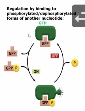
What is the diagram showing?
The top green protein is the off conformation that is bound to GDP; however, the GDP can be released, and then GTP bound to the GTP-binding protein to form the ON conformation that can regulate other proteins; the GTP bound to the GTP-binding protein can be hydrolyzed back into GDP and a free phosphate, releasing the phosphate, and the protein returns back to its inactive conformation
1. Top green protein = OFF
GDP (labeled GPP) is bound to the protein.
The protein is in its inactive conformation.
2. Left arrow going downward = Turning ON
GDP is released.
GTP (shown as GPP + P) binds to the protein.
The protein changes its conformation and becomes active.
3. Bottom green protein = ON
GTP is bound to the protein.
The protein can now interact with and regulate downstream target proteins.
4. Right arrow going upward = Turning OFF
The GTP-binding protein hydrolyzes its bound GTP into GDP + Pi.
Pi is released.
The protein returns to its inactive conformation.
What proteins facilitate the rate of hydrolysis or GDP binding to GTP-binding proteins?
GAPs (GTP-activating proteins) - increase GTP hydrolysis (GTP —> GDP) to turn the GTP-binding proteins OFF (ON —> OFF)
Guanine nucleotide exchange factors (GEFs) speed up the GTP-binding protein ability to exchange GDP for fresh GTP, turning the molecule back ON to its active state and allowing it to regulate other proteins (OFF —> ON)

Many proteins have…
many regulatory mechanisms (multiple modification/interaction sites that allows other proteins to act as molecular integrators)
proteins can have multiple regulatory/modification/interaction sites, allowing them to receive and combine signals from different sources to determine how they should function
molecular integrator
a protein that receives multiple regulatory signals and combines their effects to determine its overall activity
what are examples of molecular integrators?
cyclin-dependent kinases (CDKs) - regulate the cell cycle by phosphorylation at one site, dephosphorylation at another, binding to another protein (cyclin)
When ON, they help the cell regulate through phases of the cell cycle - they get selectively turned on during SPECIFIC phases of the cell cycle
CDKs require the binding of cyclin to be turned ON (which is why they are called cyclin-dependent kinases)
They are also called cyclins because they oscillate their amount in the cell depending on their position in the cell cycle
Big picture of modifications on proteins
Covalent modifications of amino acids play important roles in protein function
Protein function can also be regulated by liand binding (e.g. non-covalent interactions)