A&P Histology
 Call Kai
 Learn
Learn Practice Test
 Spaced Repetition
 Match
Match Flashcards
 Knowt Play
1/49

There's no tags or description
Looks like no tags are added yet.
Name | Mastery | Learn | Test | Matching | Spaced | Call with Kai | Chat |
|---|
No analytics yet
Send a link to your students to track their progress
50 Terms

Tissue, Function, Location
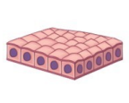
Simple Squamous Epithelium (one layer of flat cells)
Function: thin for diffusion
Location: lungs and blood vessels
Tissue, Function, Location
Simple Cuboidal Epithelium (one layer of boxed shaped cells)
Function: secretion and absorption
Location: walls of glands and in the kidneys

Tissue, Function, Location
Simple Columnar Epithelium (one layer of pillar shaped cells)
Function: absorption and secretion of mucus
Location: lining of the digestive tract

Tissue, Function, Location
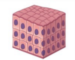
Stratified Squamous Epithelium (two ore more layers of flat cells)
Function: protection from abrasion
Location: skin, mouth esophagus, vagina
Tissue, Function, Location
Stratified Cuboidal Epithelium (two or more layers of box shaped cells)
Function: N/A
Location: in the ducts of sweat glands and mammary glands

Tissue, Function, Location
Stratified Columnar Epithelium (two or more layers of pillar shaped cells)
Function: N/A
Location: pharynx and male urethra

Tissue, Function, Location
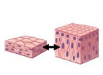
Pseudostratified Columnar (pillar shaped cells of varying heights with vary nuclei)
Function: propels particles forwards using cilia; secretes mucus
Location: trachea and fallopian tubes
Tissue, Function, Location
Transitional Epithelium (stratified epithelium with a top layer that is sometimes squamous and sometimes dome shaped)
Function: stretches easily
Location: urinary bladder, ureters, and urethra

Merocrine
Cells excrete the products via cytosis
Ex. sweat glands

Apocrine
A portion of the cell pinches off with the secreted products
Ex. mammary glands

Holocrine
Cells rupture and spill products into the ducts
Ex. sebaceous glands
Tissue, Description, Location
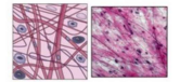
Aerolar (connective tissue)
Description: loose CT, mostly open space, holds water and salts for surrounding tissues
Location: under epithelial tissues and around organs

Tissue, Description, Location
Reticular (connective tissue)
Description: loose CT that forms a soft skeleton which supports other cells; has only reticular fibers
Location: lymph nodes, spleen, bone marrow

Tissue, Description, Location
Adipose (connective tissue)
Description: loose CT that is made of tightly packed fat cells; serves as energy reserves, insulation, and cushioning
Location: under the skin, around the kidneys and heart

Tissue, Description, Location
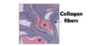
Regular dense (connective tissue)
Description: dense CT that has closely packed, parallel collagen fibers; strong when pulled in one direction
Location: within tendons and ligaments
Tissue, Description, Location
Irregular Dense (connective tissue)
Description: dense CT that has randomly arranged collagen fibers; strong when pulled in many directions
Location: in the dermis, in fibrous coverings that surround organs and joints

Tissue, Description, Location
Elastic dense (connective tissue)
Description: dense CT that has a high number of elastic fibers; allows tissues to bounce back after stretching; provides flexible cushioning
Location: walls of arteries, ligaments connecting vertebrae, lungs

Tissue, Description, Location
Hyaline cartilage (connective tissue)
Description: cartilage that is the most common in the body; few collagen fibers; firm ground substance; appears glassy
Location: larynx, bridge of nose, ribs

Tissue, Description, Location
Elastic cartilage (connective tissue)
Description: cartilage that contains many elastic fibers and is very flexible
Location: external ear, epiglottis

Tissue, Description, Location
Fibrocartilage (connective tissue)
Description: cartilage that contains rows of thick collagen fibers, allowing it to resist compression and tension
Location: intervertebral disks, cartilage of the knee
Tissue, Description, Location
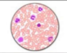
Blood (connective tissue)
Description: only liquid body tissue; has a matrix known as plasma with no fibers within it
Location: within blood vessels

Tissue, Description, Location
Bone (connective tissue)
Description: also known as osseous tissue; rigid matrix due to abundant collagen fibers and calcium salts
Location: compact and spongy bone
Simple epithelium
One layer
Stratified epithelium
Multiple layers and vary in cell shape
Pseudo-stratified epithelium
Appears to have more than one layer
Transitional epithelium
Changes its appearance as it stretches
Epithelial tissue
Lining tissue that forms the boundary between our body and the external environment. Functions include protection, absorption, filtration, secretion.
Apical surface
Free surface (top) of epithelial tissue
Basal surface
Bottom surface of epithelial tissue
Basement membrane
Membrane underneath the basal surface
Avascular
No blood vessels
Gland
A group of cells that secrete a fluid substance. Classified by complexity and how the substances are secreted (exocrine vs. endocrine).
Exocrine glands
Secrete substances outward through a duct
Endocrine glands
Ductless glands that secrete hormones through the bloodstream so they can be delivered over longer distances.
Connective tissue
Provide support and bind other tissues together. Provide insulation and protection for other tissues. Some can transport substances.
Mesenchyme
Stem cell that forms all connective tissues
Osteocytes
Secrete ground substances, fibers, collagen, and fluids. The ground substance eventually becomes calcified and hard.

Mast cells
Detect foreign substances; contain granules that secrete inflammatory chemicals

Macrophages
Blob-like cells that engulf invading substances or organisms
Red blood cells
(Erythrocytes), carry oxygen
White blood cells
(Leukocytes), provide immune response

Smooth muscle tissue
Contracts involuntarily, non-striated. Found in the walls of the uterus, stomach, intestines, bladder, arteries, veins and eyes.
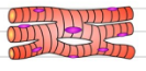
Cardiac muscle tissue
Contracts involuntarily, striated. Found in the heart and activated by the brain or hormones.

Skeletal muscle tissue
Contracted voluntarily, striated. Connected to the bones of the skeleton and controlled by somatic nervous system.

Neurons
Make up 10% of all nerve cells. Convert stimuli into nerve impulses. Pass impulses to other neurons, muscle fibers, or glands. Unable to regenerate.

Neurologia
Make up 90% of all nerve cells. Support and protect neurons. Do not generate nerve impulses. Able to regenerate after injury.
Tissue
A group of cells that work together to perform a specific job. Four major tissues include connective, epithelial, muscular, and nerve.
Loose connective tissues
Aerolar, adipose, reticular
Dense connective tissue
Regular dense, irregular dense, elastic dense
Cartilage
Hyaline, fibrocartilage, elastic cartilage