Biochem Unit 2 before quiz
 Call Kai
 Learn
Learn Practice Test
 Spaced Repetition
 Match
Match Flashcards
 Knowt Play
1/93

There's no tags or description
Looks like no tags are added yet.
Name | Mastery | Learn | Test | Matching | Spaced | Call with Kai | Chat |
|---|
No analytics yet
Send a link to your students to track their progress
94 Terms
functions of globular proteins
storage of ions and molecules- myoglobin, ferritin
transport of ions and moelcules- hemoglobin, seratoinni transporter
defense against pathogens- antibodies, cytokines
muscle contraction- acid and myosin
biological catlysis- lysosome and chymotrypsin
protein function and binding
relies on interactions with other moelcuels
binding is reversible- aka transient because it is not helpful if things are allays covalently linked- ALL REACTIONS ARE REVERSIBLE
ligand
molecule that a protein can bind to in a reversible manner- small molecule
binding site
site on protein where ligand binds- specific
binds via noncovalent forces which are thae same as the ones that dictate protein structure which is what allows interactions to be transient
2 potential methods for ligand binding
lock and key model- high specificity explained by complementary of the binding site and the lignad
complimentary in size, shape, charge, and hydrophobicity
less likely is what happens because that likely would have a rigid interaction and would not be able ot interact with each other for very long
likely true model:
induced fit mode- proteins are flexible where both the ligand and protein can change their conformations upon invading, makes binding site more complementary to ligand
bind tighter because they conformationally change to bind together
myoglobin function and structure
compact globular protein composed of a single polypeptide chain- momoner- 153 amino acids in length
carries and stores oxygen in msucle
contained a heme prostethic group- a perphoryin ring completed with an iron ion (fe2+)
oxygen binds to Mb via the heme
myoglobin structure
has eight alpha-helical regions which are designated A though H which surround the Heme group N-terminus is the A group
the Mb polypeptide chain golds to form a cradle that nestles the heme prosthetic group
protects the heme iron atom from oxidation
provides pocket into which the oxygen molecule can fit
two polar His residues are found in the interiior'; they are involved in interactions with the heme group and bound oxygen
heme
perphoryrin ring and iron
myoglobins prosttic group which is tetra dentate with a heme in the middle. without this group, myoglobin cant bind to oxygen
the Fe binds to two more things other than the amine groups on the ring which include a histidine residue-called the proximal histidine residue, and oxygen
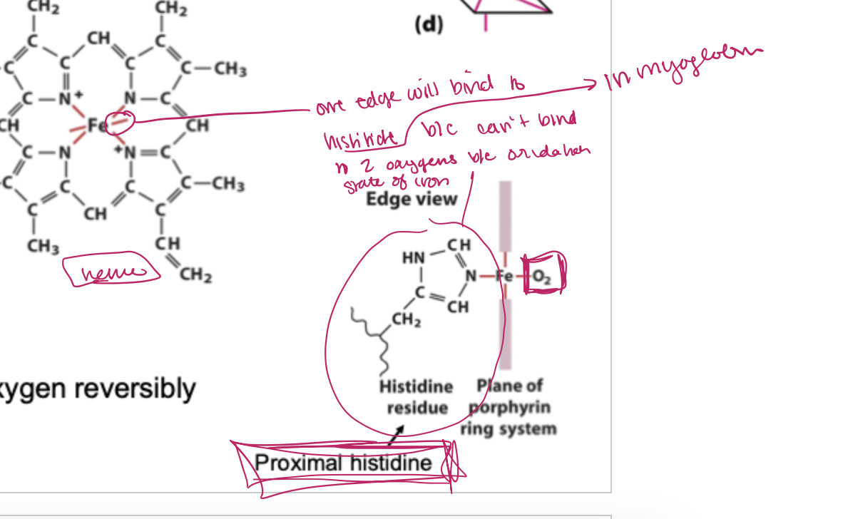
the distal histidine
statically inhibits oxygen from binding perpendicularly to the heme plane
oxygen/carbon monoxide binding to heme effect of distal histidine
o2 has a 25,000x weaker affinity for iron but because the distal histidine is interacting with it, it bends the o2. this prevents the carbon oxygen triple bond from binding because it does not want to interact with the distal histidine
not flexible triple bond
binding affinity
affinity is a measure of how tight a protein P binds to a ligand L. This is also expressed as a dissociation constant, Kd, which is simply an equilibrium constant for the dissociation reacition. Kd has units of M, the lower the value of Kd, the tighter the binding
the value of Kd is always the same for a specific protein-ligand interaction
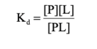
how to find Kd
when the concentration fo the protein bound to ligand equals the concentration of free protein then P/PL= 1 and the concentration of free ligand equals the Kd. therefore Kd=L at 50% saturaiton
concentration of ligand when the protein is half saturated
L=kd
L>kd
L<kd
=, half of P will be bound to L and half will be free
L>kd, P will mostly be bound to L
L<kd, P will mostly be bound to L
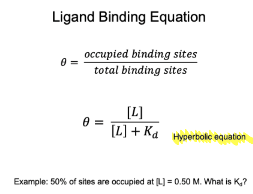
the ligand binding equation

what is the graphical trend as the Kd gets smaller
the binding curve becomes steeper
what type of grpah is it
hyperbolic
ligand binding equation
myoglobin vs hemoglobin
myoglobin- monomer
o2 storage
1 heme group so It can only old one o2
hemoglobin- heterodimer 2 alpha and 2 beta subints
function- o2 transport
4 heme groups- therefore can carry 4 oxygens
hemoglobin quaternary structure- tense and relaxed
tense state has a huge hole in the middle- T (tense) state deoxyHb- no O2 bound- is more stable (which is how o2 is able to dissociate)- lower affinity for o2- more interactins, more stable
relaxed state- R state- oxyHb- o2 bound- less stable- also almost no hole in the middle- higher affinity for 02- fewer interactions, more flexible

o2 binding triggers what conformational change
from T→R
conformational change from teh T state to the R state involved breaking ion pairs between teh Alpha 1 and beta 2 interface
as soon as hemoglobin binds to one oxygen, it wants to bind to al. of them
T versus R state conformation perforin ring
t state, perforiyrin ring is puckered. it is not in the R state so it pulls the histadine towards it which pulls the alpha helix its connected to closer which causes an overall structural change which is called cooperativity because it increases the interaction now between the rest of the hemes and oxygen
cooperatively
if the binding of one ligand molecule affects the affinity of another ligand molecule on teh same rotein
multiple subunits usually required
positive cooperatively occurs when the binding of a ligand increases the binding affinity of subsequent ligands
negative cooperatively occurs when the binding of a ligand decreases the bidning affinity of subsequent igand
Hb subunits exhibit positive cooperatively upon binding oxygen
two cooperatively models
concerted vs sequentiall
concerted is that all the subunits are either all tense or all relaxed but ligand bonding still occurs one by one
sequential- more likely- turns into a relaxed state as ligands bind
Mb binding curve- one-site binding
fraction saturation on teh side
Mb0 hypersonic binding curve for o2- insensitive to small chanfges in oxygen concentration. Mb cannot bind to O2 in a cooperative manner why? Bec cues theres only one binding site and one subunit (monomer)
the binding curves for hemoglobin and myoglobin global are consideredly more complaated due to multiple binding sites
Mb vs Hb binding curbe
Mb- hyperbolic- binding curve for O2
Hb- sigmoidal binding curve for O2- reflects a transition from low to high affinity binding this makes Hb highly sensitive to changes in oxygen concentration
sigmoidal curve purpose
has to have a low affinity in tissues and a high affinity in lungs so it can pick up the O2 at lungs (13 kpa) and dispers it in tissues(4kpa)
ligand interactions can be regulated
reversible binding of ligands is essentail for regulation
binding of ligands other than oxygen affects oxygen-binding properties of hemoglobin
protons, carbon monoxide, carbon dioxide, chloride ions, and BPG all affect the binding of oxygen by Hb
allosteric proteins w multiple binding site son the same protein
umbrella term which includes cooperativity
how binding of one ligand effects binding of another ligand to a different inning site on the same protein
does not always require multiple subunits
poistive- binding of one ligand increases affinity for the other
negative- binding of one ligand decreases the affinity for the other
bohr effect
shows how pH difference affects affinity
can be a negative or positive effector of an enzyme
for Hb, It is an antagonisht- negative effector and it decreases oxygens ability to hold on to hemoglobin in tissues- makes it give up the oxygen in tissues because you produce lactic acid in the body when you work out so you need more oxygen
2,3-bisphosphogycerate (BPG) is a negative allosteric effector of O2 binding
BPG binds to Hb and promotes oxygen release
does this by cross linking the two beta subunits which stabilizes it in the deoxygenated form (T-state)
in oxyHb, the central cavity is too small for BPG to fit
BPG shifts the oxygen saturation curve of Hb to the right, allow it to release oxygen to tissues
carbon monoxide is a positive effector
CO-colorless, odorless gas
250-fold greater affinity for Hb than O2
healthy person- 1% CO-Hb
smoker- 3-8%
china smocker- 15%
greater risk- smoker, heart/lung/blood disease/ fetus
sickle cell anemia
replacement of Glu residue at position 6 in the beta chain by Val marks the only chemical difference between Hb A and Hb S
a hydrophobic pocket forms in teh corner of each beta chain when it is in the deoxy state
these pockets nicely accommodate the val side chain of neighboring Hb S molecules leading ot aggregation into long chain polymeric structures
Hb S is less soluble than Hb A
insoluble Hb S distort red cell into elongated sickle shape
cooperatively- Kd
cooperative proteins have multiplee ligand bindings sites so the Kd becomes

the hill plot of cooperatively

key topics in protein function
reversible binding of ligands is essential
specificity of ligands and binding sites- high specificity- only certain ligands bind
ligand binding is often coupled to conformational changes, sometimes quite dramatic- induced fit
in multi-subunit proteins, conformational changes in one subunit can affect the others- cooperatitvity
interactions can be regulated
the bodies immune defenses
innate defense
what you are born with, do not have to be exposed to a pathogen to gain
external and internal innate defenses
external- skin, secretions, mucuou
internal- phagocytic cells
naturak killer cells
defensiveproteins
inflammatory response
adaptive defense
antibodies
lyphocytes
b cells
t cells
immune response
macrophage will endocytose a pathogen with a specific antigen, it will then show the antigen on its surface, and the B cell will interact with it and produce antibodies for it. the T cell then interacts with the macrophage and B cell with said antigen and targets those cells for death
memory cells
recognize different antigens through antigen receptor on cell surface.
these cells will either become memory cells which maintain antigen receptor or they will become plasma cells where they release the antibody
antigens and antibodies
antigens- foreign molecule that stimulate production fo antibodies- proteins or large polysaccharides on the surfaces of biruses or freight cells
antibodies- protiens produced by B cells that bind specifically to antigens
binding of antibodies to antigens will make the antigen for destruction or interfere with its function
a given antibody will bind to a small region of the antigen
one antigen can have several epitopes
epitopes recognized by antibody- unique shape- 5-10 amino acid sequences
one again may have more than one antibody against it, etch antibody regonizeing a unique amino acid sequence
antibodies structure and function
y shaped structure
made of four peptide cains
two identical light chains, and two identical heavy chaiins
help to gather by disulfide bonds
n- termini of light and heavy come together to form antgen binding site (unique antino amongds/epitope bind here)

structural bases of antibody diversity
immunoglobulins are divided into 5 isotopes: IgA, IgD, IgE, IgG, IgM. they differentiate in structure in the constant region and have different effector functions
IgG is the most abundant antibody in blood and lymph and I used as the prototype in illustrating antibody structure
typical immune response
following exposure to a foreign antigen, the immune sytem produces antibodies directed against the diseased antigens
body first produced antibody called immunoglobulin M (IgM)
within a week of ideates exposure IgM can be detected in blood- approximately three weeks later, IgM levels drop and IgG levels rise and then fall within a few days
the second time teh body is exposed to the same disease, the immune response is stronger and faster than the first time
this is the principal behind vaccinationand booster shops

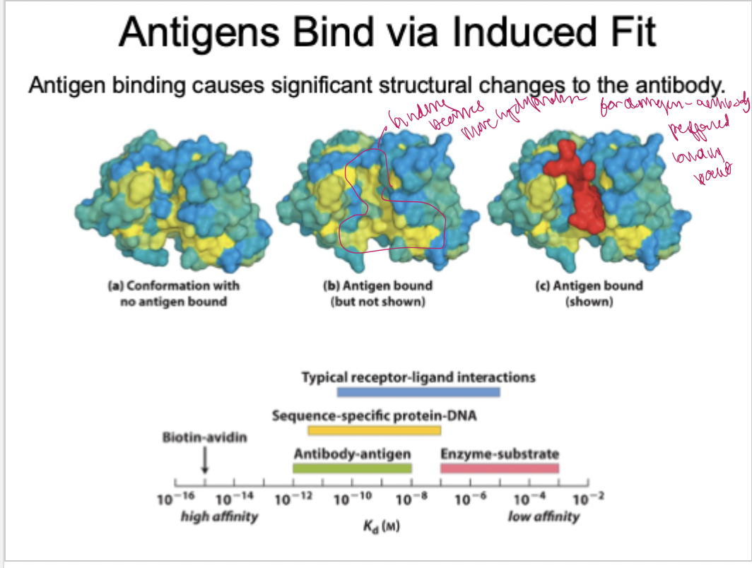
antibgens bind via induced fit
antigen binding auses significat structural changes to the antibody
Uses of antibodies in biochemistry
applications:
westernblotting protein identification following SDS-PAGE
immunoprecipitation- isolation of specific protiens and binding partners
immunoflouresence microscopy- localizatin of specific proteins in cells
ELIZA- detection of protiens in a sample
muscle proteins
msucle fiber0 large, single, elongated multinuclear cell
each fiber contains about 1,00 myofibrils which is one cell
actomyosin cycle

enzymes
systems cannot increase temperature without risking damaging structures so they use catalyssits- cah[nge the rate of a reaction without net change of catalyst, increase reationr ates without being consumed and without altering equalbrium
catalysts increase reactionaries without… (2 things)
without being consuemed
without altering equalbrium
enzyme
a substance produced by a living organism that acts as a catalyst to bring about a specie biochemical reaction; present at low concentrations
vast majority of biological catalysts
globular proteins
some RNA also catalyze reactions- ribbonymes
definition of : substratecte, product, active site
substrate- substance acted upon
product- resulting species
active site- location in enzyme where reaction occurs (extremely specific for substrates- but nonspecific for product usually)
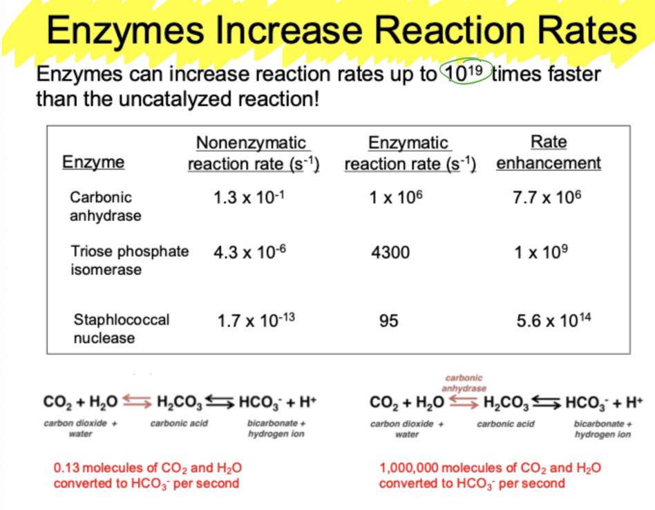
enzymes increase reaction rate
can increase up to 10^19 times faster than the uncataluzed reaction
enzyme activity
many enzymes can actin the forward and reverse reactions
specific interactions in active site determine binding affinity- shape, charge, hydrophobic
enzyme activity is regulated by
pH- can change structure by changing charges- likely in active site
temperature- can increase reactionr ate
concnetration of enzyme
concentration of sybstrate
concnettartion of cofactors or coenzymes
last 3 re more likely to regulate in vitro
cofactors
include coenzymes - organic molecules
cosubstrate- bound more loosely- consumed within reaction
proseteic groups- tightly bound, not consumed
Inorganic moleucles
1/3 of all known enzymes require metal ions- metalloenzymes
usually positively charged
app vs holo enzyme
apo is inactive
holo is active and is an app enzyme with a cofactor or coenzyme
activation energy (free energy of activation)
chemical reaction occurs when colliding molecules possess a minimum amount of energy caled the activation energy
activation energy is the difference between energy levels of the ground state and the transition state
the rate of a reaction reflects this activation energy
a higher free energy of activation corresponds to a slower paction
free energy change
the amoutn of energy available to do work
each compound in a chemical reaction contains a certain amount of potential energy related to the kind and number of its bonds, in reactions that occur spontaneously, the products have less free energy than teh reactions. thus the reaction releases free energy, which is then available to do work
many reactions that are spontaneous (neg. dG) will proceed at slow rates because they do not have the energy or correct orientation
the rate at which a spontaneous reaction occurs iimporives with increasing the temperature or using a ctalyst
spontaneous if free energy changes is negative
free energy
dG predicts the direction that a reaction will proceed
negatie dG indicates spontenaity and exergonic - rxn will go forward
positive d G indicates nonsponteanous nad endergonic- rxn will go backward
when dG is zero, at equilibrium
Is dG changed by an enzyme
not the Gibbs free energy, but the free energy of activation is changed where the activation decreases but the still total amoutn of energy release will still be the same
dg is the difference in energy between substrate and product it is not changed by an enzyme
what does change ins the free energy difference between substrate and the transition state
how? enzymes are flexible
how do enzymes increase rates
a catalystt reduces the activation energy of a reaction
the free energy of activation is the amoutn of energy needed to convert 1 molecule fo substrate from the ground state to the transition state
complementarity of active site- stickase
no enzyme- reaction will eventually happne
enzyme complementary to substtate- lock and key method
enzyme complementary to transition state- induced fit model

general acid-base catalysis
general acid- partial proton transfer from an acid lowers free energy of the reactions transition state
general base- partial proton abstraction by a base lowers free energy of a reactions transition state
covalent catalysis
covalent bond forms between enzymeand substreate
a transient covalent bond between enzyme and substrate
rchanges reaction pathway- reaction mechaism
requires a nucleophile on the enzyme
can be a reactive hydroyl, thiol, amine, or carboxyl


metal ion catalysis
involved a metal ion bound to the enzyme- metal ion interacts with substrate to facilitate binding
what metal ions can do:
help oritein substrate for reaction
stabilize negative chanrges
medicate oxidation-reduction eactions
chymotrypsin
small intestine enzyme which breaks down free protiens
protease (protein-ases)
during digestion, dietary proteins must be broken into small peptides by proteases
chymotrypsin is one of several proteases that cuts peptides at specific locations ont he ppttide backbones
likely has aromatic binding site because only cleaves next to aromatics
this protease cleave the peptide bond adjacent to (c-terminal to) aromatic amino acids
chymotrypsin is a ____ protease and requires a ____ _____
serine protease; catalytic triad
what amino acids are apart of the catalytic triad
histidine, aspartic acid, and serine
oligeomerization
heterotrimer; alpha beta and c chain
asp, his, see, and gly make up internal binding pocket
covalent catalysis using chymotrypsin; steps
bind subtrate
form covalent bond
cleave peptide bond
release 1s product (c-terminal)
break covalent bond
release product 2 (n-terminus)
enzyme is unchanged when we finish it
chymotrypsin mechanism
serine residue hydrogen bound to the histidine residue with a nonbonding but able to hydrogen bond glycine residue.
substrate then enters and aromatic ring will bind to hydrophobic pocket
when bound to pocket, c-terminal =o and hydrogen on serine is h-bonding with the nitrogen on histidine

step 2
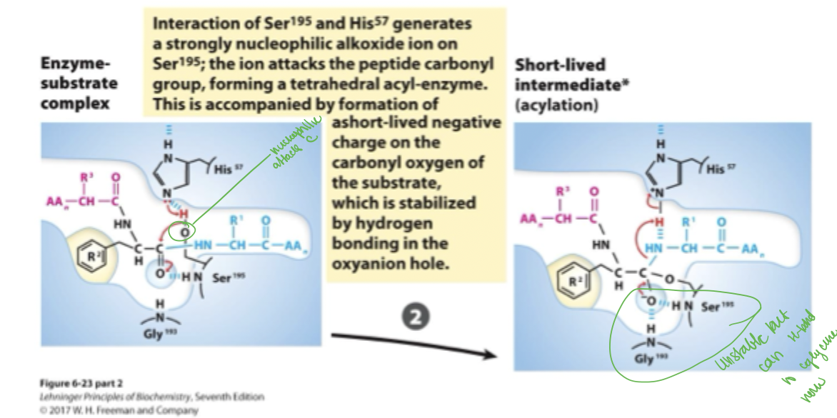
histidine takes serines proton, serines now negatively charged oxygen is now a nucleophile and attacks the carboxyl carbon which causes the =o to become a —o-
this —o- can now go back to being a =o which kicks off teh c-terminal peptide, giving the extra electrons to hydrogen
step 3
c terminus exits and n terminus is still hydrogen bound to to serine residue still

step 4
water comes in and the nitrogen on the histidine attacks the hydrogen on the water and then the hydroxide attacks the carboxyl carbon which then forms a —o- bond which can now h-bond with the glycine’s backbone. t

step 5
distance between nitrogen and hydrogen is now too far, their bond breaks because serine wants it back. serine’s O- bond with carbon also breaks because the —o- can go back to forming the =o

step 6
the product can now dissociate

six classes of enzymes
oxidoreductases
transferases
hdyrolases
lyases “synthases”
isomerases
ligases “synthetases”
oxidoreductases
catalyze electron transfer in redox reactions

transferases
transfer a funcitonal group from one molecule to another

hydrolases
cause cleavage of a bond using water- water receives the functional group being transferred- known as hydrolysis

lyases “synthases”
cleave or form a bond between two molecules

isomerases
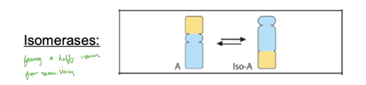
move groups within a molecule (intramolecular rearrangement)
what types of co-substrates are commonly present in enzymatic reactions
water, ATP, and NADh (e- accept or donator)
enzymes are used in cellular pathways but why isnt the rection compelted in jsut one step? why so many intermediates?
each reaction releases energy becaues it is exothermic and favorable. therefore, cannot happen all at once because cells cannot tolerate large amounts of energy or heat being released. they cannot generate a large energy burst to drive energy-requiring processes.
instead, transformations take place via sequential series of chemical reactions whose overall affect achieves dramatic energy changes, even though any given reaction in teh series proceeds with only modest input or output of energy
enzyme clinical relevance
enzymes are used clinically in three principal ways
diagnosis and prognosis- measure enzyme activity
measurement of substances (cholesterol, glucose, hormones)
therepautics