Chapter 4: Pulsed Waves
 Call Kai
 Learn
Learn Practice Test
 Spaced Repetition
 Match
Match Flashcards
 Knowt Play
1/42

There's no tags or description
Looks like no tags are added yet.
Name | Mastery | Learn | Test | Matching | Spaced | Call with Kai | Chat |
|---|
No analytics yet
Send a link to your students to track their progress
43 Terms
In Diagnostic medical ultrasound, we __________ use a sound wave that is continuous.
CANNOT
Pulsed Waves
Anatomical imaging and Doppler calculations used to measure blood flow, relying on turning the sound wave OFF and listening to the returning echos
The ultrasound system then keeps track of all the echoes, and the elaborate information in those echoes, which will then be represented in an image
Continuous Wave
two crystals in the transducer
one constantly transmits (voltage continuously applied to crystal)
one constantly receives
Echoes from every single depth (range ambiguity) are constantly received which means the machine cannot decipher depth to place echo (chaotic) so it CANNOT make an image
Used in Doppler
Not used in imaging
Pulsed Ultrasound
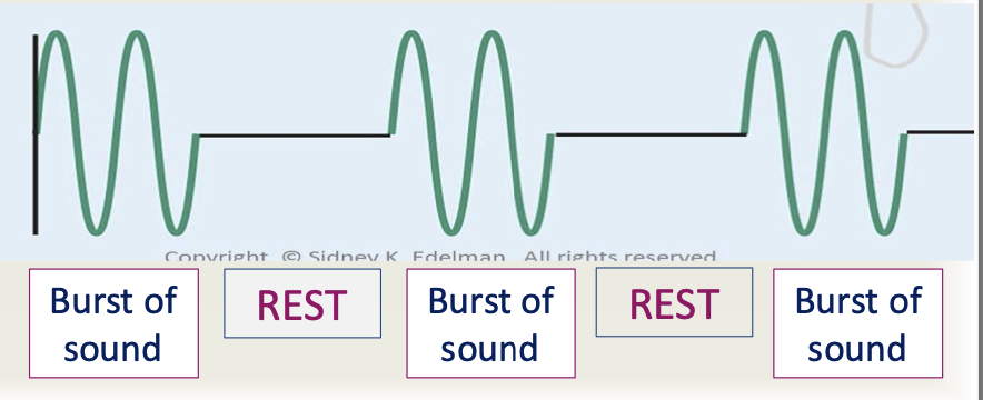
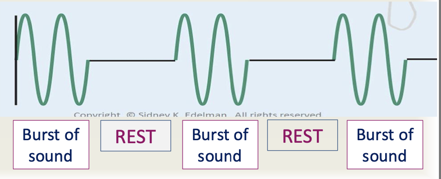
A sound wave with multiple short bursts of sound, each followed by a resting period of no sound
A set of burst of acoustic energy (voltage applied to the crystal intermittently)
Each pulse of sound has multiple cycles all traveling as one
NOT on continuously
Requires one crystal that both transmits and receives.
Why Pulsed Waves?
sound waves cannot be continuous in imaging because THEY MUST HAVE A BEGINNING AND AN END
Sound must pause for a time of listening to receive information from the sound wave
Pulsed Wave Definition
Any modality which turns the transmitter on and off periodically to reduce range ambiguity
PW Doppler, Color Doppler, 2D imaging, 3D imaging, and M-Mode
Acoustic Line
a single sound beam transmitted in a specific direction and the associated echoes
the word beam is more accurate here because line makes you think of just one dimension
Receive Line
the returning echoes registered by the system from a single direction over the time between the transmit event and the time until the next transmit occurs, as dictated by imaging depth
Display Line
(or image line) is the data displayed on the screen that corresponds to a single (or multiple) acoustic lines
Line
The general term used to refer to a beam
An Image
picture generated by sequentially transmitting many acoustic lines
also called frame, scan, or scan region
Frame Time
time required to transmit multiple beams to create an image of the desired region
Frame Rate
reciprocal of frame time, frame frequency
Sampling Rate
the frequency at which signals are detected or viewed
Parameters to Describe PULSED sound waves
Pulse Duration
Spatial Pulse Length
Pulse Repetition Period
Pulse Repetition Frequency
Duty Factor
Pulse Duration (def, units, values, determined, adjustable)
the actual time from the start of one pulse to the end of that pulse
duration = time
The ON TIME of a pulsed wave
UNITS: time (microseconds, ms, seconds)
VALUES: 0.3-2.0 microseconds
DETERMINED: by sound source ONLY
ADJUSTABLE?: No

Pulse Duration Equation
PD = #cycles x period
PD = #cycles / frequency
directly related/proportional to:
number of cycles in a pulse
period
Inversely related/proportional to:
frequency
Pulse Duration Continued
not all pulses are the same
PD varies depending on modality (2D, Doppler, etc)
The # of cycles in the pulse is part of determining the pulse duration
depends on the imaging modality (like 2D vs. Doppler)
In 2D imaging:
SHORTER pulses = BETTER image quality
PW Doppler requires a longer pulse (on time)

In pulse duration, Long duration = ???
many cycles per pulse
long periods per cycle
In pulse duration, Short duration = ???
few cycles per pulse
short periods per cycle
shorter PD = greater image accuracy
If a pulse contains 10 cycles and the period of 0.2 microseconds, what is the PD?
2 microseconds
If a pulse contains 8 cycles and the frequency is 2MHz, what is the PD?
4 microseconds
Spatial Pulse Length (def, units, values, determined, adjustable)
length or distance of a pulse
UNITS: distance (mm)
VALUES: 0.1-1.0 mm
DETERMINED: by sound source AND medium (like wavelength)
ADJUSTABLE: No

Spatial Pulse Length Equation
Spatial Pulse Length = #cycles x wavelength
Directly proportional to:
number of cycles in a pulse
wavelength
Inversely proportional to
frequency
***SPATIAL PULSE LENGTH AND PULSE DURATION MEASURE THE SAME THING IN DIFFERENT UNITS*******
In spatial pulse length, long pulses = ????
many cycles
long wavelengths per cycle
In spatial pulse length, short pulses = ????
few cycles per pulse
short wavelengths per cycle
shorter pulses = greater image accuracy
Calculate SPL for 5MHz sound in soft tissue, 5 cycles per pulse
1.55 mm/pulse
Pulse Repetition Period (PRP) (def, units, values, determined, adjustable)
time from start of one pulse to the start of the next (includes listening time)
UNITS: time, microseconds
VALUES: 100 microseconds - 1 ms
PRP is often given in sec, so you have to multiply by 1000 to get to the units of microseconds to calculate
DETERMINED: by sound source ONLY
ADJUSTABLE: Yes, when depth is adjusted, PRP is altered (only alters listening time, PD stays the same)
PRP Relationship
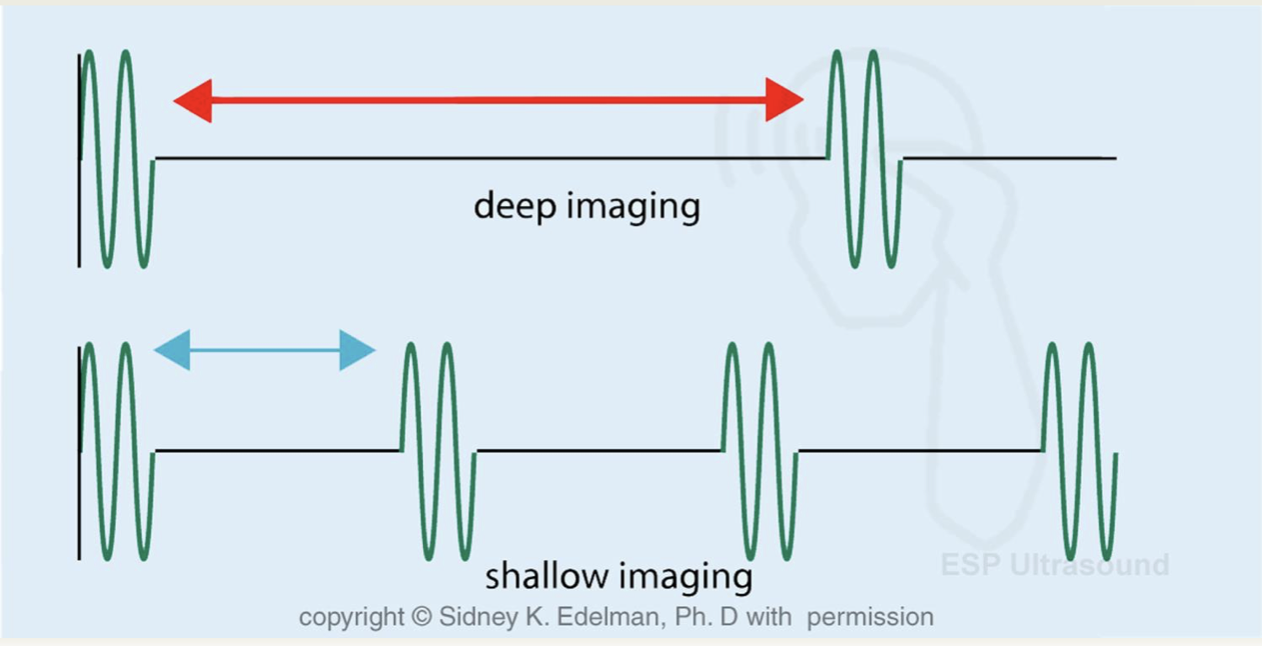
Shallow Depth Imaging
time from one pulse to the next is short
Deep Imaging
time from one pulse to the next is long
As Depth increases, PRP increases
2 Components of PRP
Transmit time - one time (blue)
Receive time - off time (black)

Pulse Repetition Frequency (PRF) (def, units, values, determined, adjustable)
number of pulses transmitted per second
UNITS: Hz or per second
VALUES: 1,000 - 10,000 Hz (or pulses per second)
DETERMINED: by sound source ONLY
ADJUSTABLE?: Yes, when depth is adjusted, PRF is altered
PRF Relationship
shallow depth of imaging
more pulses are sent, as listening time decreases
less time to wait to send the next pulse
deep imaging
less pulses are sent as listening time is longer
longer time to wait to send the next pulse
As depth increases, PRF decreases
PRF and PRP Relationship
inversely related
reciprocals
PRP = 1/PRF
PRF = 1/PRP
PRP x PRF = 1
Why is PRF so important?
PRF determines how quickly we can create an image
Duty Factor (def, units, value, determined, adjustable)
Percentage or fraction of time a pulse is being transmitted
the “on” time of a pulsed wave
UNITS: none, %
VALUES: in ultrasound, 0.2% is typical
about 500 times longer listening time than transmit time
DETERMINED: by sound source ONLY
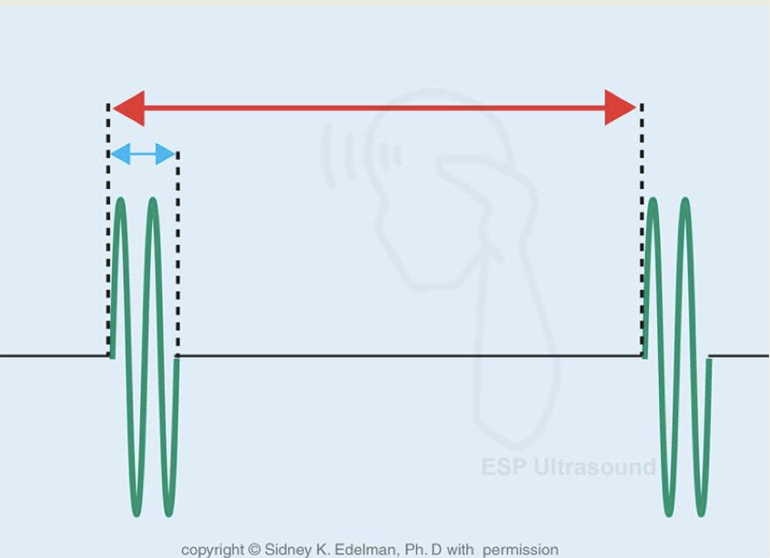
ADJUSTABLE?: Yes, changing depth changes the DF
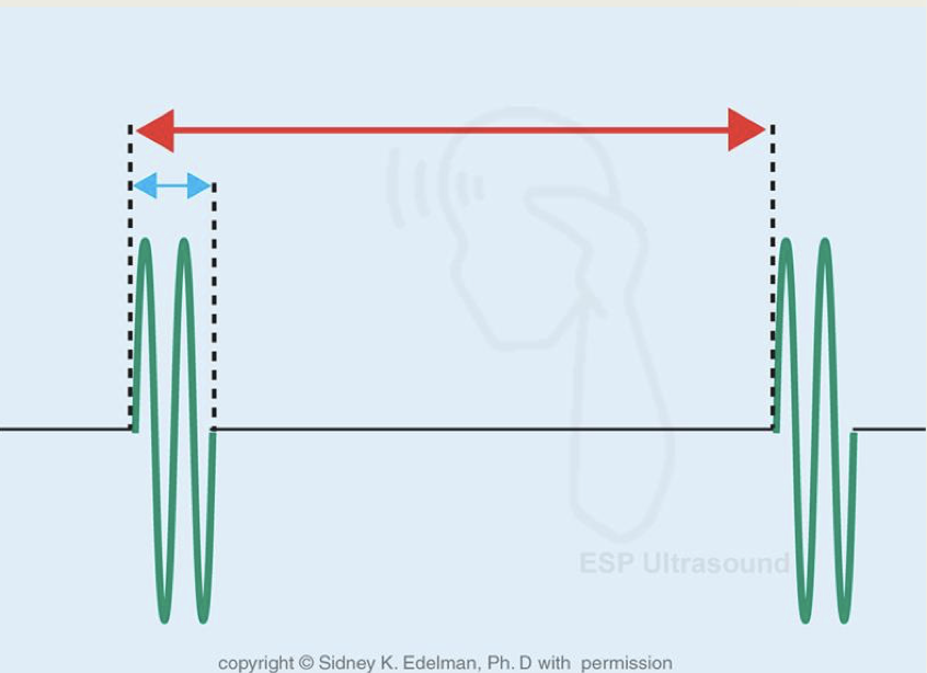
red line =
blue line =
red = PRP
blue = on time / transmit time / PD
Duty Factor Equation
DF = (PD / PRP) x 100
max value = 100% or a factor of 1
in continuous sound waves
no listening “off” time
no anatomical image can be created with continuous wave
min value = 0%
occurs when transducer is silent
ex: when we hit the freeze button
If you work 2 days out of 7 days, you work ____% of the time
29%
Duty Factor Relationship
DF is proportional to the PD
DF is inversely proportional to PRP
DF = (PD/PRP) x 100
What happens to the DF if PRP increases?
DF decreases
What happens to the DF is PD increases?
DF increases
What happens to the DF if PRF increases?
DF increases (PRP and PRF are reciprocals)
What happens to the duty factor if imaging depth increases
DF decreases