NEUR30080 L8 SF striated muscle: excitation-contraction coupling
 Call Kai
 Learn
 Practice Test
 Spaced Repetition
 Match
 Flashcards
 Knowt Play
1/66

There's no tags or description
Looks like no tags are added yet.
67 Terms
learning goals

3 types of muscle
skeletal: attached to bones and moves and supports skeleton
smooth: surrounds hollow cavities and tubes
cardiac: the muscle of the heart
skeletal muscle structure
composed of cylindrical muscle fibres (cells)
linked to bones by tendons at each end of the muscle
skeletal muscle structure image

the 3 types of muscle fibres
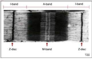
skeletal muscle fibres have a repeating, striated pattern on light and dark bands (due to arrangement of thick and thin filaments within the myofibrils)

why does skeletal muscle have a pattern of light and dark bands?
due to arrangement of thick and thin filaments within the myofibrils
skeletal excitation-contraction coupling overview
excitation (AP generation)
propagation along T-tubules
voltage sensing and mechanical coupling
calcium release from SR
muscle contraction
relaxation (calcium reuptake)
Step 1. skeletal excitation-contraction coupling
excitation (gen of AP)
Step 2. skeletal excitation-contraction coupling
propagation along T-tubules
Step 3. skeletal excitation-contraction coupling
voltage sensing and mechanical coupling
Step 4. skeletal excitation-contraction coupling
Ca release from SR
Step 5. skeletal excitation-contraction coupling
muscle contraction
Step 6. skeletal excitation-contraction coupling
relaxation (Ca reuptake)
skeletal excitation-contraction coupling overview: image

what is excitation-contraction coupling?
the process of coupling surface membrane depolarisation to Ca2+ release from the sarcoplasmic reticulum
sarcoplasmic reticulum (SR)
endoplasmic reticulum of muscle cell
stores and releases Ca2+
filaments in skeletal muscle fibre
actin-containing thin filaments are anchored to the Z lines at each end of a sarcomere
the free ends of thin filaments partially overlap the myosin-containing thick filaments in the A band at the centre of the sarcomere
filaments in skeletal muscle fibre image
actin-containing thin filaments = yellow
*myosin cross bridge IMPORTANT

filaments in skeletal muscle fibre: band overlap image
actin and myosin overlap at dark point
muscle fibre → myofibrils → sarcomeres
one muscle fibre has many myofibrils, each myofibril has multiple sarcomeres
thick and thin filaments in the sarcomere
black: Z line
yellow: actin thin filaments
red: myosin thick filaments
whole unti: sarcomere


Q:
50,000 sarcomeres
molecular mechanism of contraction
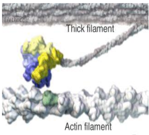
when a skeletal muscle actively shortens, the thin filaments are propelled towards the centre of their sarcomere by movements of the myosin cross-bridge that bind to actin
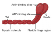
the two globular heads of each cross bridge contain a binding site for actin and an enzymatic site that splits ATP

globular head of cross bridge diagram
thin actin filament: troponin-tropomyosin
two other proteins which assist in regulating the actin-myosin relationship
tropomyosin protein coils around actin helically and attaches via troponin complex
troponin-tropomyosin complex image
each black dot is myosin binding site

which filament type is troponin-tropomyosin complex present in?
thin actin filament
Ca2+ conc role
induces contraction
myosin-binding site exposed

cross-bridge activation: summary
in a resting/relaxed muscle: attachment of myosin cross-bridges to actin is blocked by tropomyosin molecules that are in contact with the actin subunits of the thin filaments
contraction: initiated by an increase in cytosolic Ca2+ conc
the Ca2+ ions bind to troponin, producing a change in it’s shape that is transmitted via tropomyosin to uncover the myosin binding sites of actin, allowing the myosin cross bridges to bind to the thin filaments
contraction: 4 steps
cross-bridge formation: ADP + Pi → cross-bridge
power stroke: release of Pi, then ADP
detachment: ATP binding
ATP hydrolysis: ADP + Pi + energy (cocked position)
contraction: 4 steps image

actin-myosin interaction
by converting chemical bond energy into mechanical energy, myosin is able to move along the actin filament:
ATP binds to the myosin head: causes it to detach from actin
this ATP hydrolyses, removes from the myosin head and undergoes ATP → ADP + Pi rxn: releases energy, myosin now in high-energy state
myosin in it’s high energy state ‘pushes’ on the actin filament
ADP released and the process loops back around, except now the myosin is in a new position on the actin filament
actin-myosin interaction aka
actin filament sliding theory
filaments mechanism timing
filaments work out of sync with each other, preventing actin from slipping back to original position
actin-myosin interaction image
SR/transverse tubules/myofibrils
forms branching web of memb-bound tubes and sacks that wrap around myosin fibrils
SR: stores and releases Ca2+
anytime there is a Ca2+ influx, there will be a shape change
SR/transverse tubules/myofibrils image

malignant hyperthermia
excessive Ca2+ release from the SR
causes uncontrolled muscle contraction and increases body temp
Ca2+ channel blockers regulate Ca2+
contraction and Ca2+
contraction depends on a rise in Ca2+ in cytosol
this is triggered by an AP in plasma memb.
the AP is propagated into the interior of the fibre along the transverse tubules to the region of the SR, where it produces a release of Ca2+ ions from the reticulum
relaxation of a contracting muscle fibre occurs as a result of the active transport of cytosolic Ca2+ back into SR
release/uptake of Ca2+
muscle AP propagated
Ca2+ released from sac
Ca2+ binding to troponin removes blocking action of tropomyosin
myosin cross-bridge moves
Ca2+ takes back up via SERCA pumps
Ca2+ removal from troponin restores tropomyosin, blocking action
triad junction structure
AP travels to transverse (T)-tubules
results in SR Ca2+ release
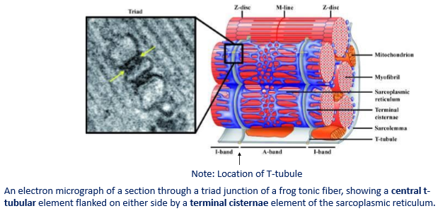
triad junction: repeating structural unit composed of 2 SR terminal cisternae facing a central T-tubule
triad junction structure image
cisternae
flattened sacks in the memb
electron MS of triads location

electron MS of triads: triads parallel to each other

transverse tubules (T-tubules)
T-tubules: the central elements of the triad form a continuous network across the muscle fibres
bring APs into interior of muscle fibre
depolarisation: voltage-dependent activation & conformational change of the subunits of the L-type Ca2+ channel or DHRP (dihydropyridine receptor)
T-tubules image

mechanical transmission at triad junction
in the early 1070s, there was much debate around the nature of transmission across the triad junction in skeletal muscle
compelling arguments were made for each of the chemical transmission, electrical transmission and for a mechanical coupling process
electrical transmission was eliminated largely because the memb. capacitance was too small to include the very large amount of SR membrane (surface area 10x larger than surface memb)
chemical transmission was argued at this time because of lack of suitable candidate
*mechanical transmission, through the junctional feet, was favoured, with a model of a series of levers connecting a voltage sensor in the T-tubule membrane with a Ca2+ release pathway in the SR memb
mechanical transmission at triad junction image

excitation-contraction coupling at the triad junction
focus on:
electrical impulse transmission (mechanical because directly acts with protein)
SERCA pump
calsequestrin (like a Ca sponge)
calmodulin (CaM) (modulates Ca)
DHPR

DHPR
dihydropyridine receptor
voltage sensor
L-type Ca2+ channel in the membrane of T-tubules
4 subunits:
alpha1S (voltage-sensing and pore-forming)
alpha2-sigma
beta1a (EC coupling, essential for surface expression of DHPRs and tetrad formation)
upsilon
DHPR image

RYR1
ryanodine receptor
Ca2+ release channel
ligand-gated (DHPR is a ligand itself & activates)
forms a homotetramer
largest known ion channel
RYR1s structures highly clustered square structures, arranged in regular rows in which corners of adjacent channels contact each other
mechanical coupling happens here
RYR1 structure image

the SR and ryanodine receptors
SR initially develops as ER containing generic ER proteins
as the cell differentiates, it becomes greatly enriched with SR-specific protiens
Ca ATPase
calsequestrin (CaSQ): low-affinity CA binding prot
RYR1
triadin and junctin: interconnectors between CaSQ, RYR1 and SR
keeps CaSQ immobilised and in close proximity to RYR1

junctional feet at SR image
focus on triad & junctional feet
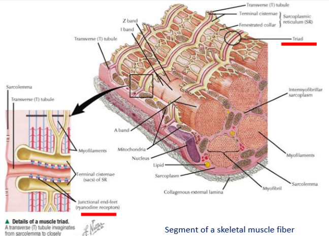
junctional feet at SR
SR vesicles contain junctional feet on outer surface
possible candidates for a role in communication between surface and internal membranes
DHPRs (voltage sensor) are grouped into tetrads
4 DHPRs composing a tetrad linked to a subunit of an underlying foot
junctional feet at SR arial view image

SERCA: reuptake of Ca2+
SERCA = sarcoplasmic endoplasmic reticulum ATPase
Ca ATPase pump (transmemb prot)
transfers Ca2+ from cytosol to sarcoplasm
SERCA: main domains
3 major domains:
phosphorylation site
nucleotide binding site
the actuator
SERCA: regulation of process
phospholamban (PLN): a 52-AA integral memb prot that regulates Ca2+ pump in skeletal muscles
sarcolipin (SLN): a small regulatory transmemb peptide that binds to and modulates SERCA pumps, reducing their Ca affinity and promoting uncoupled Ca cycling for muscle heat prod
decreases the apparent affinity of SERCA for CA
SERCA image

cardiac vs. skeletal muscle
main difference is the modulation of the extent of thin filament activation
in cardiac muscle: force development must be controlled mainly at the cellular level because each cardiac cell is activated on each beat
thus, each cell must be able to undergo the full dynamic range shown by the cardiac output
learning outcomes
understand the 6 steps

summary - 1

summary - 2

summary - 3
