PSIO 201 - Unit IV
 Call Kai
 Learn
 Practice Test
 Spaced Repetition
 Match
 Flashcards
 Knowt Play
1/245

There's no tags or description
Looks like no tags are added yet.
246 Terms
overall function of the nervous system
communication and regulation

name other more complex functions of the nervous system
rapid information transmission
regulation of body
homeostasis maintained
seat of intelligence and thought
perception
movement
behavior, learning, memory, and more!
what is the nervous system made up of?
neural tissue (neurons and neuroglia)
two basic divisions of the nervous system?
central nervous system and peripheral nervous system

what makes up the CNS?
the brain and spinal cord

what makes up the PNS?
cranial and spinal nerves
what two subdivisions make up the PNS?
somatic and autonomic
what does the somatic PNS do?
effects the skeletal muscles, voluntary movement.


what does the autonomic PNS do?
effects smooth muscle, cardiac muscle, and glands. works with involuntary movement. also includes sympathetic and parasympathetic divisions.
explain how the sympathetic and parasympathetic divisions work within the autonomic PMS
body receives dual innervation, where both of these work together to produce up and down regulations, and it is all dependent on the bodily system which is assigned a different job.
give a simple neural pathway
Sensory Receptor: sensory input —> Brain and Spinal Cord: integration
—> PNS: motor output —> Effector

flow of information in nervous system goes like…
PNS —> CNS —→ PNS
what neurons are used during sensory input?
afferent (sensory) neurons
what neurons are used during integration and processing?
interneurons
what neurons are used during motor output?
efferent (motor) neurons
sensory (afferent) nerves come from and go to?
PNS to CNS
motor (efferent) nerves come from and go to?
CNS to PNS
whats the enteric nervous system?
division of the autonomic nervous system that controls the function of the gastrointestinal tract
what kind of cells are neurons?
excitable cells
what are neuroglia’s functions?
support, protection, and nourishment of neurons.

neuron dendrite
collects signals

neuron cell body (soma)
integrates incoming signals and generates outgoing signal to axon

axon
passes electrical signals to dendrites of another cell or to an effector cell

synapse
connection point between 2 neurons

myelin
insulating material wrapped around the axon of many neurons which enhances propagation of an electrical signal
name functional neurons
afferent neurons (headed to CNS)
interneurons (integrates sensory info by analysis and storage)
efferent neurons (headed to effector through PNS)
structural types of neurons
multipolar neurons
bipolar neurons
unipolar neurons

what neuron is this?
multipolar neuron
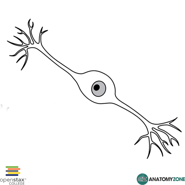
what neuron is this?
bipolar neuron

what neuron is this?
unipolar neuron
what category do bipolar neurons land in for function?
sensory
what category do unipolar neurons land in for function?
sensory
what category do multipolar neurons land in for function?
integrative and motor

astrocytes
nourish the neuron
structural support
K+ regulation
talks to neurons via glutamate
neurotransmitter uptake
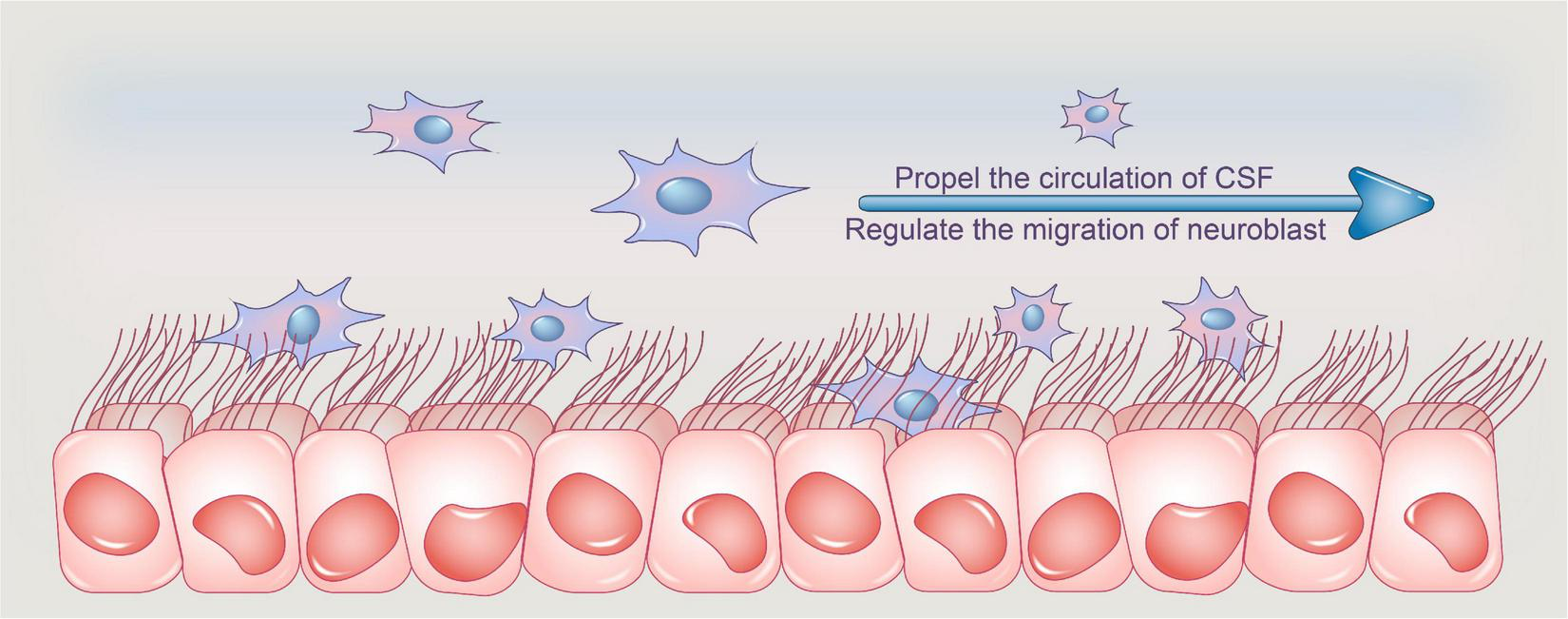
ependymal cells
line ventricles and central canal
neural stem cells
makes CSF
oligodendrocytes
insulation to speed electrical conduction in CNS
growth and repair
schwann cells
insulation to speed electrical conduction in PNS
growth and repair
microglia
immune defense cells
mobile
can prune synapses (aiding in plasticity)
cranial meninges
CT surrounding brain, protecting it and anchoring it to the cranial cavity

dura mater
“tough mother” outer layer fuses with periosteum of skull (not spinal cord) inner layers fold in cranial cavity creating sinuses.

arachnoid mater (web)
spaces for blood vessels, subarachnoid space filled with CSF.

pia mater
thin protective layer of tissue covering nerves and neruoglia
composition of cerebrospinal fluid (CSF)
plasma filtrate
glucose, O2, Na+, K+, Mg2+, other ions
functions of CSF
cushioning/shock absorption, maintain stable chemical environment, exchange nutrients and wastes.
where does CSF form?
the ventricles of the brain (1-4), numbered according to flow, and after the 4th one is emptied into the central canal of spinal cord.

choroid plexus
network of blood vessels formed from pia mater, forms CSF via filtration

CSF recycling
arachnoid granulations where CSF is absorbed back into circulatory system as fast as it produced.
what is resting membrane potential
the baseline electrical condition of all cells in the body
greater negative charge outside of the cell
greater positive charge inside the cell

with energy of 1 ATP, the Na/K ATPase pump can:
3 Na out of the cell
2 K in to the cell
what does the Na/K ATPase pump maintain?
the concentration gradients for both Na and K
at rest, the membrane is permeable to ___ through ______.
K+; potassium leak channels
what is the average resting membrane potential?
-70mV
movement of K+ ions is influenced by two competing forces:
electrical gradient (running IN to cell)
concentration gradient (running OUT of cell)
how can we change the membrane potential?
changes in membrane permeability produce large changes in the membrane potential. opening gates permits ions to move.
how do we make the membrane more permeable to K?
open K+ channels
how do we make membrane more permeable to Na?
open Na channels
what allows cells to generate electrical signals?
regulation of channel-mediated ion permeability

ion channels
integral membrane proteins
selective for certain ions
can be opened or closed
some are routinely open (ion leak channels - which are unregulated)

name this specific ion channel
chemically-gated (ligand-gated) ion channels

name this specific ion channel
mechanically-gated ion channels
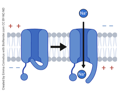
name this specific ion channel
voltage-gated ion channels
voltage-gated ion channels open when..
when the membrane potential gets less negative (depolarized)
mechanically-gated ion channels open when…
when the membrane get stretched
chemically-gated (ligand-gated) ion channels open when…
a signal molecule binds to the channel protein (e.g. ACh)
what do excitable cells do to generate an electrical signal?
change their membrane potential.
what is the principle mechanism of generating an electrical signal?
opening and closing of voltage-gated Na+ channels.
ligand gated and mechanically gate ion channels found where in relation to nervous tissue?
dendrites and cell body.
voltage gated ion channels found where in relation to nervous tissue?
axon, axon branches, and synaptic end bulbs
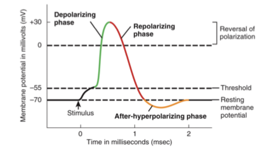
what does this graph represent?
a neuronal action potential
what are the 4 steps of generating an action potential?
local depolarization
depolarization to threshold & voltage gated Na+ channels open
voltage gated Na+ channels open
voltages gated K+ channels open
depolarization
less negative
repolarization
more negative, back to resting
hyperpolarization
most negative, past resting potential
break down of step 1 of generating an AP
local depolarization
graded potentials
local changes in cells membrane potential
2 different types
two types of graded potentials are…
IPSP (inhibitory postsynaptic potential)
EPSP (excitatory postsynaptic potential)

IPSP (inhibitory postsynaptic potential)
hyper polarizing, taking it further from an AP

EPSP (excitatory postsynaptic potential)
depolarizing, bringing it closer to excitation and AP

break down of step 2 of generating an AP
depolarization to threshold
graded potentials produce enough depolarization to reach threshold
opens voltage-gated Na+ channels
increased Na+ permeability
occurs in axon hillock
there is no turning back, action potential begins and travels all the way to the end of the axon
break down of step 3 of generating an AP
Na+ Channels Close
shortly after they open, they spontaneously close (become inactive)

break down of step 4 of generating an AP
K+ Channels Open
depolarization opens K+ channels
K rushes out making it repolarize (more negative)
brings membrane back down toward resting value

after-hyperpolarizing phase what occurs?
after repolarization but before returning to resting membrane potential
cell overshoots resting membrane potential (gets more negative)
refractory period
period when it is difficult or impossible to generate a 2nd AP
absolute refractory period
absolutely not
membrane cannot be re-stimulated to produce another AP
involves resetting of Na+ channels
relative refractory period
new AP could be produced but doing so requires a larger than normal stimulation
resetting the K+ channels
why are refractory periods important?
establishes maximum rate of AP’s
influences characteristics of AP propagation
ensures forward propagation

what is this graph showcasing?
all of the periods of an AP plus the refractory period.
if potassium channels open in a neuron what happens?
potassium rushes out of the cell, causing the inside of the neuron to repolarize
what is the threshold line at for an action potential to take place?
-55mV
at rest, na and k channels and permeability is what?
both channels are closed and the permeability is low for both.
when the neuron is depolarizing what is happening to the na and k channels and permeability?
the na channel is open and has high permeability, rushing INTO the cell. k channel is closed and has low permeability.
when the neuron is repolarizing what is happening to the na and k channels and permeability?
na channel is closed and has low permeability, k channel is open with high permeability of ions rushing OUT.
which graded potential is hyperpolarizing?
inhibitory post-synaptic potential (IPSP), it is becoming more negative, taking it further away from the threshold that shoots off an action potential.
electrical signaling occurs where and does what?
occurs in the axon and propagates info through changes in Vm.
after electrical signals, it goes to chemical signaling, where does this occur and what happens?
chemical signaling occurs in the synapses, there is a release of neurotransmitters that help transmit the info.

propagation of an action potential
entry of Na produces a local current, which spreads laterally to depolarize adjacent areas of membrane. after one action potential, the adjacent membrane area is depolarized to threshold, opening Na channels in the new region of the membrane.
once initiated, an action potential moves along the membrane of the neurons ___ until it reaches the _________.
axon; synaptic end bulbs.
what is the orientation of the propagation of an action potential?
it carries all the way to the end. Na goes first, K always follows after. Only ever moves in one direction.
why do action potentials only move in one direction?
because of refractory periods, establishing the frequency of the AP, and ensures the AP goes from the axon hillock → axon terminal

what is electrotonic conduction?
the passive spread of an electrical charge (voltage change) along a neuron's membrane or through the cytoplasm without the generation of new action potentials
what affects the velocity of propagation for an AP?
Size (in axon diameter)
myelination (insulation)