Osteoarchaeology Terminology
 Call Kai
 Learn
 Practice Test
 Spaced Repetition
 Match
 Flashcards
 Knowt Play
1/350

Earn XP
Description and Tags
with Dr Jess Beck (UCD)
351 Terms

Humerus (Image ID)

(purple zone)
Surgical neck, Humerus (easy break point)

(yellow zone)
Anatomical Neck, humerus (fusion point)

(red zone)
Head, Humerus (superior, proximal)
The Head of the Humerus articulates with:
The Glenoid Fossa of the Scapula (articulation point)

(blue zone)
Greater Tuberosity, Humerus (textured for muscle attachments. anterior, superior, and lateral)

(green zone)
Lesser Tuberosity, Humerus (textured for muscle attachments. posterior, inferior, and medial)
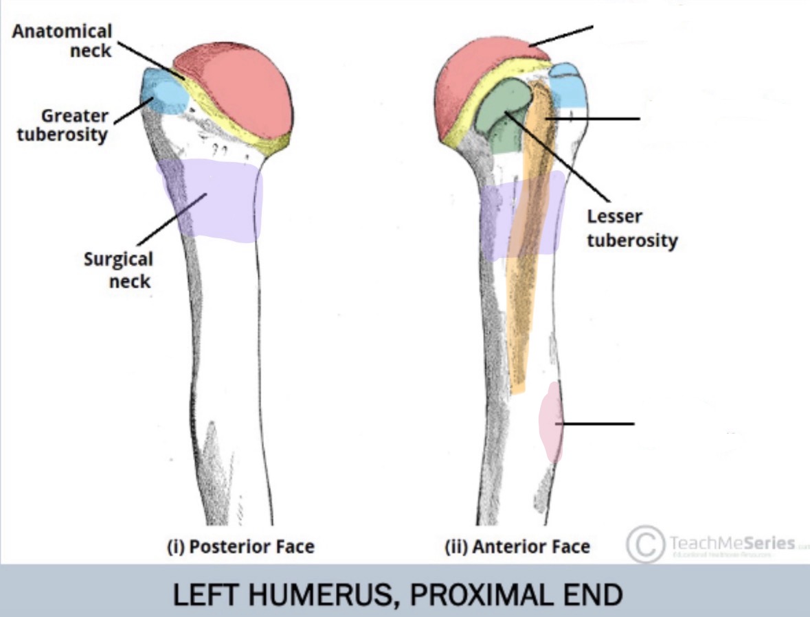
(orange zone)
Intertubercular groove, Humerus (posterior)

(pink zone)
Deltoid Tuberosity, Humerus (lateral)

(purple zone)
Medial Epicondyle, Humerus

(pink zone)
Lateral Epicondyle, Humerus

(orange zone)
Coronoid Fossa, Humerus (anterior, more medial)

(red zone)
Radial Fossa, Humerus (articulation point for radius when bending arm. anterior)

(blue zone)
Capitulum, Humerus (main articulation point for radius)

(green zone)
Trochlea, Humerus (spool shape, main articulation point for ulna)

(yellow zone)
Olecranon Fossa, Humerus (articulation point for protrusions on ulna. posterior)
The Capitulum of Humerus (inferior lateral) articulates with:
The Head of the Radius (articulation point)
The Trochelea of Humerus (inferior medial) articulates with:
The Trochlear Notch of the Ulna (semilunar) (articulation point)
In anatomical position, the radius is
lateral (towards the outside of the body)
In anatomical position, the ulna is
medial (towards the inside of the body)

Radius and Ulna (image ID)

(red zone)
Head, Radius (superior. round button)

(yellow zone)
Neck, Radius

(green zone)
Radial Tuberosity, Radius

(purple zone)
Interosseous crest, Radius and Ulna

(orange zone)
Styloid Process, Radius

(blue zone)
Ulnar Notch, Radius
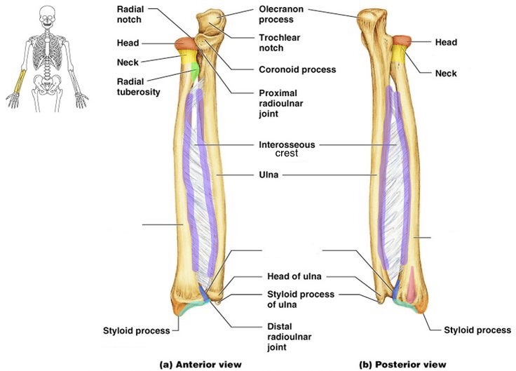
(teal zone)
Distal Articular Surface, Radius

(pink zone)
Dorsal Tubercle, Radius (posterior)
The Head of Radius articulates with:
The Capitulum of the Humerus (articulation point)
The Ulnar Notch of the Radius articulates with:
The Radial Articulation Point of the Ulna (articulation point)
The Distal Articular Surface of the Radius articulates with:
Lunate and Scaphoid carpels (articulation point)
The Trochlear Notch of the Ulna articulates with:
The Trochlea of the Humerus (articulation point)
The Radial Notch of Ulna articulates with:
The Head of the Radius (articulation point)
The Ulna does —— articulate with the the carpels
not
Radial Articulation Point of Ulna articulates with:
Ulnar Notch of the Radius (articulation point)

(blue zone)
Radial Notch, Ulna

(blue zone)
Olecranon Process, Ulna

(red zone)
Trochlear Notch, Ulna

(yellow zone)
Coronoid Process, Ulna

(pink zone)
Radial Articulation Point, Ulna

(green zone)
Styloid Process, Ulna

Carpals (image ID)

(red zone)
Scaphoid (snail like)

(orange zone)
Lunate

(yellow zone)
Triquetral

(green zone)
Pisiform (on lateral side, forms carpel tunnel)

(light blue zone)
Hamate (forms carpel tunnel, articulates with MC4 and MC5/pinky)
Hamate articulates with:
Metacarpals 4 and 5(pinky) (articulation point)

(dark blue zone)
Capitate

(purple zone)
Trapezoid

(pink zone)
Trapezium (articulates with thumb/MC1)
Trapezium articulates with:
Metacarpal 1(thumb) (articulation point)
The base of the metacarpals articulates with:
Carpels (articulation point)
The ——— of the metacarpals and metatarsals are distal
heads
The ——— of the metacarpals and metatarsals are proximal
bases
The head of the metacarpals articulates with:
Proximal Manual Phalanges (articulation point)
Distal means the section of the limb is:
towards the tips
Proximal means the section of the limb is:
towards the central body

(green zone)
MC1 (palmer view)

(blue/teal zone)
MC2 (palmer view)

(purple zone)
MC3 (palmer view)

(magenta zone)
MC4 (palmer view)

(light pink zone)
MC5 (palmer view)

(red zone)
MC1 base

(yellow zone)
MC2 base

(blue zone)
MC3 base

(purple zone)
MC4 base

(green zone)
MC5 base
The thumb has —- phalanges the than other fingers
less

(red zone)
Distal phalanges (hand)

(blue zone)
Intermediate phalanges (hand)

(green zone)
Proximal phalanges (hand)
The base of proximal phalanges
has one lobe on their articular surface
The base of intermediate phalanges
has two lobes on their articular surface

Pelvis/Pelvic Girdle (image ID)

(red zone)
Os Coxae/Innominate (x2)

(yellow zone)
Sacrum

(blue zone)
Coccyx

(pale blue zone)
Acetabulum, Os Coxae (lateral)

(green zone)
Lunate surface (of the Acetabulum), Os Coxae

(red line zone)
Obturator foramen, Os Coxae (anteriorly oriented)
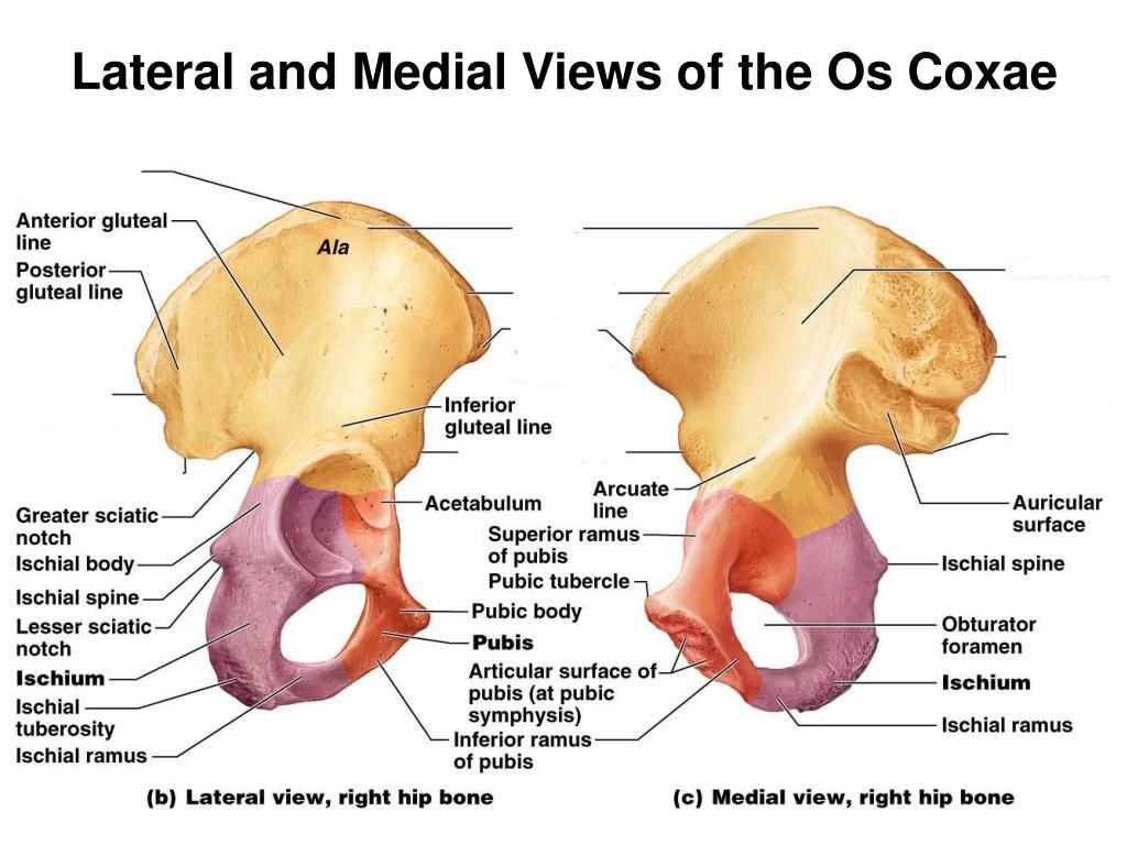
(beige zone)
Ilium, Os Coxae

(yellow zone)
Articular Surface, Ilium, Os Coxae
The Articular Surface of the Os Coxae, Ilium, articulates with:
Sacrum (articulation point)

(teal zone)
Iliac Crest, Os Coxae

(blue/purple zone)
Pubic Symphysis, Os Coxae

(purple zone)
Ischium, Os Coxae (posterior, inferior)

(red zone)
Pubis, Os Coxae (anterior, inferior)

(orange zone)
Greater Sciatic Notch, Os Coxae (posterior)

(pink zone)
Posterior Inferior Iliac Spine, Os Coxae
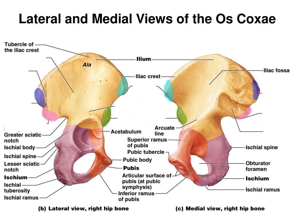
(dark blue zone)
Posterior Superior Iliac Spine, Os Coxae

(light blue zone)
Anterior Superior Iliac Spine, Os Coxae

(green zone)
Anterior Inferior Iliac Spine, Os Coxae

(teal zone)
Ischial Tuberosity, Os Coxae

Hyoid (image ID)

(green zone)
Greater Horns/Cornua, Hyoid

(yellow zone)
Lesser Horns/Cornua, Hyoid

(red zone)
Body, Hyoid
The Hyoid does not fully articulate with any bone, but is connected by ligaments to the:
Styloid Process of the Temporal Bones (articulation-ish point)