Micro Block 4
 Call Kai
 Learn
Learn Practice Test
 Spaced Repetition
 Match
Match Flashcards
 Knowt Play
1/157

There's no tags or description
Looks like no tags are added yet.
Name | Mastery | Learn | Test | Matching | Spaced | Call with Kai | Chat |
|---|
No analytics yet
Send a link to your students to track their progress
158 Terms
What are the two parts of the central nervous system?
Brain
Spinal Cord
What are the two cell types of nervous tissue?
Nerve cells
Neuroglia
What are the 2 types of nerve cells?
Neurons-in CNS
Ganglion-in PNS
What are the 4 neuroglia of the CNS?
Astrocytes
ogliodendroglia
microglial cells
ependymal cells
What are the two types of neuroglia of the PNS?
Schwann cells
Satellite cells
What are meninges?
A series of connective tissue sheaths that envelop the brain,spinal cord, roots of the peripheral nerve and optic nerve
Is the optic nerve considered CNS?
yes
What are the layers of the meninges in order from furthest outside to furthest inside?
Dura mater
Arachnoid
Pia Mater
what is leptomeninges?
the arachnoid and pia mater
What layer of the meninges contains cerebrospinal fluid?
subarachnoid space
What does pia mater adhere to?
brain and spinal cord
which layer of the meninges is highly vacular?
pia mater
when looking at meninges how tell which layer is the archnoid?
looks spider web like (and an arachnid is a spider)
Does the spinal cord have the same meninges layers as the brain?
Yes
what becomes the neural tube? what does it consist of?
The neural plate consisting of neuroectoderm
What does the neural tube give rise to?
-All cells of CNS (neurons,astrocytes,ogliodendrocytes,ependymal cells,choroid plexus cells)
-All the nerves that innervate skeletal muscle
what does the neural crest cells give rise to?
-Arachnoid and pia mater
-enteric parasympathetic ganglia
-schwann cells
-dorsal root ganglia
what does the mesoderm give rise to?
-dura mater
-connective tissue around peripheral nerves
what two terms for the forebrain embryonic brain division?
Telencephalon
Diencephalon
What derives from the Telencephalon?
Cerebrum
hippocamus
amygdala
basal nuclei
What derives from the Diencephalon?
Thalamus
Hypothalamus
what term for the midbrain embryonic brain division?
Mesencephalon
What derives from the Mesencephalon?
Midbrain
what two terms for the hindbrain embryonic brain division?
Metencephalon
Myelencephalon
What derives from the Metencephalon?
Pons
Cerebellum
What derives from the Myelencephalon?
Medulla oblongata
What are 3 structural elements of the cerebrum? describe each
Sulcus-grooves/pits
Gyrus-ridge
Fissure-deep clefts separating major regions, lobes or hemispheres
What are 2 structural parts of the cerebellum? describe
Vermis- narrow midline region
Folia-leaflike folds on cerebellar cortex
What are the 5 areas the spine can be functionally divided into?
C1-C5
C6-T2
T3-L3
L4-S1
S1-S3
What are cells of ectodermal origin very sensitive to?
Hypoxia
What are 3 neuronal cells of ectodermal origin?
neurons
astrocytes
ogliodendrocytes
what are 2 cells of mesodermal origin?
microglia
vascular epithelium
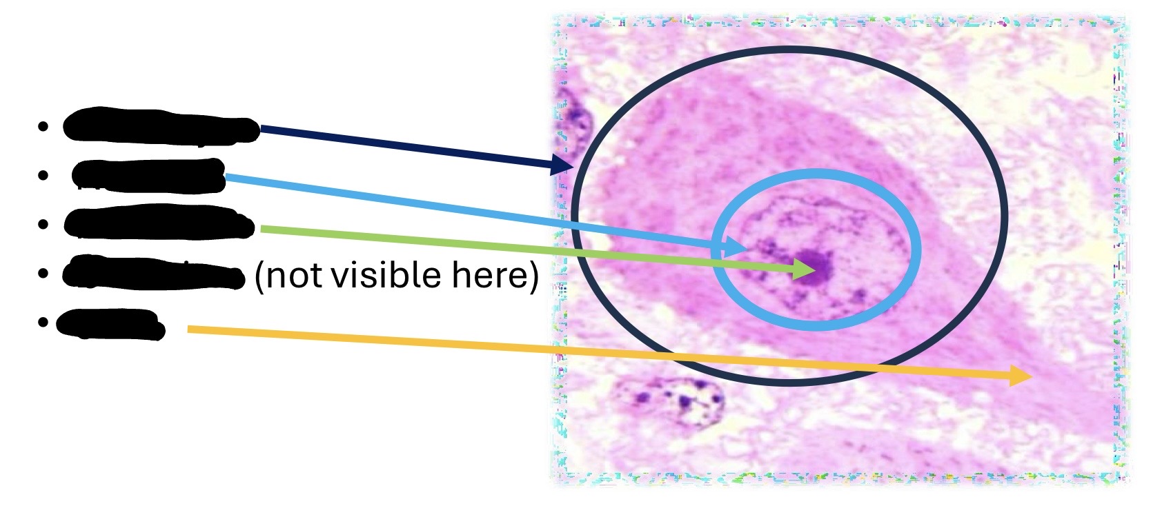
what is neuron?
Cells in CNS responsible for transmitting electrical signals throughout body
What is the general function of glial cells?
Support system in which neurons are embedded and serve many critical functions
What call neuron cell body?
Perikaryon or soma
What branch off neuron cell body?
dendrites and axon
What are 2 characteristics/properties that define neurons? describe each
irritability: the ability to generate an electrical impulse
conductivity: the ability to tansmit one such an impulse from one locality to another
what nucleus type do neurons have?
euchromatic with a prominent nucleolus
What call the basophilic cytoplasm of a neuron? what does it contain?
Nissl substance
-Rough ER and ribosomes
What is a synapse?
Specialized junction or gap where a neuron communicates with another cell
what 3 things can neurons synapse with?
nurons
muscle
glands
Because neurons are long living what can be seen inside some of them?
lipofuscin
what 3 ways can neurons be distributed?
Individually
layers
groups
where see neurons in layers?
the cerebral and cerebellar cortex
What stain use to see lipofusin?
PAS
What stains used to evaluate myelin? what do?
Luxol fast blue -stains myelin blue
Osmium tetroxide alpha naphthylamine method- stains normal myelin a brown/orange color and damaged myelin black
what call a groups of neurons in CNS? what about in PNS?
CNS: nuclei
PNS: ganglia
What are 4 glial cells of the CNS?
Ogliodendrocytes
Microglia
Astrocytes
Ependymal cells
What is functions of ogliodendrocytes?
-Support neurons
-Form myelin sheaths for several axons at once
What are oligodendrocytes best visualized with? what do stain do?
Silver stains: stain them black
Osmium tetroxide alpha naphthylamine method
Immunohistochemical stain: detect Olig2
what are the resident macropahges of the CNS?
microglia
which glial cells are the smallest?
Microglia
what are 3 functions of microglia?
-immunosurveillance (includeing antigen presentation)
-immunoregulation
-phagocytic function during inflammation and repair
what call activated microglia cells? when do they activate?
Myelophages (gitter cells)
activate during conditions causing inflammation or cell death
How do gitter cells appear microscopically?
lobular and swollen after having phagocytized debris
what call it when myelophages in a myelin sheath?
digestion chamber
What is the CNS counterpart of a fibroblast?
Astrocytes
what are the 2 types of astrocytes? where found?
Protoplasmic-grey matter
Fibrillar-white matter
how visualize astrocytes?
Use IHC that reacts with glial fibrillary acidic protein (GFAP)
What is glial fibrillary acidic protein made in abundance by?
astrocytes
What are 4 functions of astrocytes? what are its biggest 2?
-Cell communication
-Crucial component of blood brain barrier
-Transport nutrients
-antigen presentation
Describe how astrocytes, microglia and ogliodendrocytes appear on slide?
astrocytes: large circular eucharomatic nuclei and can rarely see cytoplasam or cell membrane in Hand E
microglia: very very small basophilic nucleus with little cytoplasam
ogliodendrocytes: slightly larger than microglia and have dense round nuclei surrounded by clear zone so look like a fried egg
what is white matter? what called in greek?
Accumulation of myelinated axons along with other glial cells
-Leukos
are there neuronal cell bodies in white matter?
NO
why does white matter appear white?
myelin is lipid rich giving white appearance as lots of myelinated axons in white matter
what is gray matter? what called in greek?
Neuronal cell bodies along with glial cells
-Polios
what is neurophil?
axons,dendrites, and cytoplasmic projections of glial cells that form matrix of gray matter
in cerebrum where is white matter? where is gray?
gray outside
white inside
in spinal cord where is white matter? where is gray?
white outside
gray inside
does grey matter contain axons and dendrites of neurons?
yes




which layer of the meninges contains CSF?
Subarachnoid space
What cell in the CNS myelinates axons?
oligodendrocytes
How tell you are looking at cerebellum?
has 3 layers in grey matter
what is grey matter called in cerebellum?
cortex cerebelli
What are the 3 layers in cerebellum grey matter? describe each?
molecular level-outer most layer with neurons called basket cells
ganglionic layer-border between molecular layer and granular cell layer and has neurons called purkinje cells
granular cell layer-inner layer with neurons called granule cells
what is white matter in cerebellum known as?
corpus medullare or arbor vitae
what predominates in cerebeullum white matter?
myelinated axon tracts
How to tell if medullla oblongada on slide?
Can’t really tell if white matter is on inside or outside and can’t tell if gray matter is on inside or outside
what tells you that looking at neonatal cerebellum?
Has 4 layers with its outer layer being called the exterior cortical layer
Which has three layers the cerebrum or cerebellum?
Cerebellum
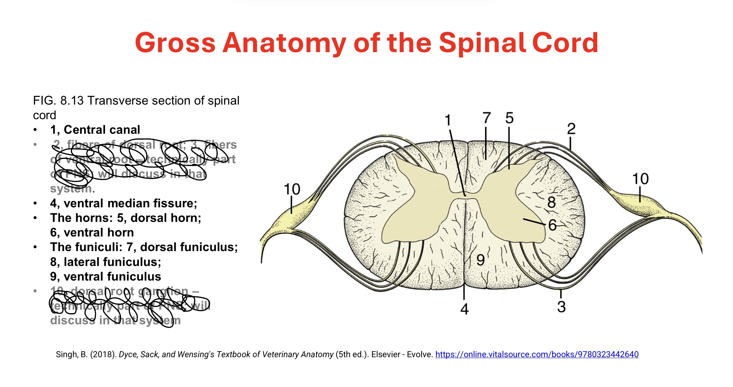
what found in ventral horns of spinal cord?
motor neurons whose axons make up the ventral roots of the spinal nerves
what found in dorsal horns of spinal cord?
sensory fibers from neurons in the spinal ganglia
when looking at spinal chord which are ventral horns?
longer wings of butterfly (on same side as longer fissure)
Are spinal cord nerves multipolar?
yes
what is ependyma?
thin epithelium that lines the fluid filled spaces of the CNA
what is ependymal cells?
the cells lining the fluid filled spaces residing on the ependyma
How do ependymal cells look?
cubordal or columnar with cilia and mircovilli
what is function of cilia on ependymal cells?
circulate CSF