Outcome 4: AV, Ao Stenosis, Ao Insufficiency, PV, and Heart Lesions
 Call Kai
 Learn
 Practice Test
 Spaced Repetition
 Match
 Flashcards
 Knowt Play
1/181

Earn XP
Description and Tags
AV, Ao Stenosis, Ao Insufficiency, PV, and Heart Lesions
182 Terms
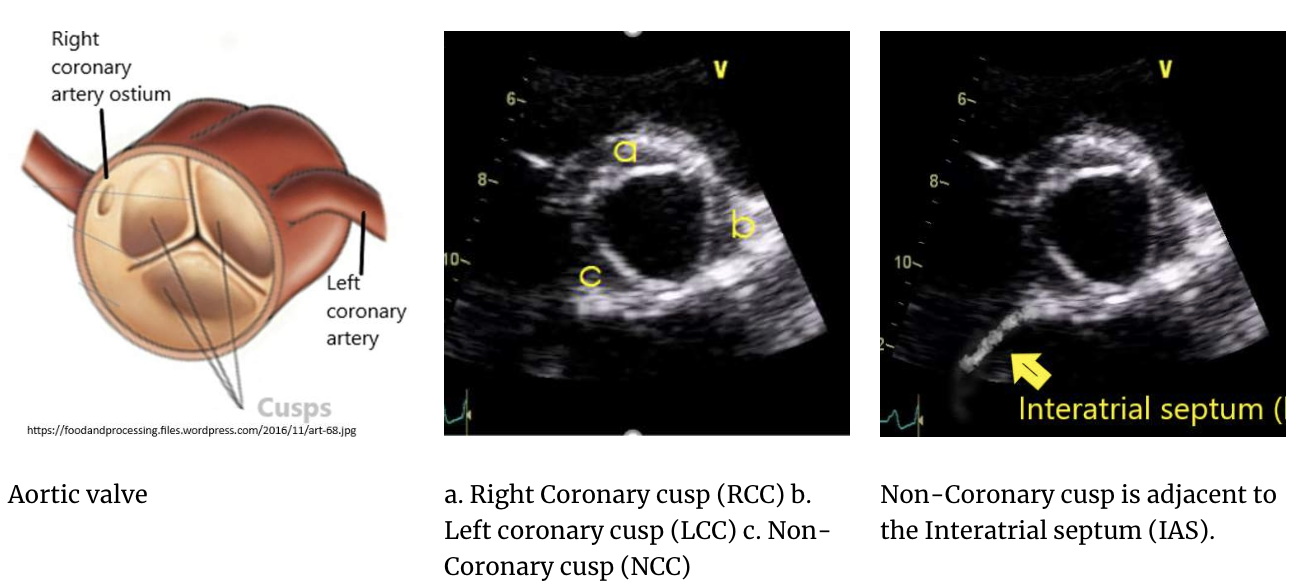
aortic valve structure
typically tricuspid (3 cusps)
each cusp has a ‘bowl-like’ shape, joined at the corners called commissures
2 cusps are found under the openings of the coronary arteries
RCC adj. to RCA opening (ostia)
LCC adj. to LCA opening (ostia)
the non-coronary cusp doesn’t have a coronary artery assoc. w/ it & is found adj. to IAS
—
the second heart sound (S2) is produced by the closure of the aortic and pulmonary valves
What are commissures in the context of the aortic valve, and what is their role?
Commissures are the points where the cusps meet, allowing the valve to open and close effectively
What is the relationship between the aortic valve cusps and the coronary ostia?
The RCC and LCC are located adjacent to the openings (ostia) of the right and left coronary arteries, respectively.

What is the typical M-mode appearance of a normal aortic valve?
On M-mode, a normal aortic valve opens with a box-like appearance with a central closure line and normal cusp separation.

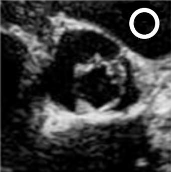
unicuspid valve
rare, severe congenital malformation where the AV cusps don’t separate from e/o
circular opening, offset to one side of the Ao root/LVOT area
can result in early development of severe aortic stenosis
can result in:
dilatation & aneurysmal changes
dissection
rupture
sono findings:
eccentric closure w/ 2D and m-mode

bicuspid valve
common congenital heart anomaly
one regular shaped cusp & one prominent cusp comprised of 2 regular-sized cusps that are fused
may or may not be a raphe present
raphe: visible fusion line on echo b/w the joined cusps
oval or football-shaped opening
can show an eccentric closure on 2D and m-mode
with time, every BAV will either become stenotic or develop severe AR
assoc. w:
CoAo
dilatation
aneurysmal changes in Ao
turner’s syndrome
PDA, VSD, ASD
sinus of valsalva aneruysms (SVA)
—
functional BAV: refers to an AV that originally had 3 cusps, but over time, the cusps calcified and fused » creating the app. of a BAV
these look diff. than a congenital BAV

quadricuspid valve
rare malformation that has 4 separate cusps present
square shape opening
assoc. w:
valve insufficiency (regurg.) NOT stenosis
VSD, ASD, PDA
hypertrophic cardiomyopathies
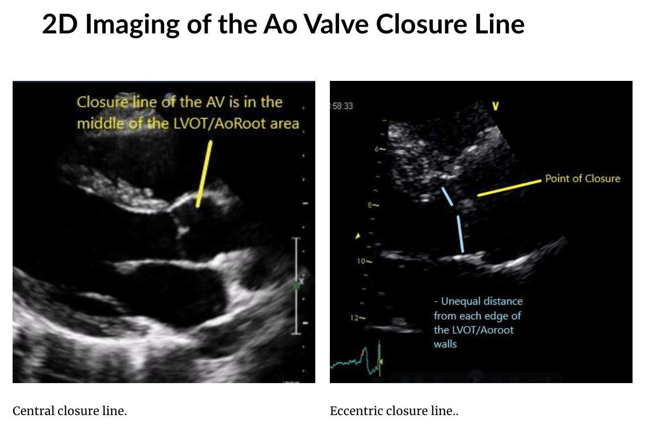
eccentric vs. central closure line
closure line = line where the Ao cusps meet when in the closed position
when the cusps don’t meet evenly in the middle of the LVOT/Ao root area on 2D or m-mode
can indicate an anomalous # of Ao cusps present (bi and uni)
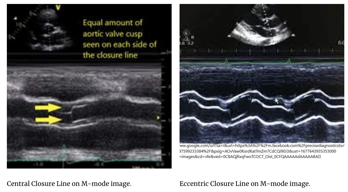
m-mode imaging of the AV closure line
openings of the cusps (RCC and NCC) creates a box-like app. within Ao root on the image
closure line will be an equal distance b/w the top leaflet & bottom leaflet opening when the AV has 3 cusps (left image)
when the closure line is closer to the top of the Ao root than the bottom edge (right image)
this eccentric closure line suggests BAV
a unicuspid valve is also possible, but will usually have more calcification & thicker valve leaflets

aortic cusp separation (ACS) measurement
measures the distance that the AV cusps move away from e/o on an m-mode image
dec. separation can indicate a dec. opening of a valve due to a bicuspid or unicuspid valve, or the presence of aortic stenosis
taken in early systole (during initial opening of the valve cusps)
leading to leading
normal = >1.5cm
—
not routinely measured anymore
congenital aortic stenosis
develops due to a congenital malformation of the AV leaflets (uni or bi), the subaortic region of the LVOT, or the Asc. Ao just distal to the AV
often found in conjunction w/ other congenital malformations
however, Ao stenosis due to BAV may also occur in isolation
2 mechanisms:
structural defect itself can cause stenosis
abnormal valve structure has a higher propensity for sclerotic changes
acquired aortic stenosis
sclerosis of the AV cusps can develop into stenosis:
sclerosis = calcification of the leaflets and valve annulus » takes time to develop
progressive sclerotic changes to the AV cusps can result in AV stenosis
most common type of Ao stenosis in adults
MV, TV, PV are less likely to undergo progressive sclerotic changes w/o rheumatic heart disease, renal disease, or carcinoid disease also being present
etiology:
sclerosis can be due to degenerative changes due to..
advanced age, high BP, and obesity
if seen in younger pts, it may be due to an abnormal valve structure or from a disease process such as rheumatic fever or chronic renal disease
echo appearance:
sclerotic leaflets appear bright and white
greater the calcification, the greater the leaflet thickness & acoustic shadowing from the calcification
complications:
calcification of the leaflet tissue can cause poor coaptation of the cusps (regurg.)
dec. leaflet excursion (stenosis)
difficulty identifying the # of Ao cusps present
retraction of the leaflets in certain diseases

sclerotic aortic valve
progressive sclerosis leads to a stenosis of the AV
leaflets becomes thickened and calcified, which impacts their mobility » leading to stenosis
clinical findings in aortic stenosis
chest pain
syncope (fainting)
SOBOE
fatigue
characteristic murmur
harsh systolic ejection murmur, systolic crescendo decrescendo murmur at the rt. upper sternal border, up to the carotids
what is a murmur
many valvular pathologies create a murmur bcuz the structural or functional change of the valve interferes w/ the normal lubb-dubb heard w/ the stethescope
hearing a murmur can be the first indication that valvular pathology is present
—
murmur = extra sound heard when listening to pt. heartbeat
can be from blood flowing past normal structures in the heart, or from abnormal pathology present
a structural change of a valve can create a murmur
if valve is stenotic, it has a murmur characteristic of the stenosis, or if the valve has regurg, it has a murmur that reflects regurg.
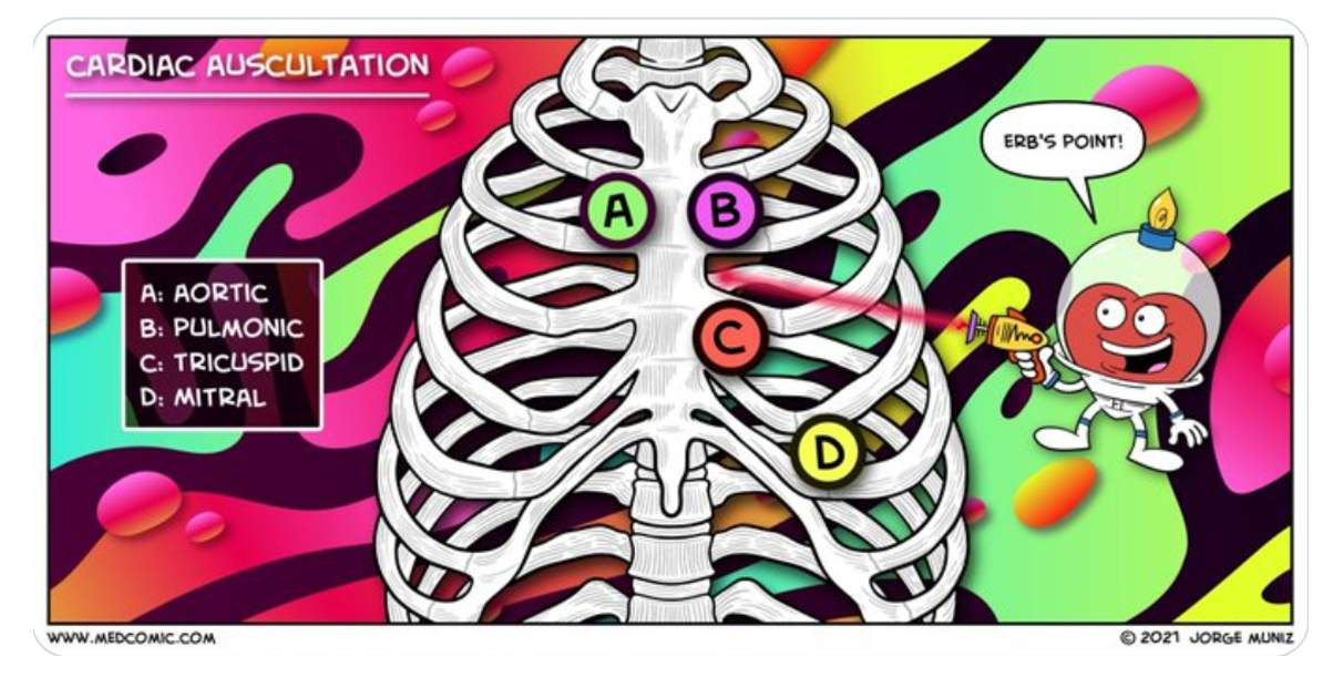
characteristics of a murmur
location:
refers to where the murmur is heard on the chest wall or the direction the sound is moving toward
timing:
refers to the stage of the cardiac cyc. in which the murmur is heard
duration:
refers to lenth of time during which a sound or murmur is heard
uses prefixes to indicate the amt. of the cyc. heard (pan—, holo—, mid—)
intensity:
refers to the loudness of a murmur
graded from I (softest) to VI (loudest)
changes in intensity:
murmurs that change over the cardiac cyc. time
ie. crescendo, decrescnedo
pitch:
refers to the freq. of the found (high, medium, low)
quality:
description that characterize the sound
ie. blow, rumbling, harsh, dull, opening ‘snap’, systolic ‘click’
components of the sound:
S2 splitting - the type of murmur that occurs normally but may be prolonged in certain disease states
valvular aortic stenosis
etiology:
ACQUIRED
progressive sclerotic changes of a normal tricuspid AV
rheumatic fever and chronic renal disease are pathologies that initiate and promote excessive sclerosis of the AV leaflets » leading to stenosis
aging
HTN
diabetes
obesity
prosthetic valve replacements
CONGENITAL
abnormal structure of a unicuspid valve » a unicuspid valve is inherently stenotic
sclerotic changes of a BAV » BAV isn’t initially stenotic but has a greater predisposition to sclerotic changes » which can progress to stenosis over time
treatment:
surgery (open heart) » a valve replacement (bioprosthetic or mechanical)
TAVR/TAVI (transcatheter valve replacement/implantation)
ideal for pts. who are unstable or have multiple comorbidities that can’t withstand open-heart surgery
balloon valvuloplasty
always done in conjunction w/ TAVR, never on its own
performed before a TAVR, so the native calcified leaflets will move easily apart against the walls of the Ao as the new prosthetic valve is positioned and expanded into place w/ a catheter
ross procedure (pediatrics)
Av of the pediatric pt. is replaced w/ the pt’s. PV
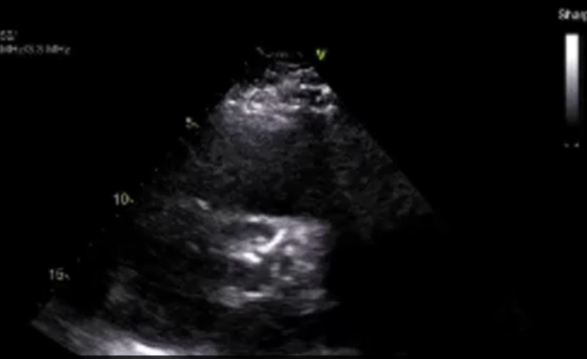
valvular aortic stenosis - echo findings
2D:
AV cusps or prosthetic valve leaflets have a dec. excursion visually, and the measured opening of the valve is small
<2cm2 if stenosis is present
aortic cusps are thickened from calcification and shows inc. echogenicity
doming of the leaflets in PSLAX view » characteristic of stenotic leaflets
LVH
post-stenotic dilatation of Ao root & Asc Ao
LA dilation
colour:
turbulent flow at valve opening
spectral:
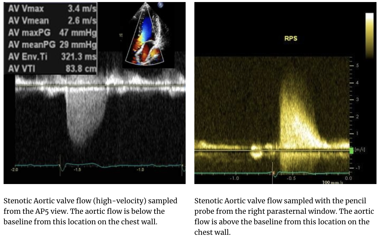
high velocity flow that begins at the AV cusps
CW used to document highest velocity
normal =/<1.9m/s
velocities b/w 2.0-2.6m/s may be elevated due to something other than valvular aortic stenosis
just bcuz velocity is inc. a little more than 1.9m/s, it doesn’t always mean aortic stenosis
RPS and SSN views are sampled w/ pencil doppler probe to capture highest velocities in calcific Ao stenosis
all assessments require 3-5 waveforms
w/ atrial fibrillation 5-10 waveforms are traced

subaortic stenosis (congenital)
etiology:
congenital malformation is a membrane or ridge of tissue that occurs before AV opening in the LVOT
can be assoc. w/ BAV, pulmonary stenosis, PDA, or VSD
2D echo findings:
demonstration of the ridge or membrane in the LVOT w/ 2D is needed
sometimes it’s not seen initially and id identified when the u/s produces confusing results
the AV is opening well, but the CW doppler velocity in the AV area is in the stenotic, high0velocity range
membrane in the image below was initially missed until the apical window when high CW doppler velocities though the AV area were found
treatment:
surgery » only if severe stenosis present
removal of obstructing membrane or removal of tissue in the LVOT


subaortic stenosis - spectral findings
colour:
turbulence in LVOT where it’s narrowed due to membrane or ridge
the accelerated flow begins in the LVOT
PW:
used to sample along IVS to localize where the obstruction in the LVOT begins
waveforms will begin to show aliasing as it meets the area of obstruction in the LVOT » bcuz the velocity is too high for the PRF of PW
CW:
used to measure the highest flow through the stenotic LVOT area, which will correlate w/ the severity of the stenosis present
stenosis velo. >2.0m/s


supravalvular aortic stenosis (etiology)
etiology:
caused by a narrowed, hypoplastic segment of the Asc Ao or by a membrane or a ridge of tissue that narrows the diameter of the vessel
this type of stenosis is just beyond the AV in the Asc Ao
this narrowing shouldn’t be confused w/ CoAo
assoc. w/ William’s disease, CoAo, and pulmonary stenosis
2D:
ridge or membrane needs to be shown
often TEE is needed to clearly visualize this area
colour:
turbulent and accelerated flow in the narrowed, stenotic area of the Asc Ao, just beyond the AV
spectral:
CW measured highest flow thru stenosed area
high-velocity flow begins after the AV, in the Asc Ao
treatment:
surgery » only if severe stenosis
removal of obstructing membrane or replacement of the AV, Ao root, and Asc Ao

complications for untreated, undiagnosed aortic stenosis
LVH
arrhythmias and sudden death
aortic aneurysm & dissection
ischemia, myocardial infarction, global systolic dysfunction
pulmonary edema
cerebral infarcts
transvalvular gradients using the bernoulli equation
doppler-derived PGs are central to grading Ao stenosis severity
simplified bernoulli equation (ΔP = 4V²) allows you to convert measured blood flow velocities into PGs across AV
—
when stenosis or narrowing is present, the flow velocity thru the narrowing will inc. proportinately to the degree of narrowing
smaller the valve opening = more significant the stenosis & the greater the velocity will be through the narrowing
—
the velocity and PGs through a stenosis are indications of stenosis severity
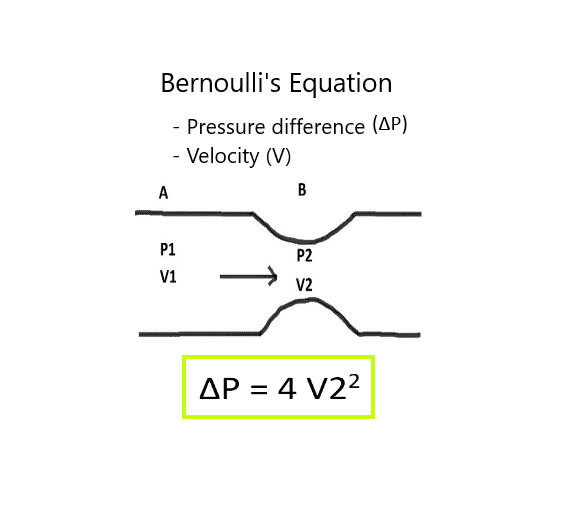
bernoulli equation
describes the indirect relationship b/w the pressure & the velocity of the blood moving through a vessel
compares two locations in the blood flow

velocity and/or trace
velocities through the Ao stenosis are measured on the spectral CW doppler, the machine calculates the respective PG for that velocity and displays it
measuring the max. and mean velocity of the blood flow on the Ao waveforms helps determine if the valve or Ao area is stenotic
CW used to sample high-velocity flow, RPS & SSN views
measurements obtained:
Maximum velocity (Vmax) (m/s) and Pressure gradient (MaxPG) (mmHg)
Mean velocity (Vmean) (m/s) and Pressure gradient (Mean PG) (mmHg)
VTI (Velocity-Time-Integral) distance (cm)

V1/V2 ratio
comparison of the velocity before a stenosis to the velocity at the stenosis
ratio assessed the severity of the stenosis
PW samples in LVOT
CW samples through stenotic AV to obtain the highest velocity waveforms
—
LVOT velocity (V1)/AV velocity (V2)
transvalvular flow velocities, PGs, and the V1/V2 ratio calculation are impacted by:
LV sys. func.
CO
presence of significant Ao insufficiency
reference ranges - Vmax, PGmean, V1/V2 ratio
max. velocity:
normal = <1.9m/s
mild AS = 2.6-2.9m/s
moderate AS = 3.0-4.0m/s
severe AS = >4.0m/s
mean PG:
normal = <20mmHg
mild AS = <20mmHg
moderate AS = 20-40mmHg
severe AS = >40mmHg
V1/V2 ratio:
normal = >0.5
mild AS = 0.4-0.5
moderate AS = 0.25-0.4
severe AS = <0.25

continuity equation for the AV area
explains the relationship b/w area and velocity while observing the Law of Energy conservation
compares the flow rate in the area before a stenosis to the flow rate at the stenosis
as blood flow approaches the area w/ a dec. radius, the velocity must inc. to maintain the same volumetric flow rate (Q) as before the stenosis
can be used to calculate the size of the narrowed or stenosed area
calculating AVA from the continuity equation and AV planimetry is considered the gold standard for grading Ao stenosis severity
—
Aortic Valve Area (AVA)cm2 = 0.785 (LVOT diameter)² x VTI LVOT (cm) ÷ VTI AV (cm)
—
images and measurements needed..
LVOT diameter
LVOT velocity time integral (VTILVOT)
AV VTI (VTIAV)
reference ranges:
normal = >2.0cm2
mild AS = 1.6-2.0m2
moderate AS = 1.0-1.5m2
severe AS = <1.0m2

planimetry to calculate AVA
tracing the largest orifice of the AV on a zoomed, 2D image, in PSSAX
provides a direct anatomical measurement of valve area
cm2
measurement may be difficult in heavily calcified or poorly visualized valves
planimetry should correlate w/ AVA calculated from continuity equation
reference ranges:
normal = >2.0cm2
mild AS = 1.6-2.0m2
moderate AS = 1.0-1.5m2
severe AS = <1.0m2
Which of the following is likely to result in an overestimation of aortic stenosis severity?
Underestimating the LVOT diameter
pitfalls in aortic stenosis assessment
reliable diagnosis of Ao stenosis severity depends not only on technical skill, but also vigilance for pitfalls & the self discipline to question discordant findings
patient conditions:
low CO (ie. HF, reduced EF)
coexistent aortic insufficiency (regurg.)
coexistent LVOTO
sonographer technical errors:
LVOT diameter
most sensitive measurement bcuz it’s squared in the continuity equation
cursor position for the PW of LVOT VTI
MR mistaken for Ao stenosis flow
Ao stenosis will have isovolumetric contraction time and MR won’t
Which of the following is a recommended strategy to ensure diagnostic accuracy in aortic stenosis assessment?
Cross-check quantitative findings with 2D imaging and clinical context.
aortic insufficiency (AI)
results from the aortic cusps not sealing together tightly or when there are perforations (‘holes’) in the valve cusps that allow the blood to flow back into the ventricle in dia
if there’s AI, we must investigate to see if there’s a structural reason for it
it could be the AV cusp structure or the aortic annulus/root may be dilated, pulling the cusps apart so they aren’t able to coapt tightly
acute onset of aortic insufficiency
presents suddenly
LV becomes hyperkinetic trying to manage the extra volume but there isn’t enough time to respond w/ dilatation
the LV end-dia press. (LVEDP) rises rapidly as a response to the inc. ventricular volume & results in dec. diastolic func. (abnormal LV filling)
echo findings:
normal sized LV cavity
hyperdynamic LV func.
inc. LVEDP
abnormal diastolic func.
mechanisms:
endocarditis of AV
Ao dissection
direct trauma
iatrogenic causes
chronic onset of aortic insufficiency
develops gradually over time
w/ significant AI (moderate and greater), the LV becomes hyperkinetic and dilates to accommodate the extra volume from the AI
the most dilated LVs are seen w/ this pathology
as the dilatation progresses, eccentric LVH develops
LV chamber has a larger volume which requires more myocardial cells to envelop it therefore inc. the LV mass
since the cells need to stretch over & cover a larger area, the myocardial wall remains normal thickness even though there have been extra myocardiocytes added
the LV can only dilate and accommodate so much volume before LV function declines
AV replacement must be done before this happens bvuz the LV func. may not return to normal
echo findings:
hyperkinetic LV which is dilated
LVH
normal LVEDP
normal LV systolic function initially, but declines if AI continues to be significant
mechanisms:
dilatation of the Ao root
calcification of the aortic cusps
aortic prolapse
flail aortic leaflet
endocarditis
masses
Left ventricular end-diastolic pressure (LVEDP)
The pressure in the left ventricle at the end of diastole; elevated in acute AI due to sudden volume overload.
Hyperkinetic LV
A left ventricle that contracts more vigorously than normal, often seen as a compensatory response to increased volume.
Eccentric hypertrophy
Increased LV chamber size, normal wall thickness, and increased LV mass. Another compensatory response to chronic volume overload.
clinical findings of aortic insufficiency
murmur:
high-pitched, blowing, diastolic decrescendo flow at the lt. sternal border
murmur w/ severe AI:
austin flint murmur is a low-pitched, mid-diastolic rumble at the apex
ONLY HEARD W SEVERE AI

central regurgitation
central regurg. jet flows straight back into the chamber it came from
jet is equal distance from the sides of the valve annulus
these jets develop from the poor coaptation of the leaflets
—
ie.
the dilatation of the Ao root will pull the cusps equally away from e/o resulting in a central jet of AI
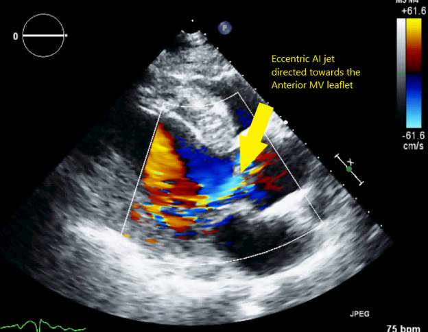
eccentric regurgitation
eccentric regurg. jets are directed to the edges of the valve and can hug the side wall of the chamber it came from
they develop from one leaflet being more affected that the others which results in unequal coaptation
the direction of the jet gives an indication of which leaflet is affected
these jets can develop from prolapsing and flail leaflets, or the presence of a VSD located adj. to one of the cusps (causing a disturbance to that cusp)
eccentric AI jets are directed to the IVS or against the anterior MV leaflet
eccentric jets may also result from the Coanda effect rather than from asymmetric leaflet pathology
high volume regurg. jets tend to adhere to the ventricular wall (Coanda effect), indicating more severe regurg.
causes/etiology of the aortic insufficiency
changes to the structure of the cusps
ie. calcification, rheumatic disease changes, flail AV cusp, BAV, AV prolapse
Ao root dilatation, sinus of valsalva aneurysm or dissection (poor coaptation of the Ao cusps)
infective endocarditis & abscess at the Ao root
non-infective endocarditis nodules on the leaflets (SLE and rheumatoid arthritis) or valvular cardiac masses
any disease resulting in Ao dilatation or dissections
ie. Marfan’s, Ehlers-Danlos, ankylosing spondylitis
VSDs adj. to AV
Flail leaflet
A valve cusp that has lost its structural support, often due to rupture or trauma, resulting in uncontrolled movement and regurgitation. Leaflet tip drops back into the LVOT.
Endocarditis
Infection or inflammation of the valve leaflets, leading to vegetations, perforations, and potential acute regurgitation.
Aortic ectasia
Dilation of the aortic root or ascending aorta, which can prevent proper cusp closure and cause central regurgitation.
Papillary fibroelastoma
A benign, small, friable tumor on the valve, increasing the risk of embolism and sometimes interfering with valve closure.
Sinus of Valsalva aneurysm
A congenital malformation or acquired via a tuberculosis or syphilis infection.
Which structural abnormality is most likely to cause an eccentric regurgitant jet in aortic insufficiency?
Flail leaflet
echo findings of aortic insufficiency
2D findings:
significant AI will cause LV to become hyperdynamic and dilated
LV is trying to cope w/ extra volume, so it contracts more vigorously
measurement of LV chamber size and LV wall thickness
visualization of the LV contractility
show structural reason for AI
M-mode:
anterior mitral valve leaflet (AMVL) flutter
AV closure line flutter
premature AV opening
premature MV closure
Which 2D echocardiographic finding is most suggestive of chronic, significant aortic insufficiency?
A dilated, hyperdynamic left ventricle with eccentric hypertrophy.

anterior mitral valve leaflet (AMVL) flutter - M-mode AI
found on M-mode of MV leaflets
jagged fluttering movement of the anterior MV leaflet in diastole
present if AR jet is eccentrically directed down onto it

aortic valve closure line flutter - M-mode AI
found on M-mode image of Ao root and LA
when Ao leaflets are closed together in diastole
the AI leaking back to the LV can cause fluttering of the M-mode AV closure line
premature aortic valve opening - M-mode AI
found on M-mode of Ao root and LA
significant AI inc. LV diastolic pressure » causing the AV to open earlier than normal
on M-mode, this is seen as premature separation of the AV leaflets during diastole
premature mitral valve closure
found on M-mode of Ao root and LA
w/ the extra blood volume of the AR helping to fill the LV in diastole, the pressure in the LV exceeds that of the Asc Ao more quickly than if the LV was filled by the MV inflow alone
therefore, the AV will open sooner and the MV closes earlier when significant AI is present
Left ventricular dilation
Enlargement of the left ventricular chamber, often seen in chronic significant AI due to volume overload.
AMVL flutter
Rapid, irregular motion of the anterior mitral valve leaflet in diastole, often caused by an eccentric AI jet striking the leaflet.
Premature Aortic valve opening
Early opening of the aortic valve during diastole, a sign of increased left ventricular pressure from significant AI.

colour doppler with aortic insufficiency
can identify the regurg. jet’s location, give info about its severity, and reveal causation (prolapse, perforation, etc.)
direction, width, and length of the colour jet can be identified from the LAX images of the AV
PSLAX view of AV w/ the colour doppler can identify where the leak occurs
qualitative and quantitative evaluation of the aortic insufficiency
evaluates 3 qualitative features of the regurg. flow
presence of proximal flow
persistence of flow
eccentricity of the colour jet
after this initial grading, refine the severity assessment using quantitative measurements
ie. vena contracta width, PW doppler flow reversal in desc. Ao, and the pressure-half of the AI on CW

vena contracta width
quantitative measurement to evaluate the severity of the AI
vena contracta is the regurg. jet’s narrowest part (the "'waist”)
this measurement is done at the valve leaflets b/w the prox. flow in the Ao and the regurg. flow in the LV/LVOT
measurement done in zoomed image of LVOT w/ colour
reference ranges:
mild = <0.3cm
moderate = 0.3-0.6
severe = >0.6cm

colour jet height/LVOT ratio
quantitative measurement
colour jet width (cm) and the LVOT diameter (cm) are measured on a colour M-mode image w/ cursor aligned through the LVOT/Ao root area from PS window
ratio calculated from these 2 distances
AI colour turbulence will appear on the closure line of the AV in diastole
it’ll be red and multi-coloured just like on the colour clip
—
on PSSAX, 2D colour image of LVOT/Ao root area » the largest amt. of AI colour is traced w/ area planimetry & then the area of the LVOT is traced
a ratio is then calculated from the two areas


diastolic flow reversal in the desc. and abdo. aorta
PW
confirmation image to take to determine if the AR is severe
sometimes it’s difficult to tell if it’s moderate, moderately severe, or severe due to imaging limitations
presence of diastolic flow reversal when sampling the desc. and abdo. aorta, confirms severe AI is present
the severity of AI inc. the further downstream the flow reversal is observed
flow reversal observed in both the desc. and abdo. aorta signifies a more severe degree of AI than then it’s limited to the desc. ao alone
the diastolic flow reversal is above the baseline when sampling the desc. ao at the SSN and below the baseline in abdo. aorta flow in subcostal

Fun Fact
Diastolic flow reversal in the descending aorta can also be present when there is a patent ductus arteriosus (PDA) present.
increased LVOT velocity
PW
AR that’s moderate or greater, will cause the AV forward flow velocity to be inc.
AR flows back into LV and adds to the stroke volume ejected w/ the next heartbeat
normal LVOT velocity is approximately 0.8-1.2m/s
in sig. AI, LVOT velocity will be greater than 1.2m/s and can measure closer to 1.9m/s or more
velo. is inc. not bcuz of valvular stenosis, but due to the inc. amt. of stroke volume ejected
this is a subtle finding and not specific to AR
all high-flow circulation states such as arterial-venous malformations, anemia, hyperthyroidism, can have higher velocity LVOT flow
decreased mitral valve deceleration time
PW
very short deceleration time correlates to inc. LV filling pressures
w/ significant AI, the ventricle is overloaded w/ too much fluid
the inc. volume adds to the LV pressure, and the diastolic function progresses to a more restrictive diastology
this feat. isn’t specific to significant AI, but will occur w/ all pathologies affecting the LV pressure & diastolic function

aortic insufficiency pressure half-time (PHT)
CW
measures how quickly the PG b/w the Ao and LV equalizes during diastole
when there’s a large amt. of insufficiency, the LV is filled by both the MV inflow & AI in diastole » this quickens the time that it takes for the LV pressure to exceed the pressure of the Ao, so the MV will close early and AV will open early
therefore, w/ a large amt. of AI, the pressure b/w the Ao and LV equalizes quickly during diastole and the velo. dec. quickly over diastole
slope is sharp and PHT value is small
if the amt. of AI is mild, the pressures b/w the 2 chambers equalize more slowly and the slope of the AI waveform will be flatter, and a large PHT
summary:
steep slope = short PHT = severe AI
flat slope = long PHT = mild AI
reference ranges:
mild AI w/ PHT is >500ms
moderate AI w/ PHT between 200-500ms
severe AI when PHT is <200ms

signal intensity/or density of aortic insufficiency waveform
CW optimized for AI flow
density and intensity of the CW doppler signal reflects the volume of regurg. flow
dense, bright AI waveform (as forward flow) suggests a large regurg. volume and more severe AI
less intense or faint signals may indicate milder AI or technical limitations such as jet eccentricity
cursor may not line up w/ eccentric jets and resultant waveform will be of lesser intensity

diastolic mitral regurgitation
CW
MR normally occurs during systole
diastolic MR can occur if the LV pressure exceeds the LA pressure during diastole
this can happen when there’s a severe amt. of AI flowing back into the LV in diastole
momentarily, the pressures invert, and diastolic MR occurs
not unique to severe AI, as it can be found in pts. w/ an AV block and atrial tachyarrhythmias
True or False: A short pressure half-time on CW Doppler always indicates severe aortic insufficiency, regardless of LV compliance.
false

proximal isovelocity surface area (PISA)
PISA calculation utilizes the physics of flow convergence to quantify the severity of AI
as blood moves towards the regurg. orifice (the small opening where the leaflets aren’t coapting tightly & the blood can leak back into the LV), a flow convergence zone develops
on colour, the flow convergence zone looks like a ‘bubble’ or dome-shaped area on the opposite side of the valve from the regurg.
this dome-shaped area consists of concentric hemispheric shells of inc. velocity
by measuring the radius of this flow convergence zone and applying mathematical formulas, the size of the regurg. orifice and the volume of regurg. can be estimated
the radius of the flow convergence is called the PISA radius
the larger the opening, the larger the flow convergence zone created & the larger PISA radius measured
reference range:
mild = no convergence zone or small radius
moderate = intermediate size radius
severe = large flow convergence and radius size

effective regurgitant orifice area (EROA)
the PISA radius is used to calculate the effective regurgitant orifice area (EROA)
EROA is the actual size of the opening that allows the regurg. to flow backward
the larger the opening, the greater the regurg. severity
EROA formula:
r = PISA radius
VN = nyquist velocity on the colour image
VAI = peak velocity of the regurg. flow
reference range:
mild = <0.1cm2
moderate = 0.1-0.29cm2
severe = >0.3cm2
—
obtaining EROA:
zoomed, colour image of AV in PS, AP5, or AP3
move colour baseline up for AI » the colour doppler baseline always moves towards the regurg. jet
by adjusting colour baseline, a noticeable flow convergence ‘bubble’ will appear on the aortic side of the AV which is then measured as the PISA radius
measure the largest PISA radius of the flow convergence dome & take note of the nyquist limit displayed on the colour » this value will be used to calculate the EROA

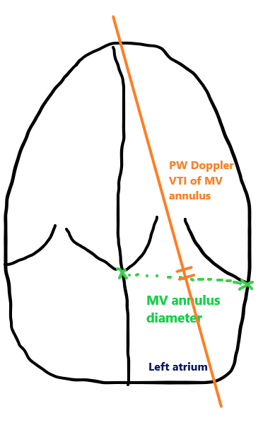
regurgitant volume and fraction
the regurgitant volume (RV) determines the volume of the regurgitant leaking back into the previous chamber
the regurgitant fraction (RF) calculates the percentage of flow thru the valve is regurgitant
the equation is based on the assumption that the stroke volume (SV) on each valve is the same (no sig. regurg. on the other valves and/or no shunt is present)
the RV and RF can be calculated for both AI and MR
regurgitant volume (RV) calculation:
calculated by subtracting the SV across the MV from the AV SV
RV = 0.785 × (LVOT diameter)² × VTILVOT – 0.785 × (MV annulus diameter)² × VTIMV annulus
regurgitant fraction (RF) calculation:
expresses the % of the total aortic SV that regurgitates back into the LV
RF (%) = RV (LVOT) ÷ SV (LVOT) x 100%
higher regurg. fractions correlate w/ more severe AI and a greater risk of adverse outcomes
reference ranges:
mild = <30%
severe = >50%
PISA (proximal isovelocity surface area)
Click to flip
The hemispheric region of flow convergence on color Doppler used to estimate the severity of regurgitation by measuring its radius.
EROA (effective regurgitant orifice area)
The calculated area of the valve opening through which blood regurgitates, reflecting the severity of the lesion.
Regurgitant Volume (RV)
The total volume of blood leaking backward through the aortic valve during diastole, measured in milliliters.
Regurgitant Fraction (RF)
The percentage of total stroke volume that regurgitates through the valve, indicating the proportion of abnormal flow.
Nyquist Limit
The velocity threshold on color Doppler imaging that distinguishes forward from reversed flow, critical for accurate PISA measurement.
complications of aortic insufficiency
HF
LV volume overload (LVVO)
LVEDP and diastolic function
LVEDP inc » diastolic function becomes compromised
treatment for aortic insufficiency
if significant regurg. is present, the pt. needs an AV replacement, either an open-heart or transcatheter surgery
if the asc. ao is also dilated along w significant AI, the asc. ao will be replaced as well
an AV with an attached conduit (bentall procedure) will be used for the replacement
valve replacements and corrective surgeries discussed in a later module
Which of the following corresponds to the 2nd heart sound?
Closing of the aortic valve
Which of the following is initially seen in the heart of a patient with long standing and/or significant aortic stenosis?
LVH
On an Aortic valve and left atrium M-mode, the closure line of a bicuspid aortic valve is usually eccentric.
true
The patient has a significant amount of aortic insufficiency (AI) when assessing the apical windows. It is unclear what to grade the AI: moderate to severe or severe. What should the sonographer do to clarify the amount of AI present?
Obtain PW Doppler image of the descending aorta
A valve needs to be sclerotic in order to be stenotic?
false
Which of the following occurs from the presence of significant aortic insufficiency?
Hyperdynamic left ventricle.
Which of the following are the most common symptoms a patient with aortic stenosis would experience?
Syncope and/or chest pain
Which of the following is the classic murmur description of valvular aortic stenosis?
Systolic crescendo decrescendo murmur at the right upper sternal border, up to the carotids.
Which of the following is an associated finding of aortic stenosis?
Dilated ascending aorta.
The velocity time integral trace calculates which of the following?
mean PG
Is it important to still use the pencil probe on a patient that you have already obtained an aortic valve velocity of 3.2 m/s from their apical windows?
true
The posterior wall of the aorta is anatomically continuous with which of the following structures?
Anterior mitral valve leaflet.
The patient has mild aortic stenosis and mild obstructive hypertrophic cardiomyopathy. Which of the following methods can be used to calculate the aortic valve area or assess the severity of stenosis? (May have multiple answers.)
None of the above
A patient has aortic valve stenosis and significant aortic insufficiency. How does this affect the amount of the aortic valve stenosis severity by the Doppler pressure gradient?
Overestimate the amount of AS present
Which of the following is premature mitral valve closure associated with?
Significant aortic regurgitation.
Flow reversal in the descending thoracic aorta, detected with pulsed-wave Doppler, may be observed in which of the following conditions?
VSD, PDA
What is doming of any cardiac valve on 2D echo consistent with?
stenosis
Aortic cusp separation (ACS) is a measurement that is performed on which of the following?
M-mode of the AV and left atrium.
On a M-mode tracing, what does fine fluttering of the anterior mitral valve leaflet in diastole suggest?
Aortic regurgitation
A patient has aortic insufficiency present. From the choices below, which velocity do you anticipate the LVOT velocity most likely to be?
1.8 m/s
Which of the following conditions is a bicuspid aortic valve associated with?
CoAo
Turner’s syndrome