3001: Fatty Acid Oxidation, Fatty Substances, Phospholipids
 Call Kai
 Learn
 Practice Test
 Spaced Repetition
 Match
 Flashcards
 Knowt Play
1/87

There's no tags or description
Looks like no tags are added yet.
88 Terms
An activated fatty acid is a fatty acyl-CoA, formed when coenzyme A attaches to the fatty acid through a thioester bond.
α-carbon: C2, immediately beside the carboxyl carbon.
β-carbon: C3, beside the α-carbon.
ω-carbon: Terminal methyl carbon farthest from the COOH group.

α-Oxidation: Primarily occurs in peroxisomes, especially in the liver, and breaks down branched-chain fatty acids.
β-Oxidation: Mainly occurs in mitochondria and generates energy.
ω-Oxidation: Begins in the smooth ER, especially in hepatocytes, with further breakdown involving peroxisomes.
BCFAs are primarily saturated fatty acids containing one or more methyl branches along their carbon chain.
They can be monomethyl, dimethyl, or multimethyl BCFAs. Examples include iso-, anteiso-, and phytanic acid.
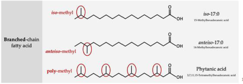
Phytanic acid has a methyl group on its β-carbon (C3), which blocks normal β-oxidation.
α-Oxidation removes one carbon, converting phytanic acid into pristanic acid, which can then undergo β-oxidation.

α-Oxidation occurs mainly in peroxisomes, particularly in liver cells.
Its primary role is detoxification and breakdown of branched-chain fatty acids. Unlike β-oxidation, α-oxidation does not directly generate significant ATP.
What is α-oxidation, and how does it allow branched-chain fatty acids to undergo β-oxidation?
Location: Primarily in peroxisomes, especially in the liver.
Function: Breaks down branched-chain fatty acids (e.g., phytanic acid) as a detoxification process.
Why needed: A methyl group on the β-carbon (C3) blocks normal β-oxidation.
Mechanism: α-Oxidation removes one carbon from the carboxyl end, shortening the fatty acid by 1 carbon and allowing subsequent β-oxidation.
Energy: According to the lecture, α-oxidation does not directly generate ATP or NADH and does not involve NADPH.
Activated form: Phytanic acid is activated to phytanoyl-CoA before α-oxidation.
Overall: Phytanic acid → α-oxidation → pristanic acid → β-oxidation → energy production.
What are the dietary sources, biological functions, and potential health benefits of branched-chain fatty acids (BCFAs), particularly in newborns?
Dietary sources: Mainly ruminant milk and beef products; lecture notes indicate they are not significant in poultry or fish. The American adult diet may contain more BCFAs than DHA.
Membrane function: BCFAs regulate membrane biophysical properties, acting similarly to cis-unsaturated fatty acids to help maintain membrane fluidity and phase-transition temperature (Tm).
Distribution: Rare in internal human organs but abundant in skin, the white waxy coating of newborns (vernix), and the intestines of healthy term newborns.
Newborn sources: May come from swallowed amniotic fluid during the last trimester, placental transfer, colostrum, and mature breast milk (not typically added to formula milk).
Absorption: In rat pups, dietary BCFAs appeared in serum and liver and were incorporated into ileal phospholipids.
Health benefits: Important bioactive lipids in early development. BCFAs have been shown to induce apoptosis (programmed cell death) in human tumour cells in experimental studies.
NEC is a severe intestinal disease mainly affecting premature or low-birth-weight newborns.
It causes intestinal inflammation, tissue damage, and necrosis. Bacteria and intestinal contents may leak into the abdominal cavity, causing life-threatening sepsis. Severe cases may require surgical removal of damaged intestine.
Refsum's disease is a rare genetic disorder caused by deficiency of phytanoyl-CoA hydroxylase, an enzyme required for α-oxidation.
Phytanic acid accumulates because it cannot be properly degraded, potentially causing neurological damage.
Thiokinase (acyl-CoA synthetase) attaches CoA to a fatty acid, forming fatty acyl-CoA through a thioester bond, between the carboxyl group of the fatty acid and the thiol group of coenzyme A
ATP is converted to AMP + PPi.
The reaction is effectively irreversible.
Activation costs 2 ATP equivalents.
There are 3 thiokinases each specific for short, intermediate and long carbon chains.
Fatty acids are activated into fatty acyl-CoA.
CPT I (carnitine acyltransferase I) transfers the acyl group from CoA to carnitine, forming acylcarnitine.
Acylcarnitine crosses the inner mitochondrial membrane through a translocase.
CPT II (carnitine acyltransferase II) transfers the acyl group back to CoA in the mitochondrial matrix.
Fatty acyl-CoA undergoes β-oxidation.
Carnitine deficiency or CPT defects can impair this process.

Dehydrogenation: Forms a double bond between the α- and β-carbons.
Hydration: Adds water, introducing an OH group on the β-carbon.
Oxidation: Converts the β-hydroxyl group into a ketone.
Thiolytic cleavage: Breaks the bond between α- and β-carbons, releasing acetyl-CoA and shortening the fatty acyl-CoA by 2 carbons.
Acyl-CoA dehydrogenase removes hydrogen atoms from the α- and β-carbons, introducing a trans double bond and forming trans-Δ²-enoyl-CoA.
FAD is reduced to FADH₂, which can ultimately generate 1.5 ATP.
Enoyl-CoA hydratase adds H₂O across the double bond of trans-Δ²-enoyl-CoA.
An OH group is added to the β-carbon, producing L-β-hydroxyacyl-CoA.
β-Hydroxyacyl-CoA dehydrogenase oxidizes the hydroxyl (OH) group on the β-carbon into a ketone, producing β-ketoacyl-CoA.
NAD⁺ is reduced to NADH + H⁺, which can ultimately generate 2.5 ATP.
Acyl-CoA acetyltransferase (thiolase) uses CoA-SH to cleave the bond between the α- and β-carbons of β-ketoacyl-CoA.
This releases:
One acetyl-CoA.
One fatty acyl-CoA shortened by 2 carbons, which can enter another β-oxidation cycle.
What are the products of one β-oxidation cycle of palmitoyl-CoA (C16), and where do they go?
Palmitoyl-CoA (C16) + H₂O + FAD + NAD⁺ + CoA-SH → Myristoyl-CoA (C14) + Acetyl-CoA + FADH₂ + NADH + H⁺.
Acetyl-CoA goes to the Krebs Cycle
Leads to 3NADH+H+ + 1FADH2 + GTP(ATP)
FADH2 and NADH + H+ go to the electron transport chain
FADH2 →→ 1.5ATP; NADH+H+ →→ 2.5ATP
How much ATP is produced from one cycle of β-oxidation and from one acetyl-CoA entering the TCA (Krebs) cycle?
1. One cycle of β-oxidation = 4 ATP
1 NADH + H⁺ → 2.5 ATP
1 FADH₂ → 1.5 ATP
2. One acetyl-CoA entering the TCA cycle = 10 ATP
3 NADH × 2.5 = 7.5 ATP
1 FADH₂ × 1.5 = 1.5 ATP
1 GTP = 1 ATP
Total = 4 + 10 = 14 ATP equivalents from one β-oxidation cycle and oxidation of its released acetyl-CoA through the TCA cycle.

Palmitoyl-CoA undergoes 7 cycles of β-oxidation, producing:
8 acetyl-CoA.
7 NADH + H⁺.
7 FADH₂.
Each cycle removes 2 carbons, and the final C4 intermediate is cleaved into two acetyl-CoA molecules.
β-Oxidation:
7 NADH × 2.5 = 17.5 ATP.
7 FADH₂ × 1.5 = 10.5 ATP.
Total from β-oxidation = 28 ATP.
TCA cycle:
8 acetyl-CoA × 10 = 80 ATP.
Gross ATP = 28 + 80 = 108 ATP.
Subtract 2 ATP equivalents for fatty acid activation.
Net ATP = 106 ATP.
Fatty acid synthesis:
Condensation.
Reduction of ketone to alcohol.
Dehydration to form an enoyl group.
Reduction of the enoyl group.
β-Oxidation:
Dehydrogenation.
Hydration.
Oxidation of alcohol to ketone.
Thiolytic cleavage.
Overall: Fatty acid synthesis builds fatty acid chains, while β-oxidation breaks them down.
ω-Oxidation is usually a minor pathway occurring in the smooth endoplasmic reticulum of hepatocytes.
It oxidizes the terminal methyl (ω) carbon farthest from the COOH group and commonly acts on medium-chain fatty acids containing approximately 10–12 carbons.
Hydroxylation: Terminal methyl group (CH₃) → primary alcohol (CH₂OH).
Oxidation: Primary alcohol → aldehyde (CHO).
Oxidation: Aldehyde → carboxyl group (COOH).
This forms a dicarboxylic acid, containing COOH groups at both ends.
What happens during step 1 of ω-oxidation, and which enzymes and cofactors are involved?
Reaction: Hydroxylation of the terminal methyl (ω) carbon of a fatty acid (CH₃ → CH₂OH).
Enzyme: Mixed-function oxidase (MFO), a cytochrome P450 monooxygenase, particularly CYP4A and CYP4F.
Cofactors: Uses NADPH and O₂.
Mechanism: NADPH donates electrons through cytochrome P450 reductase to cytochrome P450, allowing oxygen to be incorporated into the fatty acid.
Product: Fatty ω-hydroxyacid (primary alcohol), with NADP⁺ and H₂O also formed.

What happens during steps 2 and 3 of ω-oxidation, and which enzymes and cofactors are involved?
Step 2 – Alcohol oxidation:
Enzyme: Alcohol dehydrogenase.
Reaction: Fatty ω-hydroxyacid (CH₂OH) → fatty ω-aldoacid (CHO).
Cofactor: NAD⁺ → NADH + H⁺.
Step 3 – Aldehyde oxidation:
Enzyme: Aldehyde dehydrogenase.
Reaction: Fatty ω-aldoacid (CHO) → fatty dicarboxylic acid (COOH).
Cofactor: NAD⁺ → NADH + H⁺.
Overall: CH₃ → CH₂OH → CHO → COOH, forming a dicarboxylic acid that can subsequently undergo β-oxidation.
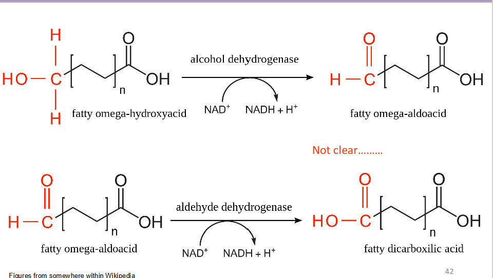
The two carboxyl groups can be activated by attachment to CoA.
The dicarboxylic acid can then undergo β-oxidation, shortening its carbon chain. The final product described in the lecture is succinate, which can enter the Krebs (TCA) cycle.
Peroxidation: A free-radical-mediated or enzymatic process in which reactive oxygen species (ROS) or enzymes oxidize polyunsaturated fatty acids (PUFAs), potentially causing oxidative degradation of membrane lipids.
Epoxygenation: Cytochrome P450 epoxygenases convert PUFAs into bioactive epoxide molecules.
Physiological levels: Controlled lipid oxidation supports cell signaling, metabolism, growth, and survival.
Excessive oxidation: Can contribute to cardiovascular disease, neurodegeneration, diabetes, and cancer.
Arachidonic acid (ARA) is an omega-6 polyunsaturated fatty acid (PUFA).
20: Contains 20 carbon atoms.
4: Contains four double bonds.
n-6: The first double bond is between carbons 6 and 7, counting from the methyl (omega) end.
Function: ARA is an important precursor for eicosanoid signaling molecules.

Humans lack Δ12 and Δ15 desaturases, meaning they cannot convert oleic acid into:
Linoleic acid (18:2n-6): Omega-6 fatty acid.
α-Linolenic acid (18:3n-3): Omega-3 fatty acid.
Plants possess these enzymes.
Therefore, both are essential dietary fatty acids and serve as precursors for longer-chain omega-6 and omega-3 PUFAs.
Linoleic acid (18:2n-6) → Δ6 desaturase (D6D) → γ-linolenic acid (18:3n-6).
γ-Linolenic acid → elongase (ELOVL5) → dihomo-γ-linolenic acid (20:3n-6).
Dihomo-γ-linolenic acid → Δ5 desaturase (D5D) → arachidonic acid (20:4n-6).
Overall: Linoleic acid undergoes desaturation, elongation, and further desaturation to produce arachidonic acid.

α-Linolenic acid (18:3n-3) undergoes Δ6 desaturation, elongation (ELOVL5), and Δ5 desaturation to form EPA (20:5n-3).
EPA undergoes further elongation, Δ6 desaturation, and peroxisomal β-oxidation to form DHA (22:6n-3).
Overall: α-Linolenic acid → EPA → DHA through elongation, desaturation, and β-oxidation.

A glycerophospholipid contains a glycerol backbone with three positions:
sn-1: Usually contains a saturated fatty acid (sometimes a MUFA).
sn-2: Usually contains an unsaturated fatty acid, often arachidonic acid.
sn-3: Contains a phosphate group attached to a polar head group.
R1 = hydrocarbon chain 1 – almost always saturated fatty acid, sometimes MUFAs
R2 = hydrocarbon chain 2 – almost always unsaturated fatty acid
Examples of head groups include choline, serine, ethanolamine, and inositol.

Arachidonic acid is commonly esterified at the sn-2 position of membrane phospholipids.
Cellular stimulation, including hormonal signaling, activates phospholipase A₂ (PLA₂).
PLA₂ hydrolyzes the ester bond at the sn-2 position.
Free arachidonic acid is released from the phospholipid.
Free ARA is rapidly metabolized into eicosanoids.
Rapidly metabolized by a series of enzymes (Cytochrome P450
epoxygenases) to generate lipid mediators (LMs) process called
ARA cascade
Note: Diacylglycerol lipase can also contribute to ARA release.
Eicosanoids are potent, short-range lipid signaling molecules produced through oxidation and peroxidation of arachidonic acid.
They regulate:
Inflammation, pain, and fever.
Blood clotting and blood pressure.
Reproduction and blood flow.
The three major groups discussed are leukotrienes, prostanoids, and EETs.
Omega-6 fatty acids: Particularly arachidonic acid, serve as precursors for many eicosanoids that generally promote inflammatory responses.
Omega-3 fatty acids: Can inhibit or compete with omega-6-derived eicosanoid production and are generally associated with less inflammatory signaling.
Overall: Omega-6-derived eicosanoids are generally more inflammatory, while omega-3 fatty acids can reduce inflammatory signaling. However, effects depend on the specific lipid mediator.
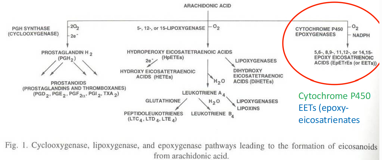
What are the three major eicosanoid pathways/arachidonic acid cascade pathways?
Cytochrome P450 (CYP) epoxygenase pathway: Produces epoxyeicosatrienoic acids (EETs).
Cyclooxygenase (COX) pathway: Produces cyclic prostanoids, including prostaglandins and thromboxanes.
Lipoxygenase (LOX) pathway: Produces hydroperoxides (HPETEs) and linear leukotrienes.
Overall: All three pathways use arachidonic acid as a precursor to produce different lipid signaling molecules.

Monooxygenases incorporate one oxygen atom from O₂ into an organic substrate, while the other oxygen atom is reduced to water (H₂O).
General reaction:
AH+BH2+O2 —> AOH+B+H2O
A reducing agent supplies the electrons required for the reaction.
How does the cytochrome P450 (CYP) epoxygenase pathway convert arachidonic acid into EETs, and what are their functions?
Precursor: Arachidonic acid (ARA, 20:4ω-6), released from the sn-2 position of membrane phospholipids by phospholipase A₂ (PLA₂).
Enzymes: Cytochrome P450 epoxygenases, mainly CYP2C and CYP2J.
Mechanism: CYP450 works with cytochrome P450 reductase. NADPH supplies electrons, allowing CYP450 to use O₂ and introduce epoxide groups into ARA.
Products: Four epoxyeicosatrienoic acid (EET) regioisomers:
5,6-EET
8,9-EET
11,12-EET
14,15-EET
Further metabolism: Soluble epoxide hydrolase (sEH) converts EETs into dihydroxyeicosatrienoic acids (DHETs), which are usually less active or inactive.
Overall: ARA → CYP2C/CYP2J → EETs (bioactive) → sEH → DHETs (usually less active).
How does the cytochrome P450 (CYP) epoxygenase pathway convert arachidonic acid into EETs, and what are their functions?
Precursor: Arachidonic acid (ARA, 20:4ω-6), released from the sn-2 position of membrane phospholipids by phospholipase A₂ (PLA₂).
Enzymes: Cytochrome P450 epoxygenases, mainly CYP2C and CYP2J.
Mechanism: CYP450 works with cytochrome P450 reductase. NADPH supplies electrons, allowing CYP450 to use O₂ and introduce epoxide groups into ARA.
Products: Four epoxyeicosatrienoic acid (EET) regioisomers:
5,6-EET
8,9-EET
11,12-EET
14,15-EET
Further metabolism: Soluble epoxide hydrolase (sEH) converts EETs into dihydroxyeicosatrienoic acids (DHETs), which are usually less active or inactive.
How does the cytochrome P450 monooxygenase system work?
Cytochrome P450 monooxygenases function with cytochrome P450 reductase.
NADPH donates electrons to cytochrome P450 reductase, which contains FAD.
Electrons are transferred to cytochrome P450, which contains iron (Fe³⁺/Fe²⁺).
Cytochrome P450 uses O₂ to oxidize an organic substrate.
One oxygen atom is incorporated into the substrate, while the other forms H₂O.
Function: Cytochrome P450 systems are important in liver detoxification and lipid metabolism.

How are EETs produced from arachidonic acid through the CYP epoxygenase pathway?
Precursor: Arachidonic acid (ARA, 20:4ω-6).
Enzymes: Cytochrome P450 (CYP) epoxygenases, mainly CYP2C and CYP2J.
Reaction: CYP epoxygenases introduce an epoxide group into arachidonic acid, producing epoxyeicosatrienoic acids (EETs).
Four EET regioisomers:
5,6-EET
8,9-EET
11,12-EET
14,15-EET
Overall: Arachidonic acid → CYP2C/CYP2J → four bioactive EETs.

How are EETs converted into DHETs, and how does their biological activity change?
Enzyme: Soluble epoxide hydrolase (sEH).
Reaction: sEH hydrolyzes the epoxide group of EETs into two hydroxyl groups, producing dihydroxyeicosatrienoic acids (DHETs).
Four products: 5,6-DHET, 8,9-DHET, 11,12-DHET, and 14,15-DHET.
Biological activity: DHETs are usually less active or inactive compared with EETs.
Overall: EETs (more active) → sEH → DHETs (usually less active).
How is linoleic acid metabolized through the CYP/sEH pathway, and what are its potential toxic effects?
Linoleic acid (18:2ω-6): An essential omega-6 fatty acid, precursor of arachidonic acid, and highly abundant in Western diets.
CYP epoxygenases: Convert linoleic acid into linoleic epoxides.
Soluble epoxide hydrolase (sEH): Converts these epoxides into linoleic diols.
Toxicity: Certain linoleic acid metabolites, particularly the diols, can cause cellular toxicity.
Health consequences: These metabolites have been associated with multiple-organ failure and acute respiratory distress syndrome in some severe burn patients.
Overall: Linoleic acid → CYP epoxygenases → linoleic epoxides → sEH → linoleic diols → potential cellular toxicity.
EETs have several physiological functions:
Stimulate peptide hormone release, making them potential drug targets.
Inhibit Na⁺/K⁺-ATPase activity in the kidneys.
Inhibit platelet aggregation.
Promote vasodilation (relaxation of blood vessels).
Inhibit cyclooxygenase (COX) activity.
They also participate in signaling involved in inflammation, pain, angiogenesis, and cancer.
How does the cyclooxygenase (COX) pathway convert arachidonic acid into prostaglandins and thromboxanes, and what are their functions?
Precursor: Arachidonic acid (ARA, 20:4ω-6), released from membrane phospholipids by PLA₂.
Enzymes: Cyclooxygenase (COX), also called prostaglandin H₂ synthase (PGHS), has two isozymes:
COX-1: Supports protective gastrointestinal mucus production.
COX-2: Produces prostaglandins involved in pain, inflammation, and fever.
Both isozymes share approximately 60% sequence identity and are located on the luminal side of the ER and outer nuclear membrane.
Two catalytic steps:
Cyclooxygenase activity: An oxidized tyrosine removes hydrogen from carbon 13 of ARA, producing a radical that rearranges and incorporates two O₂ molecules → prostaglandin G₂ (PGG₂).
Peroxidase activity: Converts PGG₂ → prostaglandin H₂ (PGH₂). Peroxidase activity is also required to initiate cyclooxygenase activity.
Products and functions:
Prostaglandins (PGD₂, PGE₂, PGF₂α): Regulate inflammation and other physiological responses.
Prostacyclin (PGI₂): Involved in vascular and platelet regulation.
Thromboxane A₂ (TXA₂): Formed by thromboxane synthase; promotes platelet aggregation and vasoconstriction, potentially contributing to atherosclerosis.
Inhibition: Aspirin irreversibly acetylates COX, while ibuprofen and naproxen reversibly inhibit COX, reducing prostaglandin synthesis.
Overall: ARA → COX (cyclooxygenase) → PGG₂ → COX (peroxidase) → PGH₂ → prostaglandins, prostacyclin, and thromboxanes.

Cyclooxygenase (COX), also called prostaglandin H₂ synthase (PGHS), has two isozymes:
COX-1 (PGHS-1).
COX-2 (PGHS-2).
Both:
Share approximately 60% sequence identity.
Are associated with the luminal side of the endoplasmic reticulum and the outer nuclear membrane.
Catalyze the same two types of reactions but have different physiological roles.
COX possesses two complementary catalytic activities at distinct sites:
Cyclooxygenase activity: Converts arachidonic acid into prostaglandin G₂ (PGG₂) using molecular oxygen.
Peroxidase activity: Converts PGG₂ into prostaglandin H₂ (PGH₂).
Overall pathway:
Arachidonic acid → COX → PGG₂ → PGH₂ → Prostaglandins and thromboxanes.
An oxidized tyrosine residue in the COX active site removes a hydrogen atom from carbon 13 of arachidonic acid.
This produces an arachidonate radical.
The radical undergoes intramolecular rearrangement.
Two O₂ molecules are incorporated, forming the cyclic endoperoxide and hydroperoxide groups of prostaglandin G₂ (PGG₂).

COX is unusual because its peroxidase activity is required to initiate cyclooxygenase activity.
Peroxidase activity helps generate the oxidized catalytic state needed for COX to produce PGG₂.
Peroxidase activity also converts PGG₂ into PGH₂.
The reverse dependence does not apply.
Overall: Peroxidase activity helps drive cyclooxygenase activity, but not vice versa.

Prostaglandin H₂ (PGH₂) is a common precursor converted by specific synthases into:
PGD₂: Prostaglandin D₂.
PGE₂: Prostaglandin E₂.
PGF₂α: Prostaglandin F₂α.
PGI₂: Prostacyclin.
TXA₂: Thromboxane A₂.
These molecules participate in inflammation, vascular regulation, platelet activity, and other physiological processes.

Although COX-1 and COX-2 catalyze the same reactions, their tissue expression results in different physiological roles.
COX-1: Expressed in many tissues, including the gastrointestinal tract. COX-1-derived prostaglandins help regulate protective mucus production.
COX-2: Expressed in several tissues and produces prostaglandins involved in pain, inflammation, and fever.
These drugs inhibit COX-dependent prostaglandin and thromboxane synthesis.
Aspirin: Irreversibly acetylates a serine residue in COX, inactivating the enzyme.
Ibuprofen: Reversibly inhibits COX activity.
Naproxen: Reversibly inhibits COX activity.
Overall: Reduced prostaglandin production helps decrease pain, inflammation, and fever

Thromboxane A₂ (TXA₂) is produced from prostaglandin H₂ (PGH₂) by thromboxane A₂ synthase through molecular rearrangement.
Functions:
Platelet aggregation: Promotes platelet clumping and blood clot formation.
Vasoconstriction: Narrows blood vessels.
These effects can contribute to atherosclerosis and cardiovascular disease when excessive.
How does the lipoxygenase (LOX) pathway convert arachidonic acid into leukotrienes, and what are their functions?
Precursor: Arachidonic acid (ARA, 20:4ω-6), released from membrane phospholipids by PLA₂.
Enzymes: Lipoxygenases (LOX), which oxidize PUFAs by introducing hydroperoxide (–OOH) groups at specific carbon positions.
Cofactor: Most LOX enzymes contain Fe³⁺ (ferric iron) in their catalytic sites, although one type uses Mn³⁺.
Types: 5-LOX, 8-LOX, 11-LOX, 12-LOX, and 15-LOX, named according to the carbon position oxidized.
Pathway steps:
Hydroperoxide formation:
5-LOX converts ARA → 5-HPETE (hydroperoxide at carbon 5).
12-LOX converts ARA → 12-HPETE (hydroperoxide at carbon 12).
Leukotriene synthesis: 5-HPETE is converted into leukotriene A₄ (LTA₄).
Further conversion: LTA₄ can form:
LTB₄, or
LTC₄ by conjugation with glutathione, followed by conversion into LTD₄ and LTE₄.
This is called the linear eicosanoid pathway because leukotrienes have linear structures, unlike the cyclic prostanoids produced by COX.
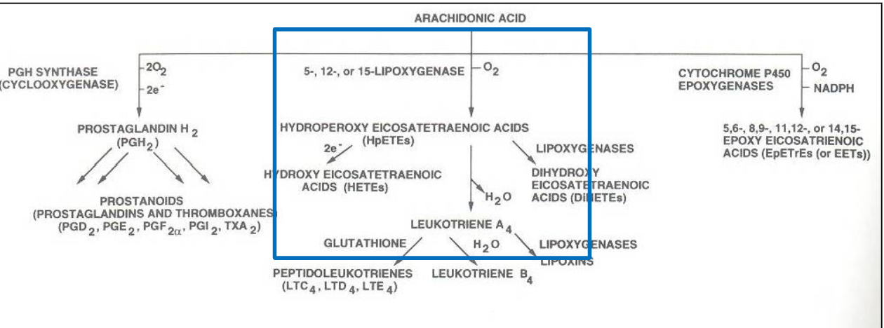
Lipoxygenases (LOX) convert polyunsaturated fatty acids into hydroperoxides by introducing oxygen at specific carbon positions.
Most LOX enzymes contain ferric iron (Fe³⁺) in their catalytic sites.
One type uses Mn³⁺ instead.
The introduction of hydroperoxides is carbon-specific.
Examples include 5-LOX, 8-LOX, 11-LOX, 12-LOX, and 15-LOX.
5-LOX: Introduces a hydroperoxide group at carbon 5 of arachidonic acid, producing 5-HPETE (5-hydroperoxyeicosatetraenoic acid).
12-LOX: Introduces a hydroperoxide at carbon 12, producing 12-HPETE (12-hydroperoxyeicosatetraenoic acid).
Overall: LOX enzymes introduce oxygen at specific carbon positions during the lipoxygenase pathway.

5-LOX converts arachidonic acid into 5-HPETE.
5-HPETE is converted into leukotriene A₄ (LTA₄).
LTA₄ can follow two routes:
Converted into leukotriene B₄ (LTB₄).
Reacts with glutathione to form leukotriene C₄ (LTC₄).
LTC₄ can be further converted into LTD₄ and LTE₄.
Linear eicosanoid pathway: Involves lipoxygenases (LOX) and produces leukotrienes, which generally have linear molecular structures.
Cyclic eicosanoid pathway: Involves cyclooxygenases (COX) and produces prostanoids, including prostaglandins and thromboxanes, which contain cyclic ring structures.
Leukotrienes are pro-inflammatory signaling molecules that generally act through G-protein-coupled receptors (GPCRs).
Their functions include:
Airway constriction: Especially pronounced in asthma.
Vasoconstriction: Narrowing of blood vessels.
Stimulation of adhesion molecule production: Helps recruit immune cells during inflammation.
Regulation of immune responses against parasites.
Overall: Leukotrienes are important mediators of inflammation, airway constriction, and immune responses.
A glycerophospholipid has a 3-carbon glycerol backbone with:
sn-1 (C1): Usually contains a saturated fatty acid, sometimes a monounsaturated fatty acid (MUFA).
sn-2 (C2): Usually contains an unsaturated fatty acid (PUFA), which creates kinks and increases membrane fluidity and flexibility.
sn-3 (C3): Contains a phosphate group attached to a polar head group.
Phospholipids are amphipathic, containing a hydrophilic head and hydrophobic fatty acid tails.

Both have a glycerol backbone, but their structures and functions differ.
Phospholipid: Contains two fatty acids and a phosphate-linked head group at C3. It is amphipathic and primarily forms cell membranes.
Triacylglycerol (TAG): Contains three fatty acids attached to glycerol. It is primarily used for energy storage.
Key difference: Phospholipids have a phosphate head group at C3, while TAGs have a third fatty acid.
Form integral components of cell membranes, maintaining their structure, fluidity, and flexibility.
Form components of plasma lipoproteins, which transport lipids in the bloodstream.
Act as reservoirs for fatty acids.
Serve as precursors for bioactive signaling molecules that regulate cellular activities.
Phospholipids are named according to the alcohol or head group attached to their phosphate group.
Choline: Phosphatidylcholine (PC).
Ethanolamine: Phosphatidylethanolamine (PE).
Serine: Phosphatidylserine (PS).
Inositol: Phosphatidylinositol (PI).
Phosphatidic acid (PA) is an important precursor for phospholipid synthesis.
Glycerol-3-phosphate is the starting glycerol backbone used to synthesize phosphatidic acid.
It comes from two pathways:
Major pathway: Dihydroxyacetone phosphate (DHAP), an intermediate of glycolysis/gluconeogenesis, is reduced to glycerol-3-phosphate.
Minor pathway: Glycerol is phosphorylated by glycerol kinase, primarily in the liver and kidneys.
Phosphatidic acid is synthesized from glycerol-3-phosphate and two fatty acids.
Fatty acid activation: Two fatty acids are activated to fatty acyl-CoA by acyl-CoA synthetase.
Fatty acid attachment: Acyltransferases attach the two activated fatty acids to glycerol-3-phosphate at C1 and C2, releasing CoA.
Product: Phosphatidic acid (phosphatidate).
ATP cost:
Each fatty acid activation consumes 2 ATP equivalents.
Two fatty acids require 4 ATP equivalents total.
Precursor for phospholipid synthesis, takes place in the endoplasmic reticulum (ER). Phosphatidate is combined with an alcohol to generate the
It can follow two major routes:
PA → Phospholipids: Used in the synthesis of phosphatidylinositol (PI) and cardiolipin through activated intermediates.
PA → Diacylglycerol (DAG): Removal of the phosphate group produces DAG, which can be converted into:
Triacylglycerol (TAG).
Phosphatidylcholine (PC).
Phosphatidylethanolamine (PE).
Phosphatidylserine (PS), through subsequent reactions.

These enzymes regulate the balance between phosphatidic acid (PA) and diacylglycerol (DAG).
Phosphatidic acid phosphatase (PAP/Lipin 1): Removes phosphate from PA → DAG + Pi.
Diacylglycerol kinase: Adds phosphate to DAG using ATP → PA + ADP.
Functions:
Regulate the amount of TAG synthesized relative to phospholipids.
Influence which phospholipids are produced.
Regulate availability of PA and DAG for cell signaling.
Location: Primarily on the cytosolic leaflet of the endoplasmic reticulum (ER) membrane.
Starting molecule: Phosphatidic acid or diacylglycerol (DAG).
Head group attachment: A phosphate-containing head group becomes attached at C3 (C-3 has OH; head group has
OH) of the glycerol backbone, forming a phosphodiester bond.