43 Neck Layers Spaces
 Call Kai
 Learn
Learn Practice Test
 Spaced Repetition
 Match
Match Flashcards
 Knowt Play
1/14

There's no tags or description
Looks like no tags are added yet.
Name | Mastery | Learn | Test | Matching | Spaced | Call with Kai | Chat |
|---|
No analytics yet
Send a link to your students to track their progress
15 Terms
Neck general boundaries
Superior
inferior margin of mandible
posterior skull
Inferior
sternum + clavicle + acromion process
thoracic aperture
Neck compartments (4)
investing compartment
contains investing fascia
trapezius m. + sternocleidomastoid m.
visceral compartment
trachea + esophagus
parathyroid + thyroid glands
vascular compartment
carotid sheath
internal jugular v.
common carotid a.
vagus n.
vertebral compartment
deep back muscles

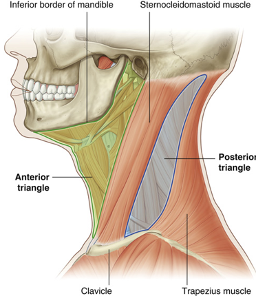
Anterior triangle of neck
Borders
anterior to sternocleidomastoid m.
inferior to mandible
Structures pathway
anterior structures pass into thorax + head

Posterior triangle of neck
Borders
posterior to sternocleidomastoid m.
anterior to trapezius m.
Structures pathway
posterior structures pass into upper limb
(eg) brachial plexus
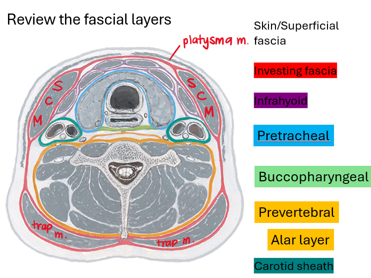
Deep cervical fascial layers (5)
investing fascia
infrahyoid fascia
pretracheal fascia
prevertebral fascia
carotid sheath

Superficial cervical fascia
subcutaneous CT: superficial fatty layer + deep fibrous layer
contains superficial nerves + vessels + lymph nodes
contains platysma m.
Investing fascia
deep cervical fascia that completely surrounds neck
arises from skull + mandible
extends inferiorly to sternum + clavicle + manubrium
surrounds sternocleidomastoid m. + trapezius m.

Infrahyoid fascia
cervical fascia that begins at the hyoid bone
forms investing fascia for infrahyoid muscles
superficial + deep layers
continuous across the midline
Pretracheal fascia
continuous fascial tube that surrounds viscera in the neck
pretracheal proper: attaches to hyoid bone above fibrous pericardium
buccopharyngeal fascia: attaches to base of skull + posterior to esophagus
covers buccinator + pharyngeal constrictors
fuses w pretracheal fascia + adventitia of esophagus

Prevertebral fascia
cervical fascia attached to the base of skull + vertebrae
encloses deep muscles
has specializations
alar fascia
axillary sheath
Sibson’s fascia
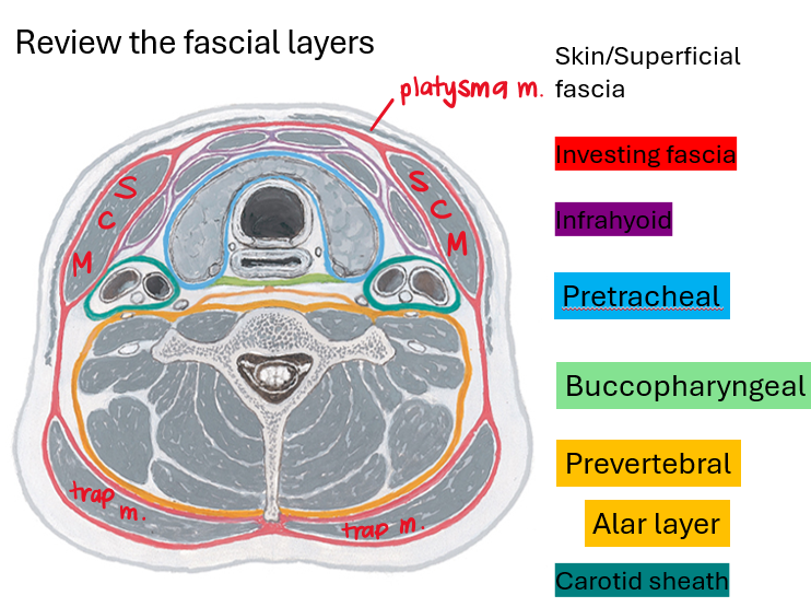
Carotid sheath contents
internal jugular v.
common carotid a.
vagus n.
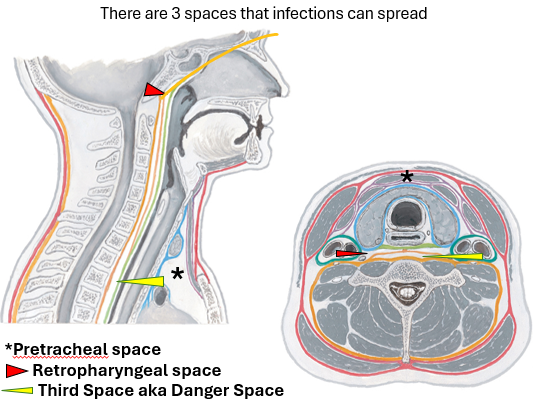
Retropharyngeal space
btwn buccopharyngeal layer of pretracheal fascia + alar layer of prevertebral fasica
spans base of skull to upper part of posterior mediastinum (sup/inf border)
infections can spread inferiorly to lower cervical levels

Pretracheal space
btwn infrahyoid + pretracheal fascia
spans betwen neck + anterior part of superior mediastinum
infections can spread inferiorly into superior mediastinum

Third space AKA danger space
within alar + prevertebral fascia
extends from base of skull towards diaphragm (sup/inf border)
susceptible to infections
can spread inferiorly to diaphragm
can spread superiorly to base of skull
air from ruptured esophagus or airway can cause pneumomediastinum
Retropharyngeal abscess
infection btwn cervical vertebrae + pharyngeal wall
Causes
oral cavity → dental abscess
nasopharynx → tonsilitis + peritonsillar abscess
retropharyngeal space infectious spread
danger space infectious spread
S/S
fever + sore throat + neck and back pain + dysphagia (difficulty + pain when swallowing) + odynophagia (painful swallowing)
Complications
airway obstruction
sepsis + mediastinitis + pneumonia + empyema (pus in pleural space)
jugular vein thrombosis + carotid artery erosion