BIOTECH 3IV3
 Call Kai
 Learn
 Practice Test
 Spaced Repetition
 Match
 Flashcards
 Knowt Play
1/70

There's no tags or description
Looks like no tags are added yet.
71 Terms
Elie Metchnikoff
a russian scientist who won the 1908 nobel prize for studying immune system
discovery:
while studying starfish larvae, he noticed moving, amoeba-like cell inside them
experiment:
poked a starfish larva w a tiny splinter and watched mobile cells swarm around the splinter and consume it
term:
Phagocytes → cells that eat
conclusion:
these eating cells protect animals by destroying invading germs
Immune system
a complex network of organs, cells, and signaling molecules work together to protect the body from harmful substances, microbes, and cell changes that make you ill
Subsystems:
Innate immunity - “first responders”
recognizes: molecular patterns DAMPs (damage-associated) and PAMPs or MAMPs (pathogen- or microbe-associated)
adaptive immunity - highly specific, has memory
recognizes: antigens
Innate Immunity
Physical barriers → initial obstacles microbes must overcome to invade the host
skin
dermis → tightly woven fibrous connective tissue
epidermis → many layers of epithelial cells
outermost = dead, filled with keratin
repels water, maintain dry envr
mucous membrane
digestive, respiratory, genitourinary tracts
constantly bathed in secretions - always wet
peristalsis of intestines and mucociliary escalator of respiratory tract remove microbes
antimicrobial substances
protect skin and mucous membranes
salt accumulates from pespiration
lysozyme degrades peptidoglycan
peroxidase enzymes break down hydrogen peroxide
lactoferrin binds iron
defensins form pores in microbial membrane
normal microbiota (flora)
production of toxic cmpds
propionibacterium degrades lipids, makes fatty acid (GI tract)
e. coli synthesize colicins in GI tract
lactobacillus in vagina produce low pH
disruption here. = predispose person to infection
Hematopoiesis
the process of making and developing blood cells
Origin: all blood cells come from hemtopoietic stem cells (HSCs) in the bone marrow
Development: signals called colony-stimulating factors (CSFs) tell stem cells to develop into diff blood cell types
Location and movement:
blood cells travel through the circulatory system
always present in normal blood, some live in body tissues
immune cell # increases during infections
Categories:
red blood cells (erythrocytes) - carry oxygen
platelets (megakaryocytes) - help with blood clotting
white blood cells (leukocytes) - function as immune cells for defence

Leukocytes
immune cells for defence
granulocytes
contain cytoplasmic granules
neutrophils → engulf and destroy bacteria
basophils → allergic rxns, inflammations
eosinophils → fight parasitic worms, allergic rxns
mononuclear phagocytes
consist of mononuclear phagocyte system (MPS)
includes monocytes and cell types that develop from them as they leave blood stream
macrophages → sentinel cell, diiferentiate from monocytes
differentiate from monocytes
dendritic cells
sentinel cells, “scouts”
engulf materials in tissues, bring to cells of adaptive immune system
develop from monocytes, also others
lymphocytes
responsible for adaptive immunity
B cells, T cells → highly specific in antigen recognition
Natural killer (NK) cells → lack specificity
Cell communication
cells send and receive signals to coordinate their responses
surface receptors
“eyes and ears”
span the cell membrane
a specific ligand binds to the receptor → triggers a cell response
adhesion molecules
help cells stick to other cells
ex. endothelial cells stick to phagocytic cells → allow to exit bloodstream
cytokines
“voices”
small signalling proteins released by cells
travel to other cells and bind cytokine receptors
can cause:
growth
differentiation
movement
cell death
work at very low conc
effects can be:
local → nearby cells
regional → cells in an area
systemic → throughout the body
Types of cytokines
chemokines → attract immune cells to a specific location (chemotaxis)
colony-stimulating factors (CSFs) → increase and help develop white cells
interferons (IFNs) → control viral infections, regulation of inflammatory response
interleukins (ILs) → produced by leukocytes, important in innate and adaptive immunity
tumor necrosis factor (TNF) → inflammation, apoptosis
Molecular patterns detected by Pattern Recognition Receptors (PRRs)
PRRs → receptors that detect signals of infection or cell damage
recognize common patterns rather than specific microbes
PAMPs/MAMPs
PAMPs = pathogen-associated
MAMPs = microbe-associated
commonly found on microbes
examples
LPS → bacterial cell walls
peptidoglycan → bacterial cell walls
lipoteichoic acid
flagellin
viral RNA
DAMPs
danger-associated
released or exposed when our own cells are damaged
Types of Pattern Recognition Receptors (PRRs)
toll-like receptors (TLRs)
where? membranes
detect what? PAMPs
plasma mem TLRs → microbes outside the cell
endosome/phagosome TLRs → materials taken into the cell
detection? sends signals to the nucleus
causes gene exp → can trigger inflammation, antiviral responses
nod-like receptors
where? cytoplasm
detect what? MAMPs
help activate protective immune response
can form inflammasome
activate strong inflammatory responses
can lead to cell death
rig-like receptors (RLRs)
where? cytoplasm
detect what? viral RNA (PAMPs/MAMPs)
dsRNA
RNA with unusual 5’ triphosphate end
detection? production of interferons
Interferons
cause cells to express inactive antiviral proteins (iAVPs)
protein kinase R, RNase L)
iAVPs are activated by dsRNA (becomes AVP) to degrade mRNA
stop protein synthesis, undergo apoptosis
The Complement System
“complements” the adaptive immune response
made of proteins in the blood and tissues
named C1-C9
can split into fragments
ex. C3 → C3a + C3b (split by C3 convertase)
all 3 pws eventually make C3 convertase
Pathways
Alternative pw
triggered by C3b attaching to a foreign cell
Lectin pw
triggered by mannose-binding lectins (MBL) binding on mannose on microbial celsl
Classical pw
triggered by antibodies bound to antigen

Complement system - 3 main outcomes
opsonization
C3b sticks to bacteria/foreign paticles
makes them easier for phagocytes to engulf
C3b = tag for eating
inflammation
C5a attracts phagocytes to the area
C3a + C5a = increased blood vessel permeability
cause mast cells to release inflammatory signals
lysis
membrane attack complex (MAC) forms holes in foreign cell mems
made from C5b, C6, C7, C8, C9
can destroy gram-negative bacteria
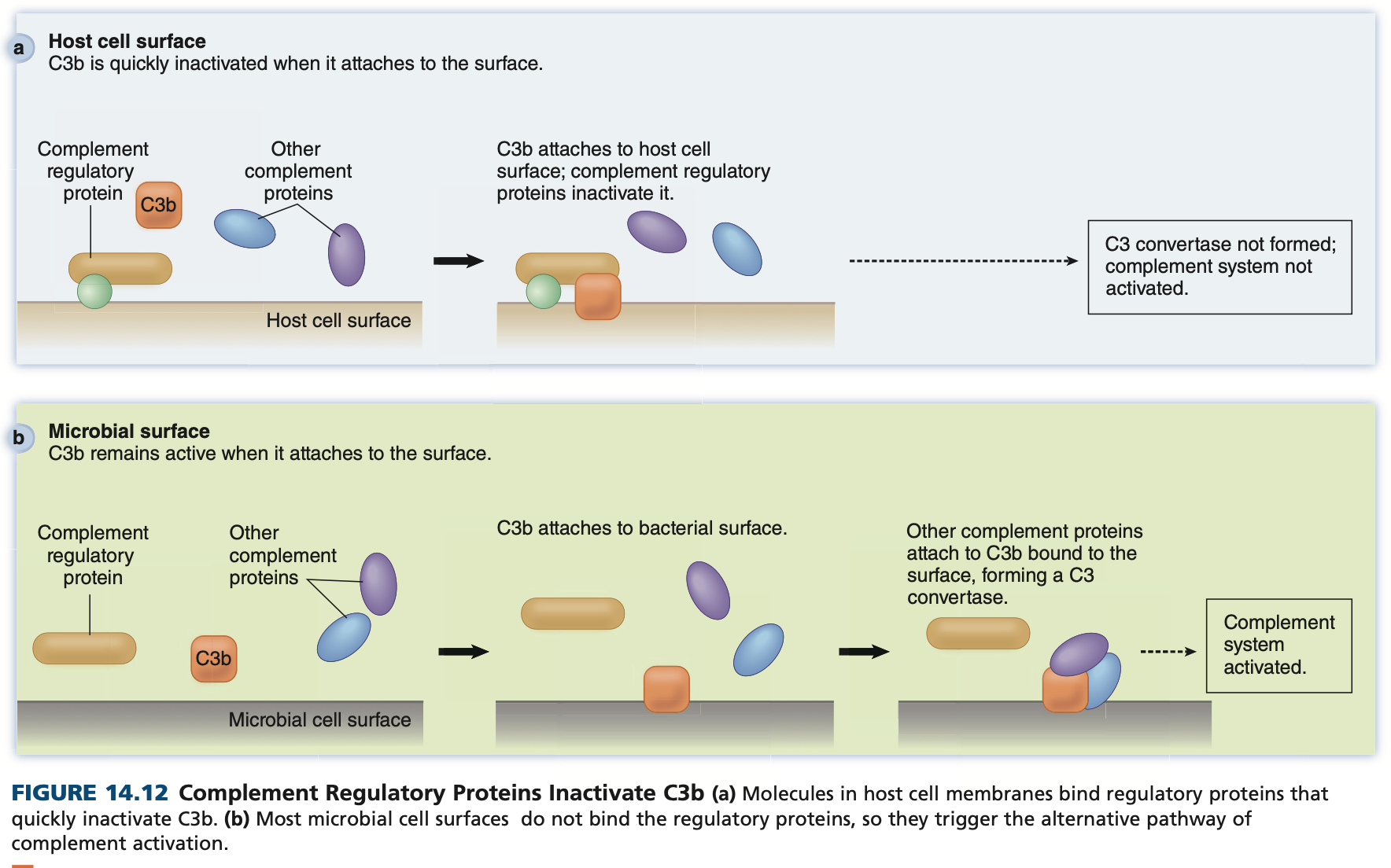
Complement system - regulation
prevents complement from attacking our own cells
host-cell molecules help inactivate C3b
prevents unnecessary opsonization and complement activation
Phagocytosis
Phagocytes engulf and digest material, pathogens
Process
chemotaxis - phagocyte recruited by chemoattractants
signals include chemokines, C5a, microbial products
recognition and attachment
direct — receptor binds mannose
indirect — binds to opsonin
engulfment
phagocyte surrounds the invader w pseudopods → forms phagosome
phagosome maturation
phagosome fuses with endosomes/lysosomes
forms phagolysosome
pH = lowers, digestive enzymes added
destruction and digestion
invader is killed and broken down
ROS + nitric oxide damage it
enzymes degrade it
defensins damage its membrane
lactoferrin binds iron needed by microbes
pH decreases
exocytosis
vesicle fuses with cytoplasm, expels remain
Attract → attach → engulf → fuse → destroy → expel
(Angela Always Enjoys Finishing Drinks Endlessly)

Types of phagocytic cells
MACROPHAGES
eat dead cells, debris, invading microbes
live for weeks to months
always present in tissues
can call in other immune cells for help
have TLRs on their surface and in phagosomes → detect invaders
release cytokines when they detect danger
can become activated macrophages → stronger
can interact with T cells → inc power
if cannot destroy something
fuse into giant cells
macrophages + giant cells + T cells = granuloma
walls of material/microbes that are difficult to destroy
NEUTROPHILS
“first responders”
arrive quickly at sites of infection/damage
important during early inflammation
MOST ABUNDANT IMMUNE CELLS
very powerful
live for 1-2 days in tissues
die after using their granules
can release DNA + granule content to form NETs (neutrophil extracell traps)
trap microbes
allow enzymes and antimicrobial proteins to destroy them
Inflammatory Response
Purpose: contain the site of damage, localize the response, eliminate invader, restore tissue function
Causes: swelling, redness, heat, pain, LoF
Factors triggering inflammation:
PAMPs, DAMPs
trigger release of inflammatory mediators
pro-inflammatory cytokines
chemicals
Inflammatory Response - Process
blood vessels dilate
vessels become wider => more and slower blood flow to the area
causes redness + heat
fluid leaks out of blood vessels
fluid with complement proteins + clotting factors enters the tissue
cause swelling + pain
stop bleeding, prevent microbes from spreading
leukocytes recruited
endothelial cells increase adhesion molecules
help grab and slow down phagocytes
phagocytes attach strongly
increase adhesion molecules
diapedesis
phagocytes squeeze bw endothelial cells
leave the bloodstream, enter the ssiue
pus formation
dead neutrophils + tissue debris + fluid accumulation = pus
dilate → leak → slow/grab → attach → squeeze out → follow signals → pus

Types of infammation
acute inflammation
short term
neutrophils and macrophages managed to control infection
chronic inflammation
if acute inflammation fails
long term (years)
macrophages + giant cells accumulate
granulomas form
Inflammatory response - Effects
Damaging effects
protects the body, but can also damage healthy tissue
phagocytes release enzymes + toxic chems → can damage nearby tissues
small/local inflammation → minor damage
inflammation in sensitive areas → more serious
ie. brain or spinal cord
Cell death & inflammation
sometimes cells sacrifice themselves to prevent spread of infection
non-immunogenic cell death
cell dies without triggering much inflammation
ie. apoptosis
“quick death”
immunogenic cell death
dying cells release DAMPs
DAMPs trigger inflammation and immune responses
ie. necroptosis, pyroptosis
“loud death”
Fever
most common host defense mechanism
strong indicator of infectious disease, esp bacterial
How it happens?
brain normally keep body temp ~37°C
during infection, pyrogens tell brain to raise temp
endogenous pyrogens → cytokines made by our cells
exogenous pyrogens → substances produced by microbes
How does it help?
slow bacterial growth
can speed up enzyme activity
increases effectiveness of immune system
inflammation, phagocyte killing, lymphocyte multiplication, neutrophil recruitment, interferon production, antibody production, release leukocytes
Antigens
Antigen → molecule can bind specifically to:
antibody
B-cell receptor
T-cell receptor
Immunogen → an antigen that can actually causes can immune response
Antigen - types
T-dependent antigens
most antigens are T-dependent
B cell needs T-helper (TH) cell help to become activated
T-independent antigen
can activate B cells without TH cell helo
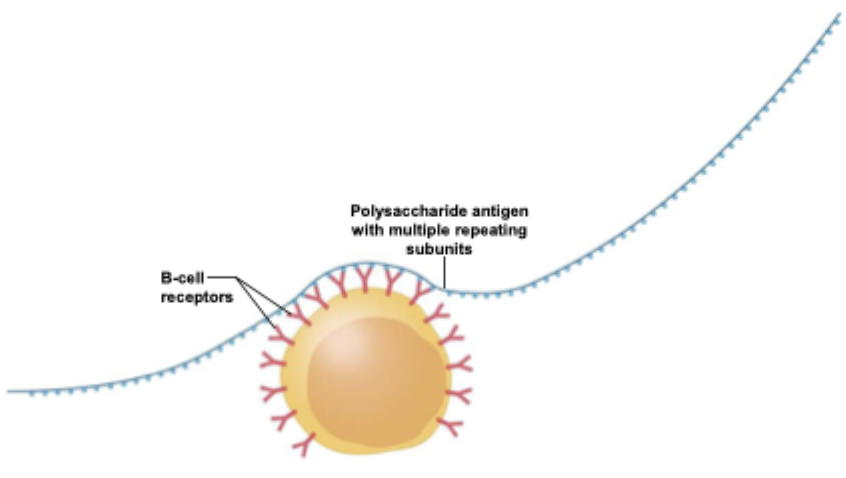
ex. LPS, molecules w repeating subunits
What Makes a Strong Antigen?
Proteins → usually strong immune responses
Lipids → usually weak responses
Small molecules → usually not antigenic
Epitopes
specific parts of an antigen recognized by the immune system
AKA antigenic determines
can be short regions of a protein
can also depend on the 3D shape of the molecule
one bacterial cell can have many different epitopes

How does the body “see” antigens
in the blood stream
antigen-presenting cells
bound by antibodies
Adapttive immunity
develops throughout life as you encounter diff antigens
develops a specific response to each invader
takes abt 1 wk or more after first exposure
has memory
first exposure → slow/weaker response
re-exposure → faster/stronger response
vaccines use this immune memory
Key features
highly specific
must distinguish
healthy self
dangerous cells/invaders
Adaptive immunity - Strategy
primary response → first time encounter
takes longer → 10-14 days
some activated B cells continue dividing
others differentiate to form antibody-secreting plasma cells
secondary response → encountered again
faster, stronger
humoral immunity
antigens outside cells
ex. bacteria, toxins, viruses in blood/tissue fluid
cell-mediated immunity (CMI)
antigens inside cells
virus infecting a cell
Antibodies
aka immunoglobulins
Y-shaped proteins
made of
2 identical heavy chains
2 identical light chains
chains held together by disulfide bonds
MAIN PARTS
Fab region = arms
binds the antigen
contains the variable region, which determines specificity
antigen-binding site fits a specific epitope
binding is reversible
uses many non-covalent bonds
Fc region = stem
contains the constant region
helps other immune system components recognize and interact with the antibody

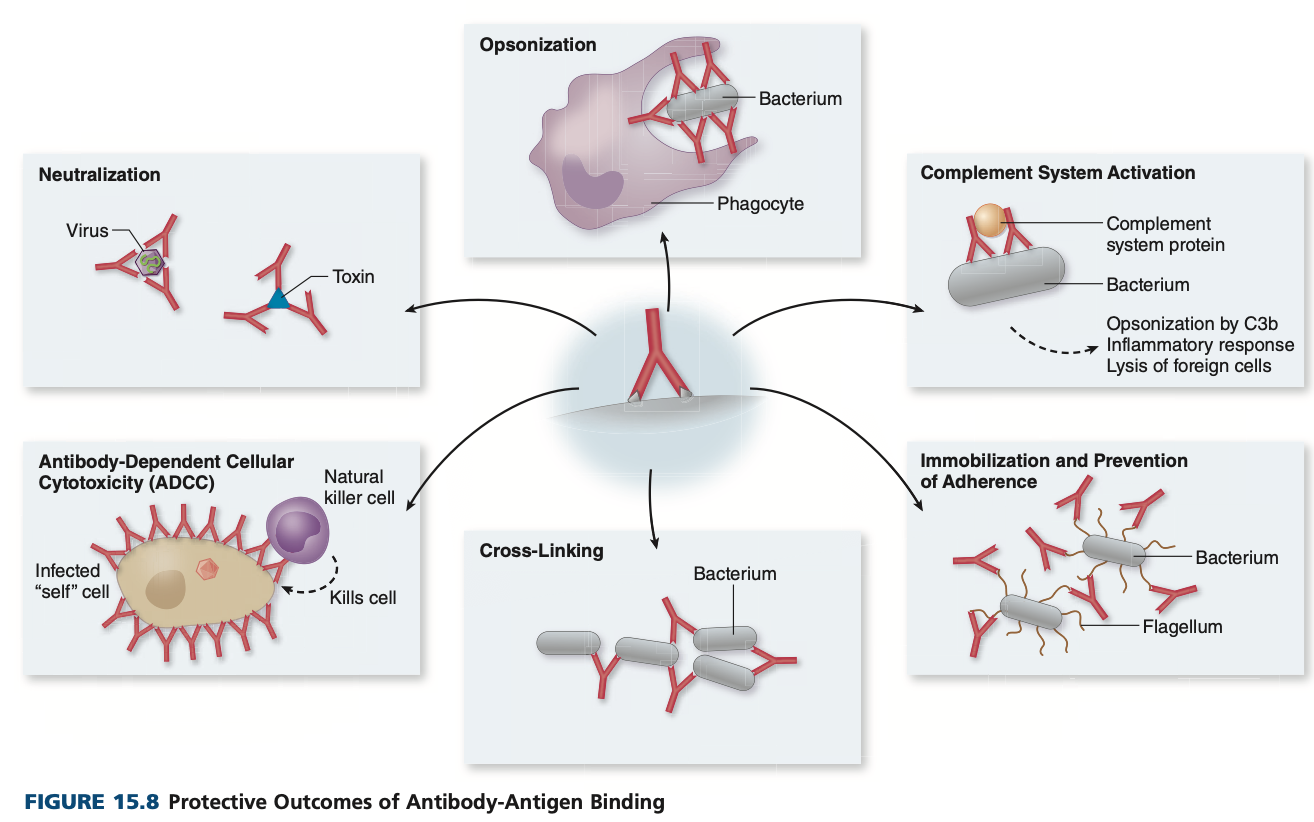
Antibody-antigen binding - protective outcomes
neutralization → prevents toxins, viruses from binding
opsonization → enhancement of phagocytosis
complement system activation
immobilization and prevention of adherence → binding to bacterial flagella or pili inteferes
aggregation → two arms of antibody bind separate antigens
antibody-dependent cellular cytotoxicity → targets cell for destruction by natural killer cells
Antibodies/Immunoglobulin (Ig) Classes
Five major classes: igM, IgG, IgA, IgD, IgE
have same basic monomeric structure
each class has different CONSTANT REGION of heavy chain
some form multimers of basic monomeric structure
each class has a distinct functions and properties

IgM

5-13% of circulating antibodies
SECRETED AFTER ANTIGEN IS ENCOUNTERED
first class produced during primary response
principal class produced in response to some T-independent antigens
pentamer
five monomeric subunits = 10 antigen-binding sites
aggregates very effectively
large size => cannot cross bloodstream to tissue => so primary role in bloodstream infections
most efficient
production begins at birth
IgG

80-85% of total serum Ig
also exits vessels to enter tissues
provides longest term protection
half-life = 21 days
first and most abundant produced during secondary response
protect via: neutralization, aggregation, opsonization, complement activation, antibody-dependent cellular cytotoxicity
transported across placenta to fetus’ bloodstream
maternal IgG protects fetus and newbord
degrade gradually over 6 mths period
infant begins producing
IgG found in colostrum (first breastmilk), absorbed by infant’s intestinal tract
Iga

monomeric form = 10-13% of serum abs
most is a dimer → secretory IgA (sIgA)
most abundant Ig class produced
sIgA → important in mucosal immunity
gastrointestinal, genitourinary, respiratory tracts
saliva, tears, breastmilk secretion
protection primarily via neutralization and interfering with attachments of microbes to host
produced by plasma cells of mucosa-associated lymphoid tissue (MALT),
secretory component (polypeptide) added to attach antibody to mucus
ATTACK BEFORE THEY EVEN GO INSIDE THE TISSUES
IgD
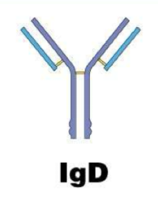
<1% of serum Ig
involved in development and maturation of antibody response
present on surface of immature B-cells
function in blood not clearly defined
IgE
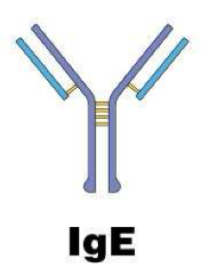
Barely detectable in normal blood
tightly bound via Fc region to basophils and mast cells
allows these cells to detect, respond to antigens
example
antigen binds to two adjacent IgE carried by mast cells → cell releases histamine and other inflammatory mediators
basophils and mast cells also release chemicals when IgE binds to normally harmless foods, dusts, pollens that can cause allergic rxns
B lymphocytes and the antibody response
Primary response
1st encounter w specific pathogen
IgM produced first, small amts
IgG produced later, large amts
slower and weaker → immune system is learning to fight the pathogen
Secondary response
2nd counter w same pathogen
faster due to memory T cells
IgG produced quickly, much higher amts
IgM produced, lower amts

Antigen-antibody interaction detection using labeled antibodies
Labeled antibodies: antibodies attached to a detectable marker (enzymes, fluorescent dyes, radioactive tags)
TYPES
direct test
detects unknown antigen (or bacteria)
antigen attached to solid surface
labeled antibodies of known specificity added
washing removes unbound, binding identifiees antigen
indirect test
detects antibodies in patient’s serum
known antigen attached to surface, serum added
unbound antibodies wash off
secondary antibodies (eg anti-human IgG) added
washing removes, binding identifies primary antibody

Common techniques using antibodies
immunofluorescence/immunohistochemistry
western blotting
ELISA
FACS
co-immunoprecipitation
Fluorescent antibody (FA) tests (immunofluorescence)
use fluorescence microscopy to locate labeled antibodies bound to antigen fixed to microscope slide
antibodies may be bound directly or indirectly

Enzyme-linked immunosorbent assay ELISA
antibodies labeled with enzyme, ie peroxidase
enzyme detected using colorimetric assay
TYPES
direct ELISA
antigen “captured” by antibodies attached to surface
enzyme-labeled antibody added to detect antigens
commercially available, including rapid grp A strep tests, home pregnancy kist
indirect ELISA
detect antibodies in human serum
eg. blood routinely screened for antibodies to HIV
may yield false positives, more reliable test
sandwich elisa
detects antigen
capture antibody attaches antigen to surface
detection antibody binds antigen
antigen is “sandwiched” bw 2 antibodies
signal = antigen present
Data analysis
typically performed at multiple dilutions/concs

Western blotting
SDS-PAGE separates proteins of antigen by size
proteins transferred to a membrane, immobilized
solutions of antibodies added
unbound antibodies washed off
labeled secondary antibodies added, unbound washed off
label is detected
Diagnostics
HIV + control proteins were separated on the gel
etc.
dark bands = locations of bound antibodies
patient a - strong reactive = lots of HIV antibodies
patient b - weak reactive = few HIV antibodies
patient c - not reactive = no HIV antibodies

Fluorescence-activated cell sorter FACS
specialized version of flow cytometer
counts, separates cells labeled with fluorescent antibodies
eg. subsets of T cells - CD4+, CD8+
label a mixture of cells w monoclonal antibodies, each w a diff dye

Co-immunoprecipitation (Co-IP)
purpose: identify protein-protein interactions
antibody binds the protein of interest (bait)
bait + its interacting proteins (partners) are pulled out of the cell lysate
agarose resin captures the antibody-protein complex
wash → removes unbound/non-specific proteins
elute → proteins are released and run on SDS-PAGE
mass spectrometry (MS) identifies the interacting proteins

Considerations for using antibodies
polyclonal vs monoclonal
mono - one specific antibody, made using hybridoma tech
poly - mixture of antibodies produced by an organism
native vs denatured protein
some antibodies recognize proteins in their native shape
others recognize proteins after they are denatured
ex. western blot = denatured, elisa = native
surface vs intracellular
is the antigen outside/cell surface or inside the cell
can the antibody actually reach and bind the antigen
Anatomy of lymphatic system
collection of tissues and organs
fluid/lymph → interstitial fluid entered a lymphatic vessel
lymphatic vessel → carry lymph
begin at peripheral tissues → connections to veins
contains WBCs and antigens from tissues
returns to lymph nodes → then empties back into circulatory system
lymphoid tissues and organs
primary = where lymphocytes form/mature
B cells mature in bone marrow
T cells migrate to thymus for maturation
secondary = where lymphocytes are activated and cloned, to contact antigens
lymph nodes, spleen, tonsils, adenoids, appendix
peyer’s patch → allow sampling of intestinal contents via specialized M cells, part of MALT
SALT - skin associated lymphoid tissue
lymphocytes → primary cells
allow body to resist or overcome infection and disease
Humoral Immunity
antibody driven
B lymphocytes or B cells = responsible
some form memory B cells → respond quickly if antigen is encountered again
develop in:
bone marrow in humans
bursa in birds
triggered to proliferate, differentiate into plasma cells in response to extracell antigens
produce antibodies
bind to antigens w high degree of specificity
have two functional regions
ARMS = bind specific antigen
amino acid at the end varies among abs = specificity
STEM = “flag” to tag antigen
recruit other immune system components to eliminate
protect directly and indirectly
directly = bind to antigen, prevent attachment to cell
indirectly = tag antigen for elimination
B-cell receptor (BCR)
membrane-bound version of B cell’s specific antibody
binding antigen triggers response
needs helper T cell, sometimes
Cell-mediated immunity
phagocytes activated → cytotoxic T cells activated
T lymphocytes/T cells → mature in thymus
cytotoxic T cells
destroy abnormal and infected cells
helper T cells
do not kill pathogens directly
“command centre”
regulatory T cells
suppress response of other T cells and B cells
to prevent immune system from mounting a response against self molecules
Helper T cells and Cytotoxic T cells
must be activated to multiply
confirms antigen signifies danger
dendritic cells activate T cells
T cell proliferates, differentiate into
effector helper T cells → orchestrate humoral and cell-mediated immunity
activate B cells, macrophages
produce cytokines to direct and support T cells
effector cytotoxic T cells → respond to intracell antigens, apoptosis
both can form memory cells
Lymphocyte receptors
BCR → specific antibody B cell is programmed to make
TCR → does not recognize free antigen, must be presented by body’s own cell

Clonal selection theory
each B cell or T cell is specific for one antigen/epitope
when that specific antigen appears, matching lymphocyte is selected and make many copies
Process
each lymphocyte has a unique BCR or TCR, each recognizes specific epitope
antigen enters
only lymphocyte w receptors that match it can recognize it
other lymphocytes stay inactive
additional signals needed to confirm the response is needed, and to prevent self-attack
clonal expansion
selected lymphocyte multiplies = many identical copies
clones become effector and memory cells
effector = actively fight pathogen
memory = remain long term, respond quickly when same antigen is encountered again
if antigen disappears = many responding cells undergo apoptosis
Types of lymphocytes
immature → antigen-specific receptor not fully developed
naive → have receptors, have not encountered appropriate antigen
activated → bound antigen, received confirmation, able to proliferate
effector → descendants of activated lymphocytes: plasma, Tc, Th
memory → long-lived descendants of activated lymphocytes, secondary response if antigen encountered again
B cell activation by T-dependent antigens

BCR binds to antigen
antigen is internalized via endocytosis
antigen gets degraded into peptide fragments
fragments delivered to MHC class II molecules for inspection by Th cells = antigen presentation
if t cell receptors binds a fragment = t cells activates B cell with cytokines, initiates clonal expansion
no Th cells recognize peptides => B cell may bcome unresponsive to future exposure of antgen → results in tolerance ot antigen
Primary antibody response
some activated B cells become plasma cells
plasma cells
generate antibodies
undergo apoptosis after a few days
as long as antigen is present, activated B cells continue to proliferate and differentiate
→ causing antibody lvls (titer) to increase
Affinity maturation
random mutations occur as B cells multiply
form of natural selection
B cells that bind antigen more strongly = likely to survive and proliferate
Class switching
TH cells can induce some activated B cells to switch
B cells → plasma cells (normally secrete IgM)
B cells (in lymph nodes) → IgG
B cells (in MALT) → IgA
Secondary antibody response
faster and stronger than primary response
pathogens eliminated before they can even cause harm
vaccination use this
memory B cells
greater numbers present
receptors fine-tuned through affinity maturation
when activated, quickly become plasma cells, produce antibodies
IgG or IA → due to class switching
proliferating B cells undergo affinity maturation =
higher-affinity antibodies after repeated exposure
T-independent antigens
can activate B cells without helper T cells
relatively few of them, but medically important
How they work
they are molecules with many identical, evenly spaced epitopes (eg bacterial polysaccharide capsules)
bind B-cell receptors at once
activate B cells
Note
not very immunogenic in young children
example: LPS
T lymphocytes
never produce antibodies
effector T cells interact directly with target cells

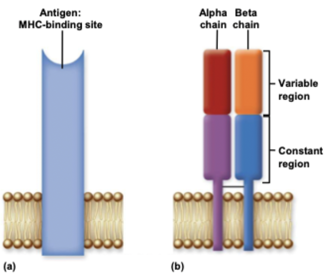
T-cell receptors (TCRs)
each T cell has many copies of its TCR
structure
two polypeptide chains
alpha + beta or gamma + delta
each has a variable and constant region
it looks like one arm of B-cell receptor
does not interact with free antigen
only antigen presented by another cell
held in major histo-compatibility complex (MHC) molecules on cell surface
MHC molecules
Types
MHC class I
carries and presents endogenous antigens
produced by all nucleated cells
MHC class II
arries and presents exogenous antigens
produced by antigen-presenting cells (dendritic, B, macrophages)

Diversity in MHC genes
ensures proper presentation and different orientations of proteins from evolving pathogen populations
inherit one set from mother, one set from father
each set has 3 genes:
HLA-B — at least 1400 diff alleles
HLA-C — at least 620 diff alleles
HLA-A — at least 890 diff alleles
Why this matters
More MHC variants = cells can present a wider range of peptides to T cells.
Across a population, a pathogen is unlikely to evade everyone’s MHC.
T-cell Activation
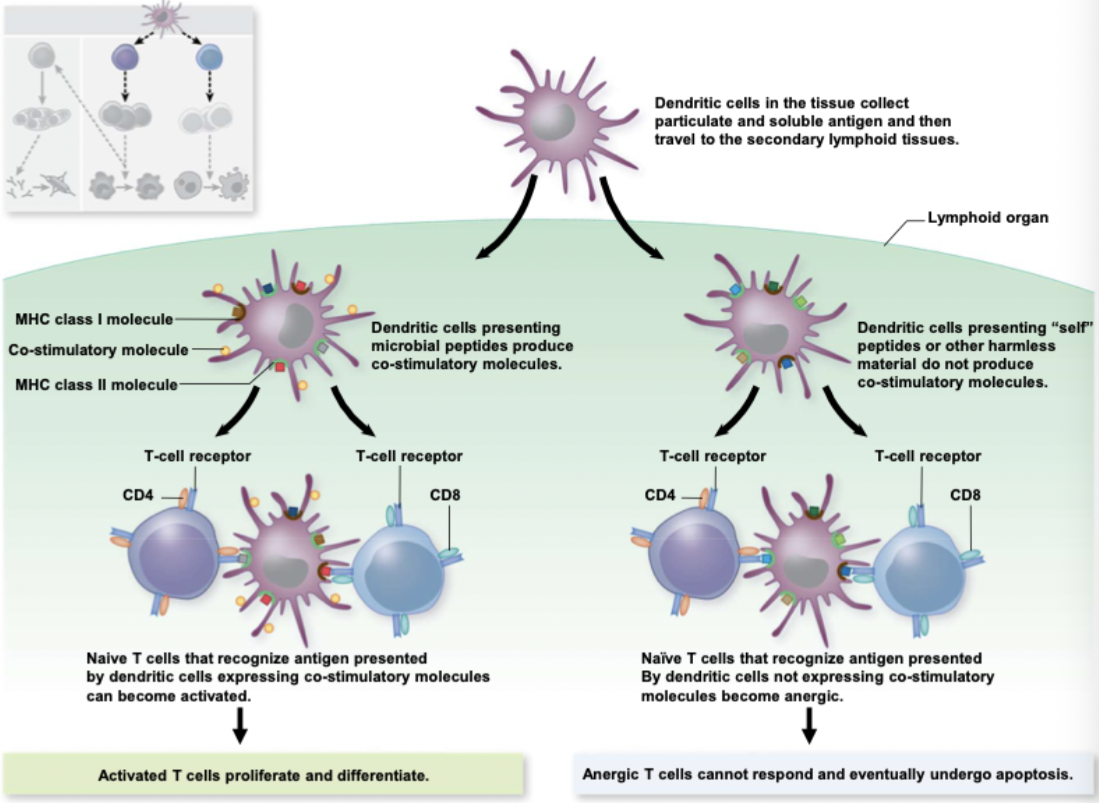
dendritic cells capture antigens
via phagocytosis and pinocytosis
detect pathogens
toll-like receptors (TLRs) recognize pathogens
trigger increased antigen uptake
travel and mature
dendritic cells migrate to 2ndary lymphoid organs and matures
present antigens
display antigens on both MHC I and II molecules
co-stimulation
produce co-stimulatory molecues that signal danger and help activate T cells
macrophages and B cells can also present antigens and provide co-stimulatory signals
NOTE
Right side: “self” peptides or harmless material
self peptides presented by dendritic cells
does not produce co-stimulatory molecules.
Naive T cells that recognize the antigen without co-stimulation become anergic.
Result: anergic T cells cannot respond and eventually undergo apoptosis.
Tc (CD8) cells — Effector functions
Tc cells check the internal proteins shown on MHC I
See something abnormal → kill the cell by apoptosis

TH (CD4) cells — Effector functions
recognize antigen presented on MHC class II from antigen-presenting cells (APCs)
release cytokines to activate other immune cells
B cells and helper T cells can recognize diff epitopes from same pathogen
Conjugate vaccines
trigger a T-dependent immune response
attach a protein to a polysaccharide capsule
B cells recognize the polysaccharide
helper T cells recognize peptides from the protein
helper T cells activate B cells → stronger immune response
Haptens
a small molecule that can bind an antibody or B-cell receptor
usually cannot trigger an immune response alone
must bind to a carrier protein to trigger an immune response
example: penicillin can act as a hapten, cause allergic rxns
TH cells in macrophage activation
recognize peptides presented on MHC class II by macrophages
release cytokines that activate macrophages
activate macrophages increase in size, metabolism, lysosome number
may fuse to form multinucleated giant cells
produce nitric oxide and other toxic cmpds to kill pathogens

Lymphocyte development
both B cells and T cells develop from hetopoietic stem cells (HSCs)
B cells mature in bone marrow
T cells mature in thymus
during development, they acquire the ability to recognize specific antigen epitopes
mature lymphocytes can participate in adaptive immune response

Gene rearrangements
creates many diff antigen receptors in B and T cells
each B cell recognizes specific epitope
the body can recognize millions of diff epitopes without the need of millions of genes
V (variable), D (diversity), J (joining) gene segments
rearrange by deleting and joining DNA sections
imprecise joining
adds or removes nts = extra diversity
in B cells, heavy and light chains form separately
combining them = more unique antigen-binding sites

Negative selection of B cells
occurs in bone marrow
B cells that strongly recognize self-antigens may undergo apoptosis or receptor editing
removes self-reactive B cells to help prevent autoimmunity
Selection of T cells
occurs in thymus
positive selection
T cells must recognize the body’s own MHC molecules to survive
negative selection
T cells that strongly recognize self-peptides presented on MHC = eliminated by apoptosis
over 95% of developing T cells die during thymic selection