BIOL302- Fundamentals of Genetics and Genomics
 Call Kai
 Learn
Learn Practice Test
 Spaced Repetition
 Match
Match Flashcards
 Knowt Play
1/183

There's no tags or description
Looks like no tags are added yet.
Name | Mastery | Learn | Test | Matching | Spaced | Call with Kai | Chat |
|---|
No analytics yet
Send a link to your students to track their progress
184 Terms
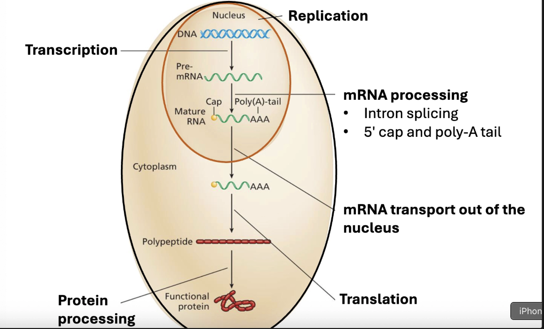
Central dogma of Biology
DNA to RNA to protein

DNA structure
Antiparallel double helix, AT double bonded, CG tripple bonded- semiconservative replication, with errors which can be propegated
Start codon
AUG = Met
Stop codons
UAA UAG UGA
Prokaryotic gene structure
Cis regulatory sequence, promoter region where transcription factors bind, termination sequences

Eukaryotic gene structure
Intron & exon, exon is the coding sequence that gives rise to a protein- introns spliced during transcription
Cis-regulatory elements
are specific DNA sequences located on the same molecule as the gene they regulate, whereas trans-regulatory elements are diffusible molecules (like proteins or RNAs) that can travel through the cell to regulate genes anywhere in the genome
Chromosome compaction
DNA around histones, nucleosomes- further compaction of proteins, tightly looped, and coiled into x shape
Affects availability of DNA for TF access
Euchromatin
loosely wrapped regions are accessible to tfs
Heterochromatin
tightly compacted, inaccessible
Loci of regulation
Allele
alternative forms of a gene- wild type v
Genotype
genetic constitution of an organism
Phenotype
An organism’s observable traits
Pleiotropy
a genetic phenomenon where a single gene or genetic variant influences two or more seemingly unrelated phenotypic traits
Polygenic trait
a characteristic controlled by two or more genes working together
Impacts on phenotype
Genotype and External environment, and internal environment of the cell
Phenylketonuria
Example of Phenotype is not affected solely by genotype:
Lack enzyme to convert phenylalanine to tyrosine
Can treat with low phenylalanine diet
What does genetic variation arise from?
Mutation
New variation of genes = new alleles
Recombination
Different combinations of alleles
What are mutations
are a change in DNA seq. that can lead to:
Altered gene coding/regulatory sequence
Altered phenotype
Mutant is an individual with altered phenotype compared to an often arbitrarily defined form
Somatic mutations
Non heretible
Happen in body tissue
Population of cloned mutant cells
Germ line mutations
Passed to approximately half of next gen
Bc mutations Only affects one copy of a gene
Point mutations
change/add/del of one or few nucleotides
Insertional mutations
insertion of large chunks of DNA- transposable elements
Chromosomal mutations
losing gaining, or swapping large bits of chromosomes
Trisomy 21
Synonymous point mutations
silent mutation
Codons specify the same amino acid
Eg. AGG → CGG both coding for Arg
Missense point mutations
altered codon specifies another aa
Conservative
Chemically similar aa
AAA (Lys(basic)) → AGA (Arg(basic))
Nonconservative: chem. dissimilar aa
Consequences depend on location
A functional domain will have greater weight
Nonsense point mutation
aa specifying codon replaced by a stop codon
Truncated proteins-
Mutation location matters- if early in the sequence, likely strong loss of fxn
Frameshift mutation
Addition or deletion alters downstream codons by putting them out of frame
Chance of having premature stop codons leading to truncation
Regulatory sequence
Promoter
Polyadenylation
Splice site
DNA replication
Base substitutions
swapping of a base pair
Transition
Purine to purine; pyrimidine for pyrimidine
Transversion
purine to pyrimidine, pyrimidine to purine
What if a mutation falls outside the coding region
If it falls within an important cis sequence, the mutation can affect transcription of a gene
Wrong place, wrong time
A mutation that affects a splice junction can retain introns or splice out part of an exon
Spontaneous mutations
Spontaneous- ‘background’ level of mutations, no purposeful or accidental exposure to mutagen
Mispairing
Tautomeric shifts allow base mispairing
Bc of altered physical form, mispairing happens
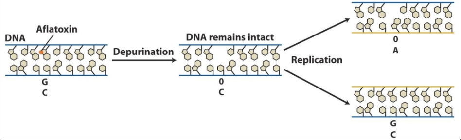
Depurination
lose a purine group-
Repair enzymes will slot something in -
If not detected and repaired- bp substitutions can occur before replication
Deamination
loss of nitrogen group
C → U is easy to spot and repair
DNA methylation can cause problems
Locations of 5 methylcytosine are mutational hotspots

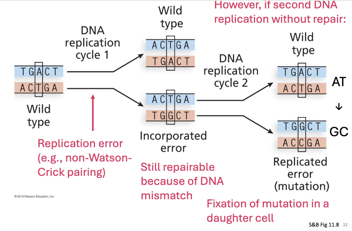
Fixation of spontaneous mutation
Fixation of mutation is when second replication round occurs without repair- the mutation is fixed in the genetic code
Replication slippage- usually associated with repeated DNA sequences
Can lead to additions and deletions

Base analogs
Can be integrated into DNA in place of nucleotides, less stable forms than bases; shift base-pairing affinities
eg. 5 bromouracil (5BU)
Causes A/T to G/C or vice versa
Base alteration
(eg. alkylating agents)
Eg ethylmethane sulfonate, adds ethyl group
G/C to A/T

Intercalating agents
mimic base pairs / integrate into double helix
Eg provlavin, ethidium bromide
In our environment
Radiation - eg uv radiation and x rays
Chemicals - cigarette smoke, barbecuing, benzoyl peroxide
Infectious agents- HPV, helicobacter pylori
Base damage- uv
UV light can cause interactions between adjacent pyrimidines
Creates this different linking , typically between adjacent thymines
Creates bulky bend in DNA that can cause issues in replication

Base damage - other mutagens
aflatoxin B1, leads to apurinic site via depurination event
Ames test
Whether or not a compound is mutagenic/ poses mutagenic risk
Start with a initial mutation in bacteria that’s hist- , place it on a medium that contains histadine- it shouldn’t grow
Then you place another potential mutagenic compound, and look at how many reversion compounds you get
REVERSION is the central concept.
True reversionf
Get a reversion of function- revertant codes for the same aa

Intragenic reversion
restores the reading frame

How to test mutagenic potential in animals
is add enzymes- see if the compound is mutagenic AND if the metabolic compounds may be mutagenic as they’re broken down
Rat liver enzymes (S9) use to see if chemicals that are n
How do organisms deal with mutations?
Very efficient DNA repair systems
Proofreading subunit of DNA polymerase repairs many
Reverse mutations
Remove altered nucleotides
Detect and remove mismatches
Paste together broken DNA molecules
Direct reversal (not in humans)
Photorepair of pyrimidine dimers
Performed by photolyases which detects bulky distortions in the helix
Base excision repair
Removal triggers the AP repair system
Ap endonuclease cuts at the AP site
Nucleotide excision repair
DNA damage where no glycosylase to detect that type of damage
Pyrimidine dimers from uv damage
Cuts made either side to remove a stretch of 30 nt around damage
DNA polymerase uses the template strand to repair the strand
Mismatch repair
In short window of time post replication, when parent/daughter strands don’t line up-

What happens if dna replication stalls?
SOS repair/translesion repair- bulky additions and/or pyrimidine dimers can lead to stalling of DNA polymerase
To let replication to continue, error - prone ‘repair’ polymerase is activated that puts random nucleotides opposite the lesion (leads to mutation itself
Goes until regular polymerase can rejoin.
Nonhomologous end joining
Ends trimmed/ligated back together, which causes deletions
Error prone
Helpful when no template to guide correct repair is available
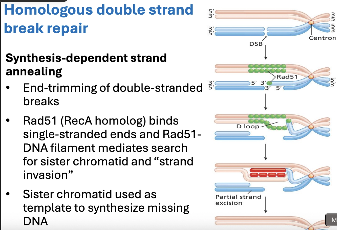
Homologous double strand break repair
Allele
alternative form(s) of a gene
Genotype
genetic constitution of an organism
Phenotype
observable traits (genotype + environment)
How is dominance/recessiveness determined?
Dominance of one allele over another is determined by the
protein products of the allele and the way protein products
of alleles work to produce the phenotype
Muller’s morphs
a set of terms created by geneticist Hermann J. Muller to classify mutant alleles based on how their function changes compared to the normal (wild-type) gene
either loss of function or gain of function
Null mutation
LOF Muller-
protein is non-functional or not made

Leaky mutation
LOF muller
protein retains some function or only some protein made

Dominant negative mutation
LOF Muller
non-functional protein affects
function of normal protein

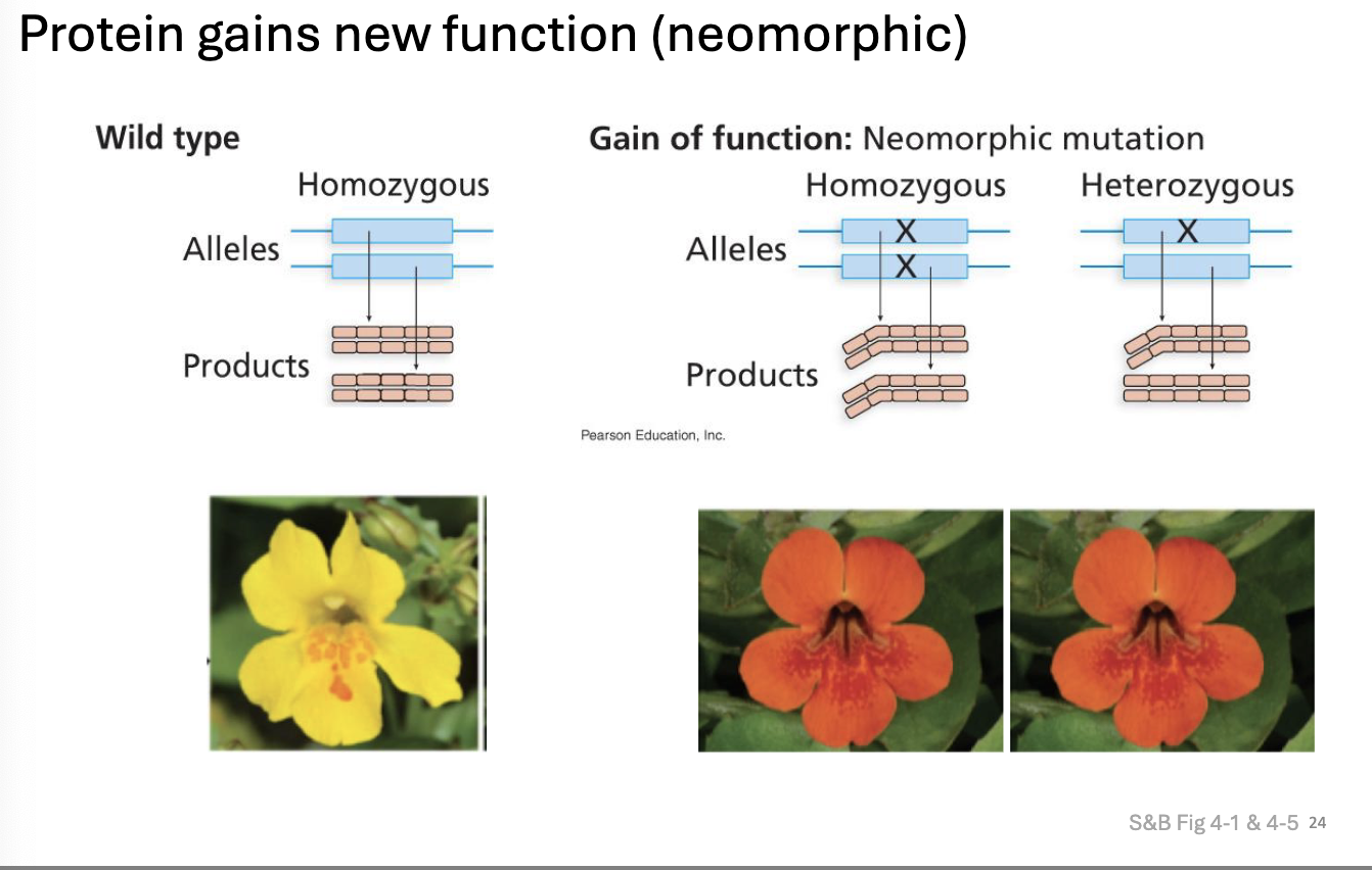
Gain of fxn mutation
Protein is hyperactive
• More protein made or made in wrong time/place
• Protein gains new function (neomorph)
17
Hypermorphic mutation

Neomorphic mutation
Protein gains new fxn
Dominance vs recessiveness
Is one copy of the wild type ‘enough’ to do the job?
Haplosufficiency
If one copy of wt allele is enough, mutant allele is recessive
Haploinsufficiency
If one copy ISN’T enough- mutant allele is dominant
Incomplete dominance
Eg. ‘bully’ whippets

Codominance

Blood type codominance

Penetrance

Expressivity

Mitosis rev.

Meiosis in germ cells

Meiotic basis for Mendel’s law of segregation

Mammals Sexual alleles
XX = female, XY = male (hemizygous)
Hemizygous, either have the wild type or otherwise
Y determines maleness
Pseudoautosomal regions
mediate XY pairing in meiosis- contain few genes that Act like autosomal gnes

Experimental methodology for determining if Y is responsible for maleness
look at XXY and XO progeny
Drosophila sex alleles
XX = female, XY = male
However, sex determined by ratio of X chromosomes to autosomes
Female if X:autosome ratio is 1:1
XXY(female), XO (male)
Bird sexual alleles
ZW = female hemizygous, ZZ= male
Still unclear how maleness v femaleness is determined
Could be dosage or W dominance
How do organisms deal with gene dosage for genes on the x chromosome?
Drosophila: alter gene expression levels, genes on the single X chromosome are hyperactivated in males
Mammals: One of the x chromosomes is randomly inactivated in somatic cells
Mammal random x inactivation
Can see in nucleus as structure (get condensed into Barr body)
Happens early in development, 2 wk post fertilization
Can see patches of expression if heterozygous for a gene carried onto the X chromosome

Differences in Sex development
Way to characterize genes and hormones in reproductive development- sex dev is different than most others
3 categories of differences in Sex development
Sex chromosome, XY, XX
Sex chromosome dsd
abnormality in number of sex chromosomes
Klinefelter syndrome (XXY)
Turner syndrome (XO)
XY DSD
affects genetic males
Atypical teste development
insensitivity to male hormones
XX DSD
affects genetic females
ovarian development issues
excessive exposure to male hormones
Can lead to ambiguous gneitalia or masculinisation of genitalia
Physical basis of Mendelian law of heredity
Chromosomes (diploid case; 2n).
Aberrant euploidy
change in the number of the complete set
chromosomes (e.g., 3 copies instead of 2 → triploid)
Aneuploidy:
plus or minus specific chromosomes in a set (e.g.,
2n+1 → Trisomy 21, Down’s syndrome)
Autopolyploid
Extra set of chromosomes from same species
Allopolyploid
extra set from a different species
Triploids

Causes of autopolyploidy
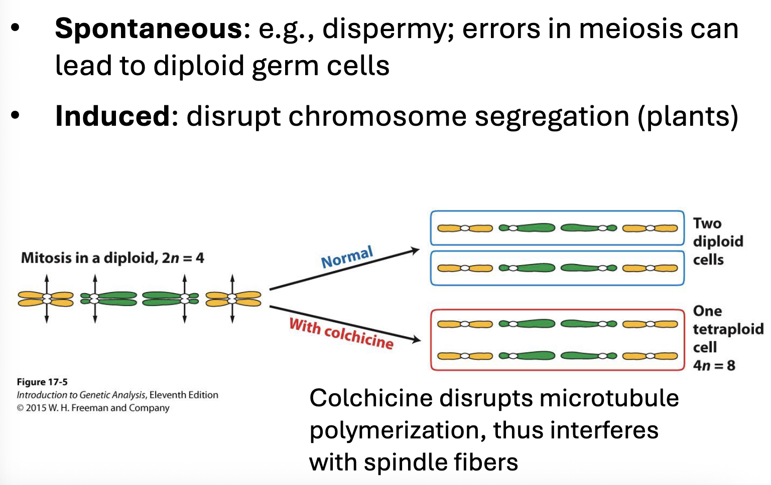
Somatic consequences of autopolyploidy
Or- larger size, which is common in plants

Germline consequences of autopolyploidy
Infertility in some cases, eg triploid bananas having no seeds