Clinical Pulm. Physical Therapy
 Call Kai
 Learn
Learn Practice Test
 Spaced Repetition
 Match
Match Flashcards
 Knowt Play
1/99

There's no tags or description
Looks like no tags are added yet.
Name | Mastery | Learn | Test | Matching | Spaced | Call with Kai | Chat |
|---|
No analytics yet
Send a link to your students to track their progress
100 Terms
Functions of pulmonary system
Exchange gases between tissue, blood and air; maintain acid-base balance; temp. homeostasis; metabolism and excretion of toxins
rib numbers
12 pairs, 7 true, 3 false, 2 floating
head of rib articulates with
thoracic vertebra
inspiratory muscles
diaphragm, intercostals, accessory mucles (SCM, scalenes, others)
expiratory muscles
at rest its passive, forced is intercostals and abdominals
origins of diaphragm
xiphoid, ribs 11-12, lumbar vertebrae, arcuate ligaments
insertion of diaphragm
central tendon
diaphragm innervation
left and right phrenic nerves
These structures pass through the diaphragm at what level? : aortic, esophagus, IVC
T12, T10, and T8 respectively
diaphragm excursion
quiet breathing: 2/3 inch
maximal ventilation: 2.5-4 inches
the right lung has ____ segments, the left lung has ___ segments
10, 8
conducting airways
mouth/nasal cavity, pharynx, larynx, trachea, bronchi, bronchioles
respiratory airways
respiratory bronchioles, alveolar ducts, alveolar sacs - gas exchange occurs here
hilum
place where blood vessels, nerves, ducts, and lymphatic vessels enter and leave
pleura of lungs
serous membranes, visceral to parietal and pleural space in middle (in to out)
suprasternal notch corresponds to …..
T2- clavicle articulates with the manubrium at lateral border of manubrium
Sternal angle of loius
inferior aspect of manubrium at sternal angle - palpable as a ridge, corresponds to T4/5 and second rib/second intercostal space
sternal angle is location of carina - bifurcation into L and R mainstem bronchi
carina
bifurcation into L and R mainstem bronchi, at T4/5 or rib 2 by sternal angle
visceral pleura is ________
not well innervated, why lung cancer not painful until later stages
there is significant potential space in lungs pleura for
negative pressure, which keeps lungs expanded
Bronchopulmonary segments (numbers)
10 in right lung (upper lobe 3, middle lobe 2, lower lobe 5) and 8 in left lung (upper lobe 4, lower lobe 4)
Name each segment starting at 1 going clockwise

L apico-posterior, L anterior, L superior lingular, L inferior lingular, L anterior basal, L lateral basal, L posterior, R medial basal, R posterior basal, R lateral basal, R anterior basal, R medial, R lateral, R posterior, R anterior, R apical
Name each segment starting at 1, going clockwise
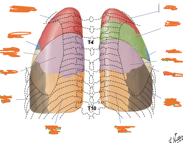
R apical, R posterior, R anterior, R superior, R lateral, R lateral basal, R posterior basal, L posterior basal, L lateral basal, L superior, L singular lingular, L anterior, L apico-posterior
lambert canals connect __________ nearby alveoli
terminal bronchioles
Pores of Kohn
connect adjacent alveoli to one another
alveolar type 1 and 2 cells are _____ in number, but type ____ cells make up 95% of surface area of alveoli
equal, 1
what can type 2 alveloar cells do that type 1 cannot
proliferate and restore both types via cell division, and secrete surfactant
tidal volume
the amount of air that moves in and out of the lungs with each normal, resting breath, usually 500mL
Functional residual capacity (FRC)
the volume of air left in the lungs after a normal, passive exhalation, aka buffer volume, 2.6-3.4 L (mixes with air coming in helps dilute)
Inspiratory reserve volume (IRV)
the maximum extra volume of air that can be inhaled forcefully after a normal, quiet tidal volume inhalation, 1.9-2.5L
Expiratory Reserve Volume (ERV)
the extra amount of air you can forcibly exhale after finishing a normal, quiet tidal breath, 1.1-1.5L
residual volume (RV)
the amount of air that stays in your lungs after you breathe out as hard as you can, 1.5-1.9L
Total Lung Capacity (TLC)
maximum volume of air the lungs can hold after a maximal inhalation, IRV, TV, ERV, RV) 4.9-6.4L
Inspiratory capacity (IC)
total amount of air you can breathe in after a normal breath out (TV + IRV) 2.3-3.0L
Functional Residual Capacity (FRC)
the volume of air remaining in the lungs after a normal, passive exhalation, (ERV + RV) 2.6-3.4L
Vital Capacity (VC)
the maximum amount of air a person can exhale from their lungs after taking a maximum inhalation (ERV + TV + IRV) 3.4-4.5L
capacity
sum of 2 or more volumes
ventilation
exchanges air between lungs and atmosphere so that O2 can be absorbed and CO2 eliminated, mostly changes PCO2
oxygenation
the addition of oxygen to the body, mostly changes PaO2
minute ventilation equation
= respiratory rate (rr) x tidal volume (Vt)
hypoventilation
CO2 increases
hyperventilation
CO2 decreases
as you breath in, the chest _______, the diaphragm _________ and the pressure in the lungs ________
expands, drops and contracts, decreased
when you breath out, the chest _______, the diaphragm _________ and the pressure in the lungs ________
deflates, rises passively, increases
compliance equation
= change in volume/ change in pressure
if change in pressure is small and change in volume is large, compliance is……
high, lungs inflate easily
if change in pressure is large and change in volume is small, compliance is….
low, lungs are stiff
normal infant airway diameter is _____, adult is _______ Edema for each is:
4mm, 10 mm (2mm, 8mm)
resistance equation
1/radius^4, so if diameter is ½ the resistance is 16x!
airway compression
occurs during forced exhalation in healthy individuals bc muscles, but MUCH more in people with obstructive diseases like emphysema as airways collapse more
ventilation and perfusion are both greater in the _______ than in the ______
apices, bases
Zone 2
in upper lung of healthy, ventilation in excess of perfusion
Zone 4
in lower lung of healthy, perfusion is excess of ventilation
Zone 1
dead space, not found in healthy lung
at the top of lung V/Q is ________ and excess _______, at bottom of lung V/Q is _____ and excess _______, and around middle is _____ with 1.0
3.0 ie there is ventilation in excess of perfusion
0.7 ie, there is perfusion in excess of ventilation
first rib is 1.0
at apex of lung, is ventilation or perfusion in excess?
ventilation
at base of lung, is ventilation or perfusion in excess?
perfusion (bc blood more dense than air)
physiologic dead space
ventilation with no perfusion bc no blood flow (ex. pulm embolism)
shunt
Perfusion with no ventilation bc airflow is blocked (ex. alveoli fluid filled from pneumonia)
gas exchange in lungs/tissues is mostly ______, bc of ________
passive via diffusion, large surface area small distance and gradients present
alveolar gas values
O2 100-105, CO2 40
venous blood in pulmonary capillaries blood gas
PO2 40, PCO2 46
arterial blood in pulmonary veins blood gas
PO2 100-105, PCO2 40
cells and tissues blood gas
varies, usually PO2 <40 (mitochondrial <5) and PCO2 >46
respiratory center includes the
dorsal respiratory center (DRG), ventral respiratory center (VRG), apneustic center, and pneumotaxic center
dorsal respiratory center
active during inspiration, makes connections with skeletal muscle motor neurons
ventral respiratory center
active during forced respiration
apneustic center
signals to the DRG to delay the switch off signal provided by pneuomtaxi ccenter
pneumotaxic center
Inhibitory input to DRG, during expiration, PTC is active shutting off DRG neurons
peripheral chemoreceptors
aortic and carotid bodies located in aortic arch and at bifurcation of carotid artery, moniter arterial PO2, pH, maybe PCO2, homeostatic negative feedback
irritant receptors
-increase frequency of breathing
-decrease tidal volume
-limit inspiration of irritant molecules
juxtacapillary receptors (J receptors)
-located in alveolar walls
sensitive to congestion, pneumonia, interstitial fluid
produce increased respiratory rate with decreased tidal volume
bradycardia, hypotension, dyspnea
stretch receptors
in airways
-responsible for the Hering-Breuer reflex
-stretch during large inspiration inhibits DRG resulting in expiration and loss of stimulus for inspiration
-shuts off inspiration during generation of high minute ventilation
obstructive disease
a flow limitation due to increased resistance to expiratory flow from reduced diameter of airways
characteristics: obstructed/narrowed airways, loss of normal structure/retention of secretions
presentation: SOB, dyspnea with exertion, prolonged expiration, cough/secretions, postural deficits, wheezing
examples of obstructive disease
COPD (chronic bronchitis, emphysema, asthma), bronchiectasis, cystic fibrosis
chronic bronchitits
inflammation of bronchi causing irritation and productive cough on most days for 3 months for 2 consecutive years, may have hyperinflation, primary in airways
emphysema
irreversible enlargement of airspaces distal to terminal bronchioles with wall destruction. primary in walls
common x-ray findings
air is dark from hyperinflation, horizontal ribs less movement as limited joint mobility, flattened diaphragm disadvantage and blunted costophrenic angles, barrel chest
COPD treatment
stop smoking, medications, oxygen therapy (if PaO2 <55, SaO2 <89%), surgery, pulm rehab
asthma
obstructive, inappropriate smooth muscle contraction, acute inflammatory, immune system hypersensitivity
Bronchiectasis
obstructive, irreversible dilation of one or more bronchi with chronic inflammation and infection, usually from infections or inhalation of toxins, wall injury
cystic fibrosis
obstructive, males infertile, chromosome 7 CFTR protein
Restrictive dysfunction
a volume limitation, abnormal reduction in ventilation due to restriction of expansion of chest wall or lungs, NOT a disease itself
common causes of restrictive dysfunction
birth, pneumonia, connective tissue disorders, trauma, theraputic, occupational disorders
pathophysiological changes of restrictive
decreased lung volume and capacities, increased rr, dyspnea, cough, decreased vent-perf match, compliance sighing
signs of restrictive
tachypnea, hypoxemia, decreased breath sounds, crackles, decreased diffusing capacity, cor pulmonale, dyspnea, dry cough, anorexia
cor pulmonale
an alteration in the structure (enlargement or hypertrophy) and function of the right ventricle of the heart caused by a primary disorder of the respiratory system or lungs
restrictive treatments
primary treat of cause, supplemental O2, antibiotics, vent, secretion clearance, nutrition
idiopathic pulmonary fibrosis
insidious progressive restrictive disorder, honeycombing on scans, supportive treatment like lung transplant
obstructive disease affects the ______ while restrictive affects the ______
airways, lung parenchyma and thoracic pump
obstructive is difficult with ______ and restrictive with ______
expiration, inspiration
pathophysiology of obstructive is _______ while restrictive is ______
increased airway resistance, decreased lung/thoracic compliance
useful measurement for obstructive is ____ while restrictive is
flow rates, volumes/capacities
pneumonia
ususally RLD, 4 types, Inflammation of parenchymal structures of the lung such as alveoli, bronchioles
tuberculosis
leading cause of death from single infections agent, strict aerobe so likes lungs,
lung cancer
non-small cell is more common (squamous, adenocarcinoma, large cell) and small cell ,
adeno, squamous cell tend to have better prognosis because of tendency to remain localized longer.
pleural effusion
restrictive, collected fluid in pleural space, exudate (protein), hemothorax (blood),
pneumothorax
collapsed lung, open means are can move in and out of pleural space frely, closed means air enters during inspiration but cannot exit…this is medical emergency
acute respiratory distress syndrome
3 phases (exudate, cellular proliferation, fibroproliferation)
broncho dilators
beta agonists, xanthine derivatives, theophylline, anticholinergics, glucocorticoids, cromones, leukotriene inhibitors