bio 201 lab exam 2
 Call Kai
 Learn
 Practice Test
 Spaced Repetition
 Match
 Flashcards
 Knowt Play
1/140

There's no tags or description
Looks like no tags are added yet.
141 Terms
apex

oxygenated vs deoxygenated
oxygenated: red arteries
deoxygenated: blue veins
base

pericardium
Fibrous pericardium
Serous pericardium
Parietal layer
Visceral layer (epicardium)
Pericardial cavity
fibrous pericardium

Serous pericardium
parietal pericardium
visceral pericardium (epicardium)
parietal pericardium

parietal cavity

endocardium
The thin inner lining of the heart chambers

myocardium
The thick muscular middle layer of the heart wall that contracts to pump blood

epicardium
The thin outer layer of the heart wall that covers the heart’s surface.

left atrium
Mostly on the posterior/back of the heart; not very visible from the front.

right atrium
Forms most of the right border of the heart.
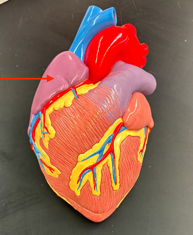
right ventricle
Forms most of the anterior/front surface of the heart.

right ventricle (deep)

left ventricle
Forms the apex and much of the left/inferior border.

left ventricle (deep)

right auricle
Small flap extending from the right atrium, visible on the front.

right atrium (deep)

left atrium (deep)

left auricle
Small flap extending from the left atrium, visible on the front.
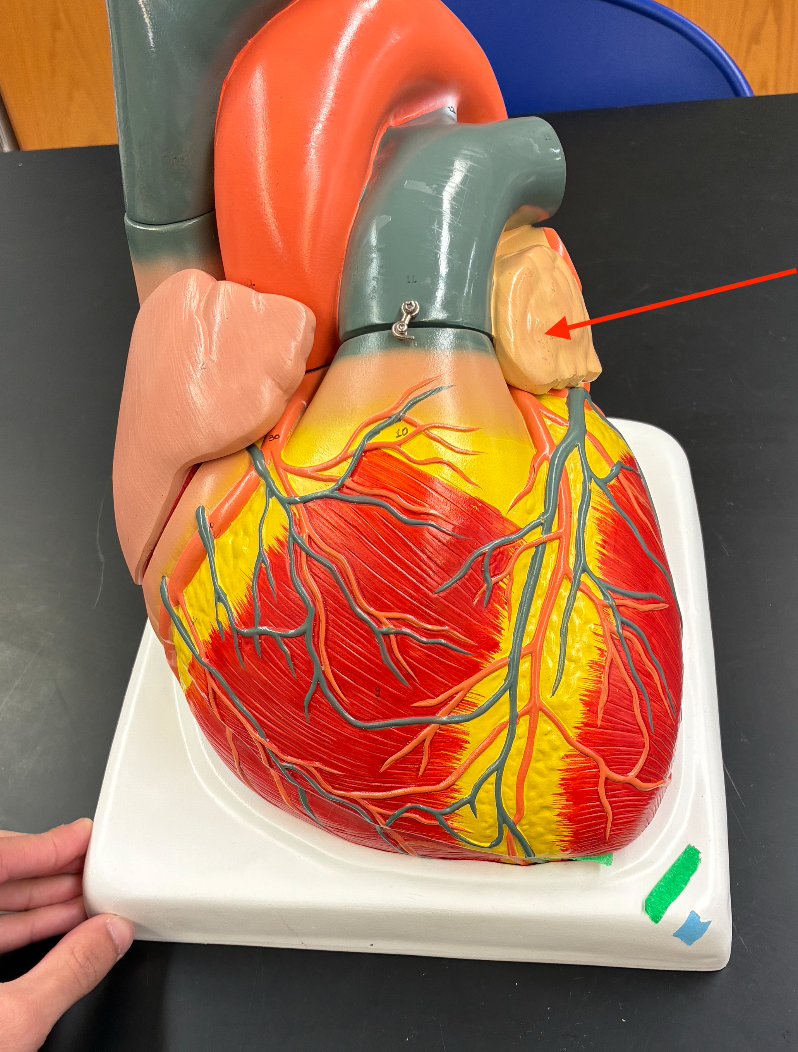
coronary sulcus
Groove that separates the atria from the ventricles.

Anterior interventricular sulcus
Groove on the front separating the right and left ventricles.
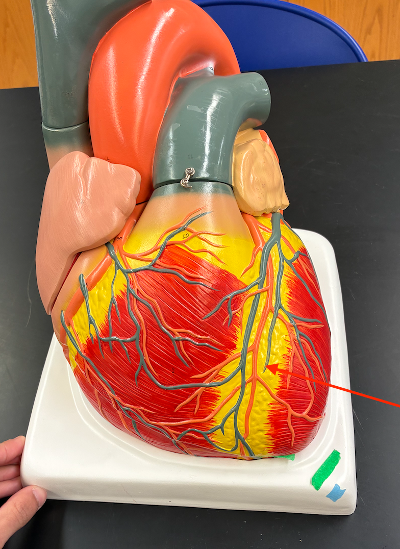
Posterior interventricular sulcus
Groove on the back separating the right and left ventricles.

interventricle septum
Internal wall between the right and left ventricles.
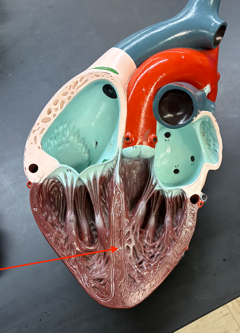
interatrial septum
Internal wall between the right and left atria.
Superior vena cava
Large vessel entering the top of the right atrium.

superior vena cava
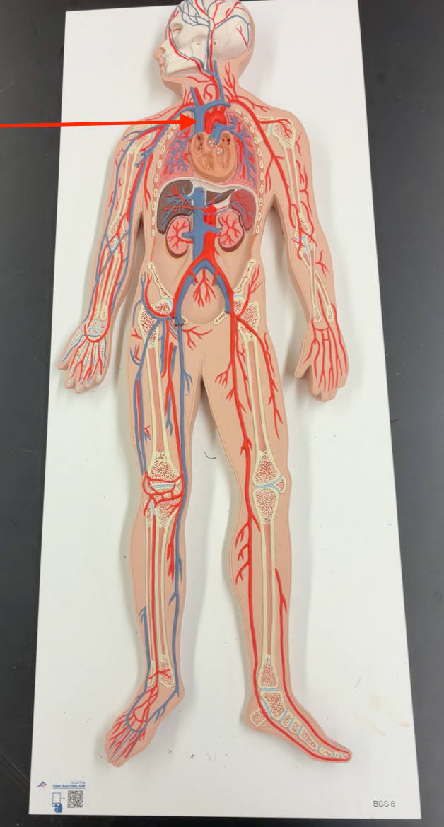
inferior vena cava
Large vessel entering the bottom of the right atrium.

Pulmonary trunk
Large vessel leaving the right ventricle, located toward the front/top of the heart.
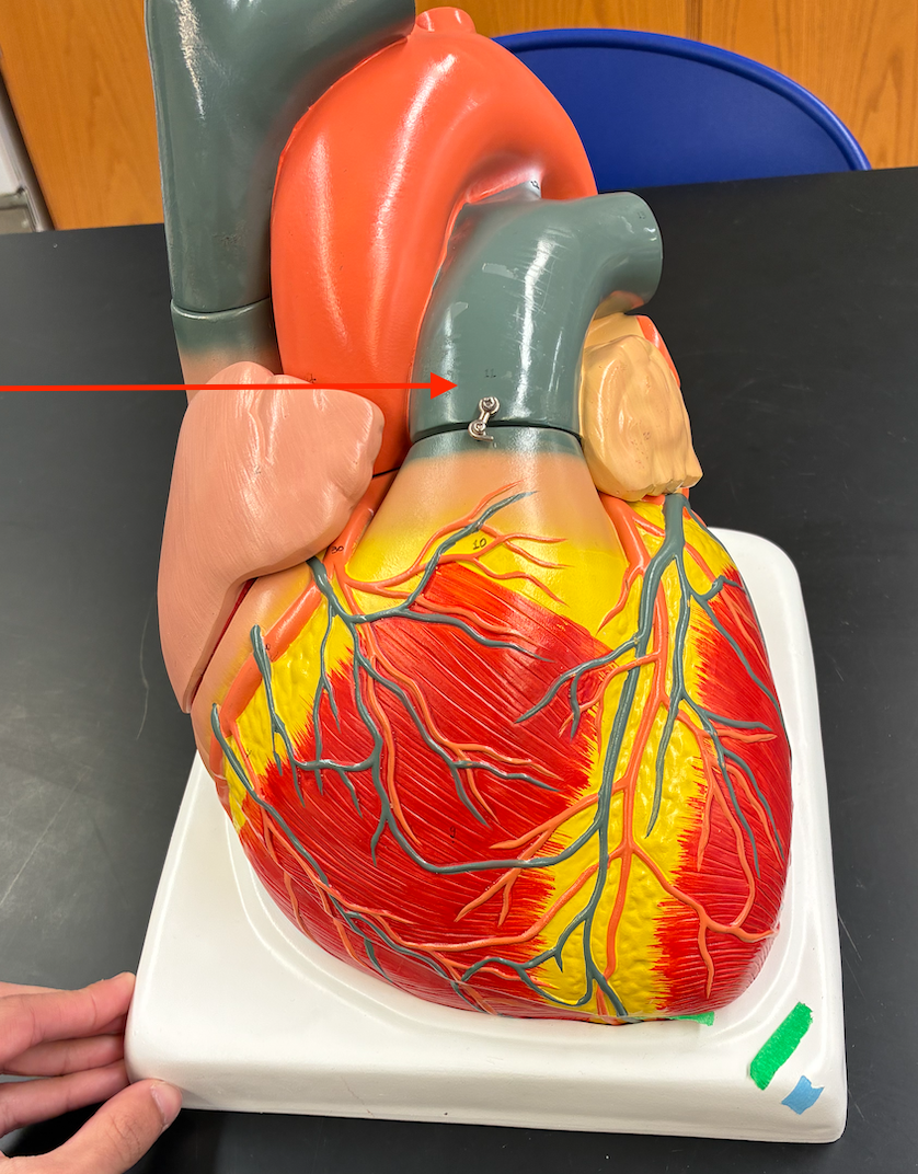
pulmonary trunk
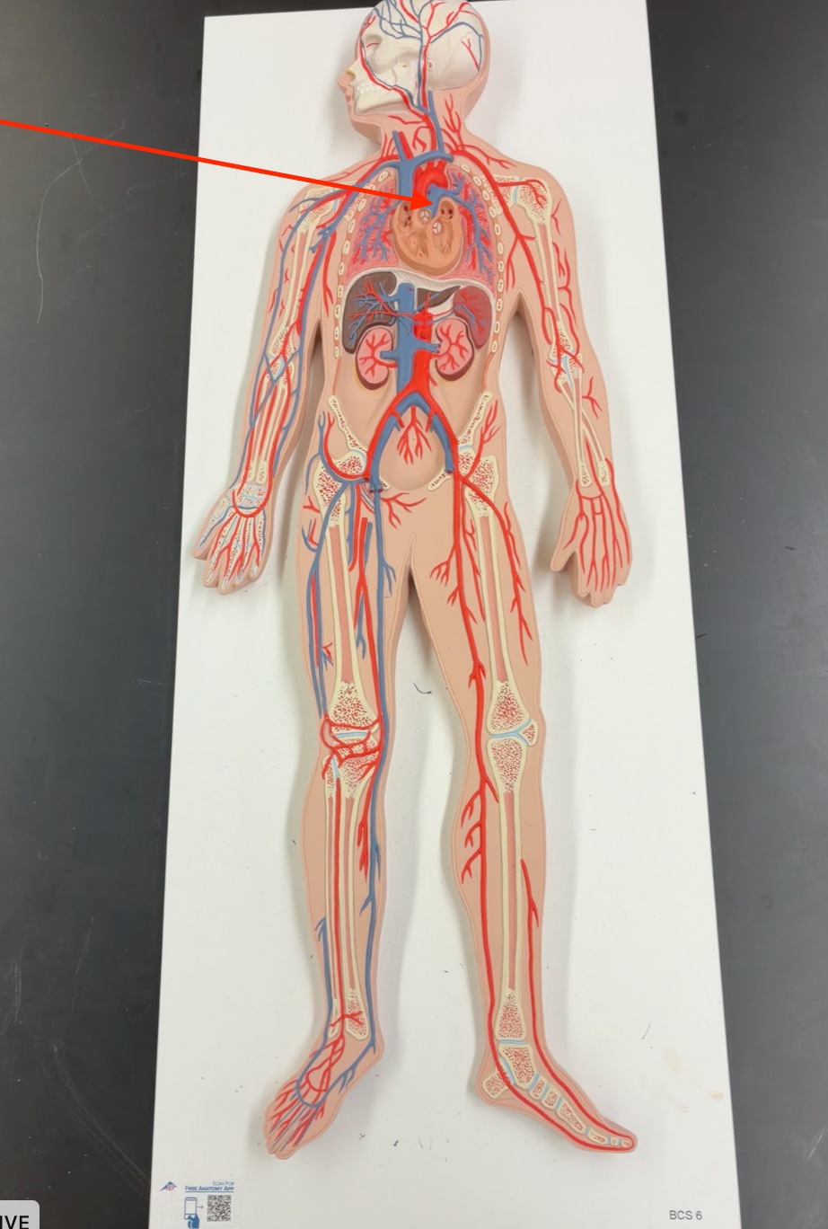
right pulmonary artery
Branches of the pulmonary trunk carrying blood from the heart toward the lungs

left pulmonary artery
Branches of the pulmonary trunk carrying blood from the heart toward the lungs
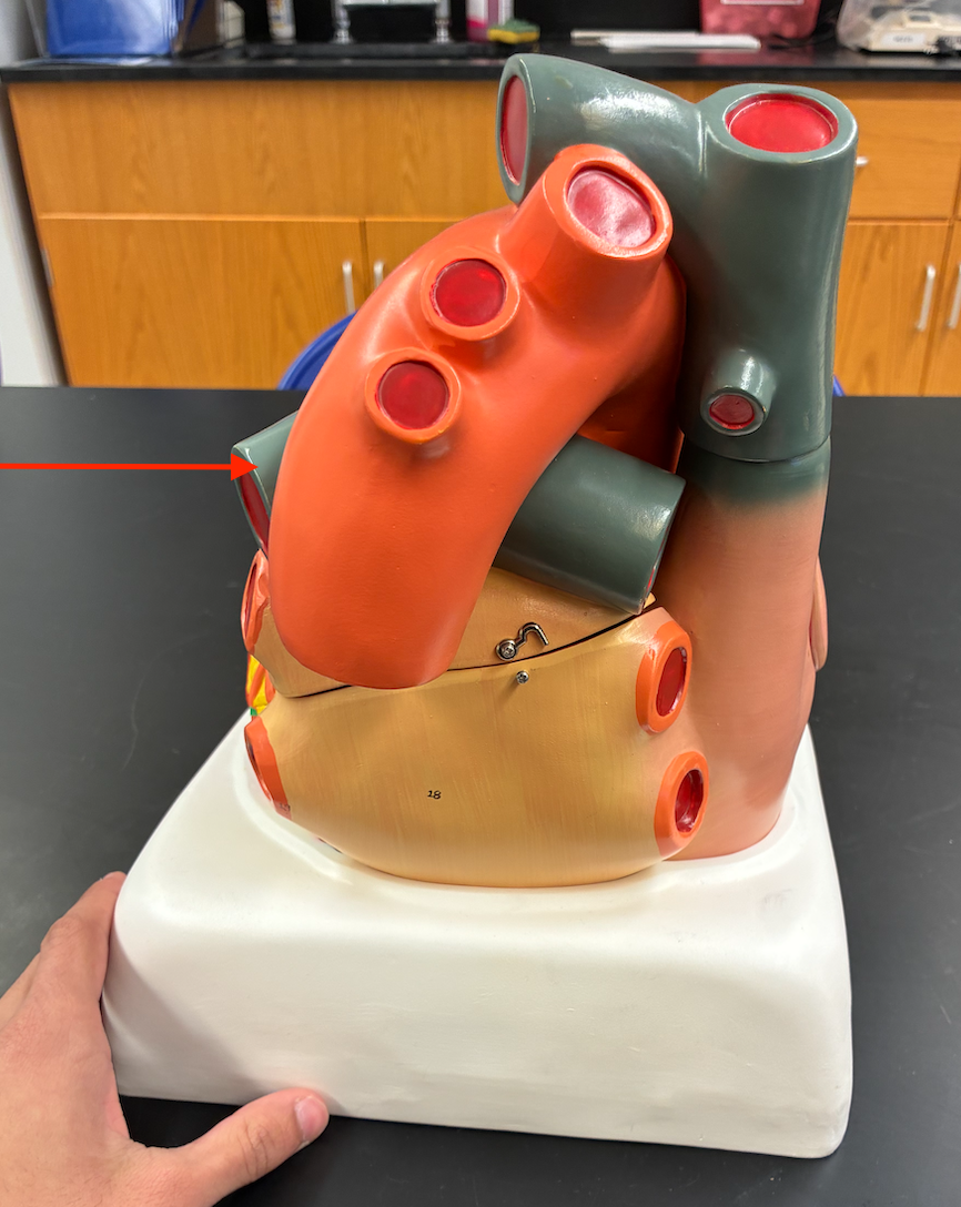
right pulmonary veins
Vessels entering the left atrium from the lungs.
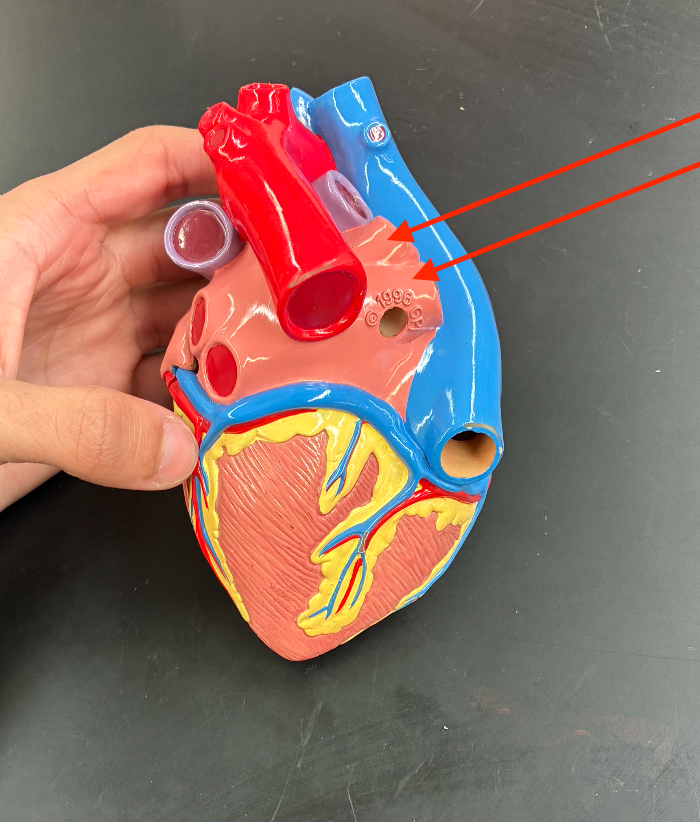
left pulmonary veins
Vessels entering the left atrium from the lungs.

aorta
Large artery leaving the left ventricle and arching upward

tricuspid valve (Right atrioventricular valve)

bicuspid, mitral valve (Left atrioventricular valve)

Pectinate muscles

papillary muscle

Aortic semilunar valve

pulmonary semilunar valve

right coronary artery

left coronary artery

Anterior interventricular branch (LAD)
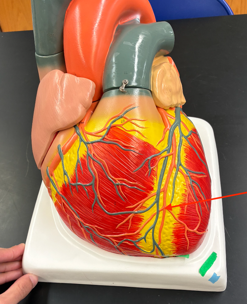
Circumflex branch

Posterior interventricular branch
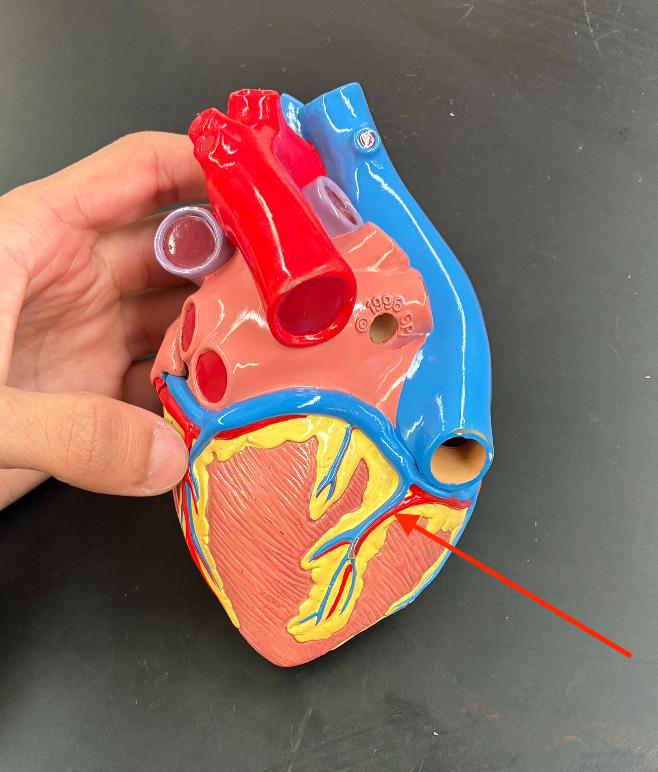
Marginal branch
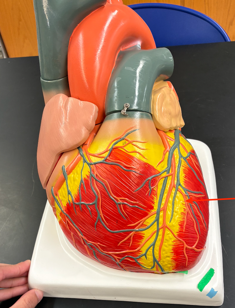
Great cardiac vein

Middle cardiac vein
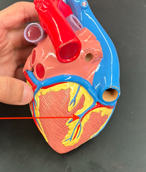
Coronary sinus

What is the complete pathway of blood through the heart?
SVC/IVC → Right atrium → Tricuspid valve → Right ventricle → Pulmonary semilunar valve → Pulmonary trunk → Pulmonary arteries → Lungs → Pulmonary veins → Left atrium → Mitral/bicuspid valve → Left ventricle → Aortic semilunar valve → Aorta → Body
blood-flow pathway (picture)

What are the four chambers of the heart in order of blood flow?
Right atrium → Right ventricle → Left atrium → Left ventricle
What valve is between the right atrium and right ventricle?
Tricuspid (right AV) valve
What valve is between the right ventricle and pulmonary trunk?
pulmonary semilunar valve
What valve is between the left atrium and left ventricle?
Mitral/bicuspid (left AV) valve
What valve is between the left ventricle and aorta?
Aortic semilunar valve
Where does deoxygenated blood enter the heart?.
Through the superior and inferior vena cava → right atrium
Where does deoxygenated blood go after the right ventricle?
Through the pulmonary semilunar valve → pulmonary trunk → pulmonary arteries → lungs.
Where does oxygenated blood from the lungs enter the heart?
Through the pulmonary veins → left atrium.
Where does oxygenated blood go after the left ventricle?
Through the aortic semilunar valve → aorta → body.
What are the two AV valves?
Tricuspid valve on the right; mitral/bicuspid valve on the left
What are the two semilunar valves?
Pulmonary semilunar valve and aortic semilunar valve.
What is the simplest way to remember the two sides?
Right side → lungs; Left side → body.
systole
The phase when the ventricles contract and eject blood.
diastole
The phase when the heart chambers relax and fill with blood.
What is the cardiac cycle?
One complete heartbeat, including systole and diastole.
What happens during ventricular systole?
The ventricles contract, AV valves close, and blood is ejected through the semilunar valves.
What happens during ventricular diastole?
The ventricles relax, semilunar valves close, and the ventricles fill with blood.
What causes the first heart sound (S1, “lub”)?
Closure of the AV valves (tricuspid and mitral).
What causes the second heart sound (S2, “dub”)
Closure of the semilunar valves (pulmonary and aortic).
Where do you auscultate (listen to with stethoscope) the aortic valve?
2nd intercostal space, right sternal border.
Where do you auscultate the pulmonary valve?
2nd intercostal space, left sternal border.
Where do you auscultate the tricuspid valve?
Left lower sternal border, around the 4th–5th intercostal space.
Where do you auscultate the mitral valve?
5th intercostal space, left midclavicular line (apex).
What is a pulse?
The pressure wave felt in an artery as the heart ejects blood.
What is pulse rate/heart rate?
The number of heartbeats (or arterial pulses) per minute.
What is pulse pressure?
Systolic pressure − diastolic pressure.
What instrument measures blood pressure?
A sphygmomanometer (blood pressure cuff), used with a stethoscope for auscultation.
What is systolic pressure?
The highest arterial pressure during ventricular contraction.
What is diastolic pressure?
The lowest arterial pressure during ventricular relaxation.
What are the sounds of Korotkoff?
Sounds heard through a stethoscope as the blood pressure cuff is deflated
What does the first Korotkoff sound indicate?
Systolic blood pressure.
What does the disappearance of Korotkoff sounds indicate?
Diastolic blood pressure.
What is mean arterial pressure (MAP)?
The average pressure in the arteries during one cardiac cycle.
What is the formula for MAP?
MAP = DBP + ⅓(SBP − DBP)
What is the approximate MAP for 120/80 mmHg?
93 mmHg → 80 + ⅓(40).
basilar artery

Internal carotid arteries

External carotid arteries

common carotid arteries

Vertebral arteries

Brachiocephalic trunk

Ascending aorta
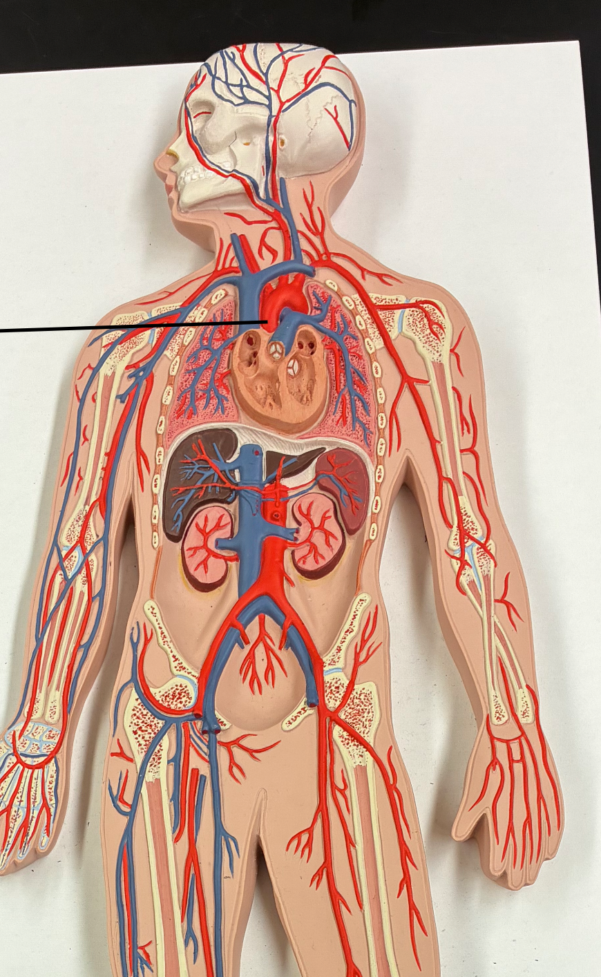
thoracic aorta

abdominal aorta

pulmonary trunk + arteries (are blue even tho artery)

Subclavian arteries

Axillary arteries
