Heart Anatomy | A&P II
 Call Kai
 Learn
Learn Practice Test
 Spaced Repetition
 Match
Match Flashcards
 Knowt Play
1/46

There's no tags or description
Looks like no tags are added yet.
Name | Mastery | Learn | Test | Matching | Spaced | Call with Kai | Chat |
|---|
No analytics yet
Send a link to your students to track their progress
47 Terms

____ receives deoxygenated blood from the body
Superior & inferior vena cava

From the superior & inferior vena cava, deoxygenated blood flows into the:
Right atrium
The ___ connects the right atrium to the right ventricle
Tricuspid valve
The ____ connects the right ventricle to the pulmonary trunk
Pulmonary valve
Deoxygenated blood flows from the pulmonary trunk to the ____
Pulmonary arteries
The pulmonary arteries flow to the ____
Lungs
Oxygenated blood flows from the lung tissue to the ____
Pulmonary veins
Oxygenated blood flows from the pulmonary veins to the:
Left atrium
The ____ valve connects the left atrium to the left ventricle
Bicuspid/mitral
The left ventricle is connected to the aorta by the:
Aortic valve
What are the 3 parts of the aorta, in order of bloodflow?
Ascending aorta, aortic arch, descending aorta
What are the semilunar valves?
Pulmonary and aortic
What are the atrioventricular (AV) valves?
Tricuspid and bicuspid/mitral
Blood flows from the aorta into either _____ or ____ circulation
Systemic; coronary
The _____ circuit starts at the right ventricle and ends in the left atrium
Pulmonary

The ____ circuit starts at the left ventricle and ends at the right atrium
Systemic


In a fetus’ heart, the _____ provides a bypass between the left and right atria.
Foramen ovale

This vestigial structure in the right atrium is the:
Fossa ovalis
The equivalent to the fossa ovalis in the left atrium is the:
Fossa lunata
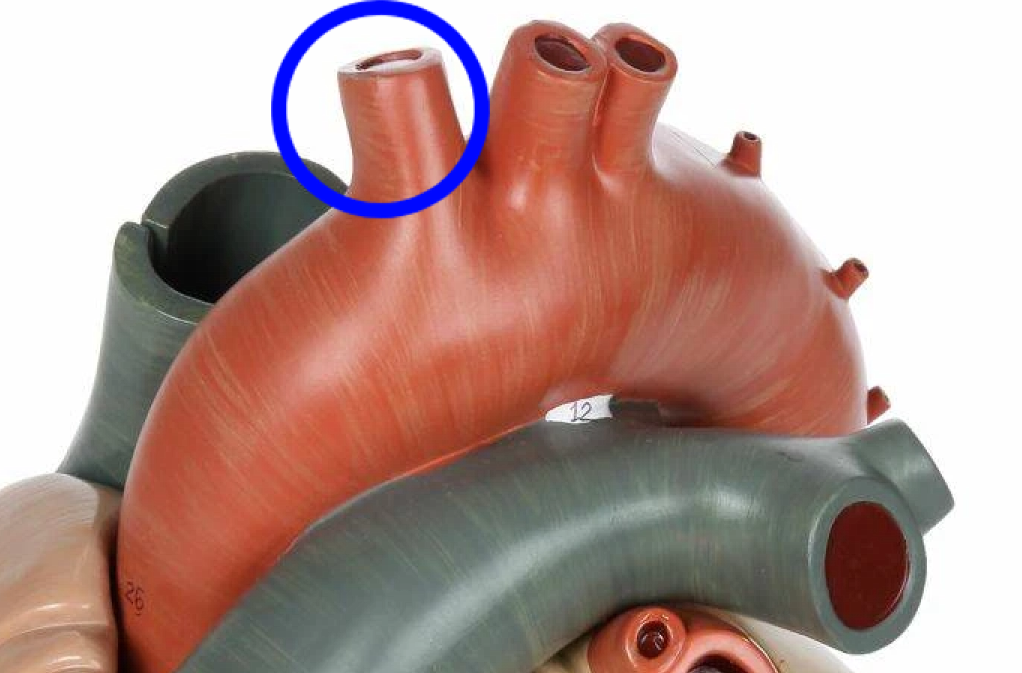
The (anatomically) rightmost branch of the aorta
Brachiocephalic artery

The middle branch of the aorta
Left common carotid artery

The (anatomically) leftmost branch of the aorta
Left subclavian artery

The right subclavian & right common carotid arteries branch off of the:
Brachiocephalic artery

The fibrous band connecting the pulmonary artery to the aorta is called:
Ligamentus arteriosus

In a fetus’ heart, the _____ provides a bypass between the pulmonary artery and the aorta.
Ductus arteriosum

This layer protects the heart and is composed of dense irregular connective tissue
Fibrous pericardium
The serous membrane of the heart is composed of the ____ (outer) and ___ (inner) layers
Parietal; visceral
The heart lies in the:
Mediastinum
The visceral layer of the serous pericardium (membrane) is also called the:
Epicardium
The muscular layer of the heart
Myocardium
The inner lining of the heart
Endocardium
The ____ is the serous fluid filled space between the parietal & visceral pericardium
Pericardial cavity
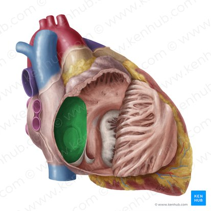
Separates the right & left atria
Interatrial septum

Separates the right & left ventricles
Interventricular septum

The groove separating the atria and ventricles
Coronary sulcus aka atrioventricular groove

This groove is the:
Anterior interventricular sulcus

This groove is the:
Posterior (or inferior) interventricular sulcus

Strong fibers that attach to the tricuspid & bicuspid valves to prevent backflow
Chordae tendineae

The _____ connect to the chordae tendineae to prevent backflow
Papillary muscles

Muscular ridges in the ventricles that reduce adhesion of blood to ventricular wall
Trabeculae carneae

Four fibrous bands around heart valves & bases of pulmonary trunk and aorta:
Cardiac skeleton

The natural pacemaker of the heart, located in the right atrium
Sinoatrial node
The cardiac muscle cells that control and coordinate heartbeat
Conducting system
The cardiac muscle cells that produce contractions
Contractile cells
These structures interconnect cardiac muscle cells
Intercalated discs
Intercalated discs are strongly anchored by
Desmosomes
____ allows from substances to pass between cardiac cells
Gap junctions