BMOL30090 L2 epithelial barriers & immune reservoirs
 Call Kai
 Learn
Learn Practice Test
 Spaced Repetition
 Match
Match Flashcards
 Knowt Play
1/26

There's no tags or description
Looks like no tags are added yet.
Name | Mastery | Learn | Test | Matching | Spaced | Call with Kai | Chat |
|---|
No analytics yet
Send a link to your students to track their progress
27 Terms
L2-6 learning objectives

First line of defence?
prevention (forming a barrier against entrance of pathogens)
body is in constant threat of infection by microbes in our env.
surfaces of contact: eyes, GI tract, lungs
gut epithelial cells
monolayer
absorbs O2 and monosaccharides

lung epithelial cells
pseudostratified (large airways)
absorption (O2)
gas exchange (release CO2)

skin epithelial cells
multi-stratified
low absorption (well-sealed)


Q: what type of epithelium is this?
lung
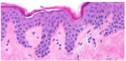
Q: what type of epithelium is this?
skin

Q: what type of epithelium is this?
gut
epithelial barriers: multiple layers of defense
some are:
physical (e.g. goblet cells)
biochemical (e.g. paneth cells)
help the IS (e.g. microfold cells)
goblet cells
example of physical function
intestine and lung
produce mucins (many mucins for mucus, mucus covers external surface of epithelium and traps microbes)

mucins
proteins packed into goblet cell granules
mucin can be memb-bound or gel forming
Muc2: most abundant gel-forming mucin in intestines
Muc5A & 5B: most abundant mucin in airway
mucin —> mucus
goblet cells secrete granules, releasing mucin monomers, which oligomerise to form the mucus
mucins expand to form mucus - process requires Ca2+ and increased pH
creates an extra layer of protection, reducing direct contact between microbes and epithelium
mucus in the skin?
NO mucus in the skin, instead there is a cornified envelope
infectious agents and mucus
infectious agents often use specialised mechanisms (e.g. flagella) to ‘swim’ through mucus and reach epithelium
Q: mucus is found in and produced by _____
goblet cells in the airway and intestine
paneth cells
biochemical functions
NOT epithelian stem cells (SCs), but help them by providing signals:
bactericidal mediators (e.g. lysozyme and defensins) to kill certain bac. types
SC-stimulating factors to promote proliferation and differentiation
located in intestinal crypts surrounding SCs

stem cell progenitors
migrate and differentiate into diff types of epithelium
self renewal and repair:
homeostasis - replace old cells
disease - heal wounded tissue, replace cell loss
microfold (M) cells
immune supportive functions
aid in exposing local IS to antigens & surveyal of potential harmful agents that could cause IR
in intestine and airway, in prox. to MALT
M cells do not present antigens, but capture & establish a controlled flux for recogn. by APCs

mucosal-associated lymphoid tissues (MALT)
multifolicular structures that can be found in diff. tissues
cells present in MALT typically:
antigen-presenting mononuclear phagocytes (DCs and Macs)
B and T lymphocytes
MALT and M cells process
M cells allow controlled pass of microbial components from exterior into MALT
APCs capture these & present them on surface
lymphocytes recognise these to learn about potential threats
Q: which of these is an example of a physical epithelial barrier?
goblet cells
Q: which of these is an example of an immune supportive epithelial barrier?
M cells
Q: which of these is an example of a biochemical epithelial barrier?
paneth cells
MALT location and structure
mucosa beneath epithelium is rich in scattered immune cells
structures rich in lymphocytes and APCs called MALT
MALT located in intestinal tract (Peyer’s patch), pharynx (tonsils), Bronchial-Assoc. Lymphoid Tissue (BALT)
epithelial localisation near MALT function
makes M cells antigen ‘shuttles’
M cells provide controlled flow of antigens to Malt to be recognised
summaries of epithelial cells with protective roles

barrier defects: cause or consequence of disease
certain infections, e.g.:
S. aureus exploits damaged skin barrier
S. typhi ‘swims’ through mucus, bypassing epithelium
chronic inflam diseases:
IBS: loss of epithelium
Psoriasis: excess./unorg. keratinocyte growth
asthma & eosinophilic esophagitis: hypertropic/unorg. epithelium