Anatomy 2300 Unit 4: Ventricles, CSF & Arteries of the Brain
 Call Kai
 Learn
 Practice Test
 Spaced Repetition
 Match
 Flashcards
 Knowt Play
1/40

There's no tags or description
Looks like no tags are added yet.
41 Terms
Cerebrospinal Fluid (CSF) (Function?)
A clear fluid filtered from blood that surrounds the brain and spinal cord, filling the ventricles, central canal, and subarachnoid space.
It is continuously produced by the choroid plexus, flows through the ventricles into the subarachnoid space, and is reabsorbed through arachnoid granulations into the dural venous sinuses.
Functions: Acts as a shock absorber, transports nutrients, removes waste, and maintains ion balance in neural tissue.

Subarachnoid Space
The space between the arachnoid mater and the pia mater, directly on the surface of the brain and spinal cord.
It is filled with CSF and holds the cerebral arteries and veins. CSF leaves the ventricles into this space and is drained from it.
(J)

Ventricles of the Brain (Name all?)
Spaces inside the brain that contain and direct the flow of CSF.
There are four: two lateral, the third, and the fourth. They are linked by small channels.

Choroid Plexus
A structure in all the ventricles that continually produces CSF by filtering blood. It is the source of all CSF. The slides mark it in the lateral, third, and fourth ventricles.

Lateral Ventricles (Drains into?)
Two C-shaped ventricles (right and left), one deep in each cerebral hemisphere. They are the largest and most superior ventricles, arching from the frontal lobe back toward the occipital lobe and curving down into the temporal lobe.
Each drains into the third ventricle through an interventricular foramen.

Interventricular Foramen (Connects?)
A small opening connecting each lateral ventricle to the third ventricle. There is one on each side. It is the first "doorway" in CSF flow.

Third Ventricle (Drains into?)
A single, narrow midline ventricle located below and between the lateral ventricles, in the center of the brain.
It receives CSF from both lateral ventricles and drains through the cerebral aqueduct.

Cerebral Aqueduct (Connects?)
A thin channel running through the midbrain that connects the third ventricle to the fourth.
Because it is so narrow, it is an easy place for CSF flow to get blocked.

Fourth Ventricle (Drains into?)
A single, diamond-shaped ventricle in front of the cerebellum and behind the pons and medulla. It receives CSF from the cerebral aqueduct.
From here, CSF goes two places: out to the subarachnoid space through the median and lateral apertures, or down into the central canal of the spinal cord.

Central Canal
A narrow, CSF-filled channel running down the center of the spinal cord.
It is continuous with the fourth ventricle at the top, and CSF can move in both directions between them. It is the spinal cord's version of the ventricles.

Median Aperture
A single opening in the midline of the fourth ventricle. It lets CSF escape into the subarachnoid space.

Lateral Apertures
Two openings, one on each side of the fourth ventricle. Together with the median aperture, they are the only exits from the ventricles into the subarachnoid space.

Arachnoid Granulations
Small projections of arachnoid mater that poke through the meningeal dura into the dural venous sinuses, mainly the superior sagittal sinus at the top of the brain.
They are the main site where CSF re-enters the venous blood (reabsorption).

List the overall CSF circulation and drainage.
Quick check: production = choroid plexus, reabsorption = arachnoid granulations into the venous system:
Lateral ventricles (choroid plexus makes CSF)
Interventricular foramen
Third ventricle
Cerebral aqueduct
Fourth ventricle
* Median and lateral apertures
Subarachnoid space (some CSF also goes down the central canal)
Arachnoid granulations
Dural venous sinus (into venous blood)

Hydrocephalus
Obstruction of CSF circulation, so fluid builds up and compresses the brain.
Production keeps going while drainage is blocked. The block can be in the ventricular pathway (for example, the narrow cerebral aqueduct) or at reabsorption.
In infants, whose skull bones have not fused, the head visibly enlarges, as in the slide photo.

Vessels of the Head and Neck (path?)
Pathway: Aortic arch → brachiocephalic trunk (right side only) / directly from aortic arch (left side) → common carotid aa. → internal and external carotid aa.
Subclavian aa. → vertebral aa. → basilar a. → posterior cerebral aa.

Ascending Aorta
The first part of the aorta, leaving the left ventricle and heading up. It gives off the coronary arteries and continues into the arch.

Arch of Aorta
The curved part of the aorta that turns backward over the heart. It gives off three branches in this order: brachiocephalic trunk, left common carotid, left subclavian.

Brachiocephalic Trunk (Splits into?)
The first branch of the aortic arch, found only on the right.
It splits into the right subclavian a. (supplies the right arm and gives rise to the vertebral a.) and right common carotid a. (supplies the head and neck).

Left and Right Subclavian Artery (gives rise to?)
Supply the upper limbs and give rise to the vertebral arteries, which contribute to the posterior circulation of the brain.
The right subclavian artery branches from the brachiocephalic trunk, while the left subclavian artery branches directly from the aortic arch.

Vertebral Artery (Travels through? Gives rise to?)
Branches from the subclavian a., travels through the cervical vertebrae's transverse foramina, and enters the skull through the foramen magnum.
The two vertebral aa. join to form the basilar a., which gives rise to the cerebellar aa. and terminates as the right and left posterior cerebral aa., supplying the brainstem, cerebellum, and posterior cerebrum.

Left and Right Common Carotid Artery (Splits into?)
Ascend the neck to supply the head and neck before dividing into the internal carotid artery, which supplies the brain, and the external carotid artery, which supplies the face and superficial structures of the head and neck.
The right common carotid artery branches from the brachiocephalic trunk, while the left common carotid artery branches directly from the aortic arch.

Internal carotid artery (Passes through? Supplies?)
One of two branches of each common carotid (the other is the external). It passes through the carotid canal and supplies the brain and eye (see above).

External Carotid Artery (Stays? Supplies?)
The other branch of the common carotid. It stays outside the skull and supplies the face, scalp, and neck through branches such as the superior thyroid, lingual, facial, occipital, maxillary, and superficial temporal arteries.
It does not supply the brain.

External Jugular Vein (Drains what? Empties into?)
A superficial neck vein running down over the surface of the neck muscles. It drains the scalp and face and empties into the subclavian vein.

Internal Jugular Vein (drains into? Drains via?)
The major deep vein of the neck, running alongside the common/internal carotid artery.
It drains the brain (via the dural venous sinuses, where CSF is also reabsorbed), plus the face and neck. It joins the subclavian vein to form the brachiocephalic vein.

Subclavian Vein (drains into?)
The large vein draining the upper limb, running behind the clavicle. It joins the internal jugular vein to form the brachiocephalic vein.

Brachiocephalic Vein (drains into?)
Right and left veins, each formed by the union of an internal jugular and a subclavian vein. The two join to form the superior vena cava.

Superior Vena Cava (drains into?)
A large vein formed by the union of the two brachiocephalic veins. It returns blood from the head, neck, and upper limbs to the right atrium of the heart.

Arteries of the Brain (path?)
vertebral aa. → basilar a. → posterior cerebral aa. (PCA).
Internal carotid aa. → anterior cerebral aa. (ACA) and middle cerebral aa. (MCA)
The anterior and posterior circulations connect through the Circle of Willis.
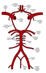
Vertebral Artery (Branches off? Travels through? Gives rise to? Later Supplying?)
Branches from the subclavian a., travels through the cervical vertebrae's transverse foramina, and enters the skull through the foramen magnum.
The two vertebral aa. join to form the basilar a.,, later supplying the brainstem, cerebellum, and posterior cerebrum.

Basilar Artery (Formed by? Runs along? Supplies? Gives rise to?)
A midline artery formed by the union of the two vertebral aa. It runs along the anterior pons and supplies the brainstem and cerebellum.
Gives rise to the cerebellar aa. and small pontine branches (non-terminal), then terminates as the right and left posterior cerebral aa. (terminal)

Cerebellar Arteries (Branches off? Supplies?)
Three paired arteries that branch off the vertebral aa. and supply the cerebellum (do not need to know the branches)

Right & Left Posterior Cerebral Artery (PCA) (Branches off? Supplies? Connect to?)
Terminal branches of the basilar a.
They curve around the midbrain to supply the occipital lobes and inferior temporal lobes. They connect to the internal carotid circulation through the posterior communicating aa.

Posterior Communicating Artery (Links?)
A branch of the internal carotid that runs backward to join the posterior cerebral artery. There is one on each side.
It links the anterior (carotid) circulation to the posterior (vertebral/basilar) circulation and forms the side walls of the circle of Willis.

Internal carotid artery (In brain) (Branches from? Gives rise to? Connects to?)
Branches from the common carotid a. and enters the skull to supply the brain.
Gives rise to the anterior cerebral a. (ACA) and middle cerebral a. (MCA) and connects to the posterior circulation through the posterior communicating a.

Middle Cerebral Artery (MCA) (Branches off? Travels and Supplies?)
The largest terminal branch of the internal carotid a.
It travels through the lateral sulcus and supplies the lateral surfaces of the frontal, parietal, and temporal lobes, covering most of the cerebrum's outer surface.

Anterior Cerebral Artery (ACA) (Runs along? Branches off? Supplies?)
A terminal branch of the internal carotid a. that runs along the midline between the cerebral hemispheres, supplying the anterior and medial surfaces of the cerebrum.
The two anterior cerebral aa. connect through the anterior communicating a.
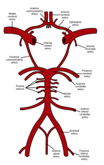
Anterior Communicating Artery (connects?)
A short, single artery in the midline that connects the right and left anterior cerebral arteries. It forms the front of the circle of Willis and allows blood to cross from one side to the other.

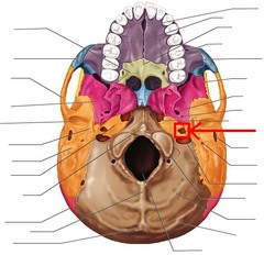
Carotid Canal (Enters via?)
A bony tunnel in the temporal bone at the base of the skull. It is the entry point of the internal carotid artery into the cranium, seen from below as a small opening to the side of the foramen magnum.
Circle of Willis (Components? Clinical Importance?)
A ring of arteries at the base of the brain that connects the internal carotid and vertebrobasilar circulations. It consists of the:
anterior communicating a.,
two anterior cerebral aa.,
two internal carotid aa.,
two posterior communicating aa., two posterior cerebral aa.
The middle cerebral aa. and basilar a. connect to the circle but are not part of it.
Function: Provides collateral circulation, allowing blood to reroute if an artery is blocked. It is also a common site for aneurysms.
