Lesson 60: Disorders of the Tonsils, Pharynx and Esophagus
 Call Kai
 Learn
 Practice Test
 Spaced Repetition
 Match
 Flashcards
 Knowt Play
1/37

There's no tags or description
Looks like no tags are added yet.
38 Terms
Tonsils: hyperplasia
diffuse "tonsillar" hyperplasia common in foals
exposure to the environment and bacteria & viruses → chronic irritation reaction
Disorders of the tonsils: Lymphoid necrosis
feline panleukopenia
canine parvoviral infection
canine distemper
BVD
Tonsillitis
red, swollen
causes: systemic disease (e.g., ICH), local infection
Tonsils: neoplastic dieseases
Lymphosarcoma (L)
Squamous cell carcinoma (R)

Clinical signs of pharynx disorders
Signs of respiratory distress
Stridor, coughing, dyspnea, Gagging
Exacerbated by excitement, heat, stress, or exercise
Pharyngitis causes
trauma, bacterial & viral agents (as for nasal cavity), neoplasms, cysts, lymphoid hyperplasia
Lesions associated with pharyngitis
serous, catarrhal, suppurative, fibrinous, fibrinonecrotic, hemorrhagic or granulomatous inflammation
Pharyngeal Obstruction and Perforation
Intraluminal foreign bodies: medicament boluses, apples, or potatoes
can move down and obstruct the larynx and trachea
Sites of lodgement of FBs
Anywhere in the pharyngeal region
location and size determine the occurrence of dysphagia, regurgitation, dyspnea, or asphyxiation
Pharyngeal obstruction: Masses in the surrounding tissue
Neoplasms
thyroid gland, thymus, parathyroid glands
Pharyngeal Obstruction and Perforation: Nonspecific insults can cause lesions
perforation of the caudodorsal wall of the pharynx from the improper use of drenching or balling guns in sheep, cattle, and pigs
choking injury because of the use of collars in dogs and cats
shearing forces of bite wounds
Pharyngeal obstruction and perforation: Sequelae of trauma
Minimal: local edema and inflammation
Serious: complete luminal obstruction by exudate
Pharyngeal diverticulum in pigs: in the pharyngeal wall rostral and dorsal to the
esophageal entrance
barley awns may lodge in the diverticulum, inflammatory swelling that affects swallowing
The diverticular wall may be perforated by awns or drenching syringes,
exudate can extend down: the tissue planes between muscles of the neck, even into the mediastinum
Pharynx of the dog: trauma
trauma from chicken bones, sticks, and needles → pharyngeal abscessation
Dysfunction/Responses to Injury: esophagus
healing is relatively rapid → Normal epithelial turnover rate is 5 to 8 days
breach of the epithelial barrier resulting in ulceration (because of acid reflux or mechanical damage) → Can lead to circumferential scarring → Permanent narrowing of the lumen → predisposition to future episodes of obstruction
Esophagus: Portals of Entry/Pathways of Spread
Materials, caustic chemicals, some parasites, gastric reflux
thoracic cavity: penetration or obstruction by foreign objects
Iatrogenic puncture of the esophagus
Esophagus defense mechanisms
Stratified squamous epithelium: Keratinized in pigs, horses, and ruminants, Nonkeratinized in dogs and cats
Healing is relatively rapid
Muscularis: Striated in ruminants and dogs, Smooth in horses
Submucosal mucus glands: Present throughout the esophagus (pigs, dogs) or pharyngeal junction (cats, horses, ruminants)
Megaesophagus (esophageal ectasia)
dilation of the esophagus: insufficient, absent, or uncoordinated peristalsis in the mid- and cervical esophagus
Dogs, cats, cows, ferrets, horses, and New World camelids
Causes of megaesophagus
Innervation or denervation disorders
Partial physical obstructions and stenosis 2º secondary to: inflammatory diseases of esophageal musculature, persistence of the right aortic arch (PRRA)
Idiopathic (in aged camelids)
Congenital megaesophagus
Partial blockage of the lumen of the esophagus by a persistent right fourth aortic arch (PRAA) → vascular ring forms around the esophagus and trachea, preventing full dilation of the esophagus
Aorta, pulmonary artery, and ductus arteriosus form the ring
Image of congenital megaesophagus

Acquired megaesophagus (aka. esophageal achalasia)
failure of relaxation of the distal esophageal (cardiac) sphincter of the stomach obstruction and thus dilation occurs cranial to the stomach
But does not involve the cardiac sphincter
Causes of acquired megaesophagus
Idiopathic
Secondary to: Polymyositis, Esophagitis, Hypothyroidism
Myasthenia gravis (disorder of acetylcholine receptors of the neuromuscular junction)
Recurrent gastric dilation. Increased risk in dogs is seen in German shepherds, Golden retrievers, and Irish setters.
Clinical signs of megaesophagus
Regurgitation after ingestion of solid food → congenital megaesophagus is often recognized at weaning
Animals are thin, ± Aspiration pneumonia.
Radiographically: Esophagus is dilated anterior to the lesion and retains radiopaque dyes
Esophagus: neoplasia
Generally rare
Bracken fern (Pteridium aquilinum) consumption ± papilloma virus infection
Squamous cell carcinomas in cattle
Signs of neoplasia or tumors in the esophagus
Dysphagia, regurgitation, weight loss, dilation of the esophagus proximal to the mass
Tumors of the esophagus are occasionally palpable but are most often intraluminal rather than mural
Epithelial - Papillomas, and Squamous cell carcinomas → wide metastatic potential
Fibrosarcomas of dogs: often develop associated with Spirocerca lupi infestation
Leiomyoma, Lymphosarcoma
where does Choke (Esophageal obstruction) occur
mostly in anatomic locations in which the esophagus cannot fully expand
Dorsal to the larynx, cranial to the first rib at the thoracic inlet, base of the heart, diaphragmatic hiatus
If foreign bodies are lodged against the epithelium for longer than 2 days it leads to?
circumferential pressure necrosis of the esophageal mucosa strictures during healing
reflex regurgitation after ingestion of food → starvation or aspiration pneumonia
Choke initial signs
Hypersalivation (ptyalism), Coughing, Repeated attempts to swallow, Nasal discharge of feed material and saliva (This is a key indicator)
Choke progression of signs
Gagging retching, and Regurgitation, Dysphagia, Decreased Appetite and Weight Loss, Lethargy, Anxiety and Restlessness, Pain with Movement
Other possible signs of choke
Abdominal Distension, Bloat, Smacking/Licking Lips, Pawing at the Mouth or Face, Pacing/Restlessness.
Esophagus: erosion and ulcers causes
Reflux of stomach acid
Improper use of stomach tubes: linear scraping on the crests of the longitudinal folds of the esophageal mucosa
Foreign bodies, Infectious diseases, mucosal injury in other locations as well
Esophageal parasites: Spirocerca lupi of dogs pathogenesis
most pathogenic of the esophageal parasites
penetrate through the gastric mucosa → adventitia of arteries → abdominal aorta → caudal thoracic aorta → granuloma in the adventitia
passage forms between the esophageal lumen and the granuloma containing the parasite
Spirocerca lupi clinical signs
dysphagia, aortic aneurysms, hemothorax, and rarely esophageal fibrosarcomas or osteosarcomas
Chronic aortic granulomas: spondylosis deformans adjacent to the aortic granulomas
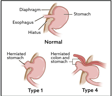
Esophagus: Hiatal hernia
Protrusion of the abdominal esophagus and cardia of the stomach through the diaphragm into the thoracic cavity
Inversion is generally into the esophageal lumen
can lead to gastroesophageal intussusception (sometimes)
Images of hiatal hernia